Ultrasound
The ultrasound (from the Greek «ἠχώ» [ēkhō] 'echo', and «γραφία» [graphy] 'write'), also called ultrasonography or echosonography, is a diagnostic procedure used in hospitals and clinics that uses ultrasound to create two- or three-dimensional images. A small instrument very similar to a "microphone" called a transducer emits ultrasound waves. These high-frequency sound waves are transmitted to the area of the body under study, and their echo is received. The transducer is responsible for sending small pulses of high frequency acoustic waves, inaudible to the human ear, which go inside the body. These will bounce off organs, tissues, or fluids, and the device will record minute changes in sound. A computer converts this echo into an image that appears on the screen. This process occurs thanks to the so-called piezoelectric effect.
Ultrasound is a simple procedure, despite the fact that it is usually performed in the radiodiagnosis service; and because of its simplicity, it is often used to visualize developing fetuses as well as musculoskeletal ultrasound in addition to many other uses. It is a non-invasive, low-cost and risk-free test, unlike other diagnostic procedures or imaging tests such as radiography, in which nuclear radiation is used. When undergoing an ultrasound exam, the patient simply lies on a table and the doctor moves the transducer over the skin over the part of the body to be examined. Before it is necessary to place a gel on the skin for the correct transmission of the ultrasounds. However, one drawback is that the ultrasound is an operator-dependent imaging method and this requires a long learning period in order to obtain and correctly interpret the images. It has the advantage that the ultrasound equipment is mobile, so it can be taken to the patient's bed if he or she is immobile.
Ultrasound could be divided into two groups, with contrast or without contrast, normally most ultrasounds are with contrast, this consists of stabilized gas microbubbles that present the resonance phenomenon thus increasing the signal received by the transducer. This contrast method is capable of differentiating between normal and diseased tissues, those diseased areas will look brighter when doing the exam, but above all the experience of the doctor doing the exam is essential to be able to interpret the images correctly. For example, if there is a tumor or cancer, as already mentioned, it will be seen on the monitor brighter due to the increase in blood flow.
For most ultrasound exams, the patient will be placed face up on a stretcher, it will also be possible to move the patient on their side or face down, but in principle it will depend on each type of exam to be performed. When performing the test, a water-based gel must be placed that will help the transducer make safe contact with the patient's body, therefore this process is based on breaking the air molecules that can form and prevent the passage of particles. sound waves towards the tissue, organ, etc.
If necessary before starting the exam, depending on the area that you would like to see, an injection will be made that will be applied with an intravenous catheter with the contrast material, since the area to be studied is probably difficult to visualize through the monitor, it will be applied with an intravenous catheter.
History
Ultrasounds were discovered by Lazzaro Spallanzani, while he was developing his work as a biologist in 1794 studying bats.
In 1880 in Paris Pierre Curie and his brother Jacques discovered the piezoelectric effect.
In 1881 Gabriel Lippman discovered the reciprocity of the piezoelectric effect which allowed the possibility of the reception and emission of ultrasound.
In 1914 the first sonar was built.
In 1935 the first radar system was invented by physicist Robert Watson-Wat.
In 1940, American acoustic physicist Floyd Firestone created the first ultrasound imager using echo, he called it the “Supersonic Reflectoscope”. In this same year, ultrasonic energy was applied to the human body for the first time solely for medical purposes, in Maryland, United States.
In 1941, in Austria, psychiatrist Carl Theodore Dussik attempted to identify the cerebral ventricles by measuring ultrasound attenuation through the skull.
In 1947, Dr. Douglas Howry detected soft tissue structures by examining the reflections produced by ultrasound at different interfaces.
In 1949, a pulsed echo technique was published to detect intracorporeal stones and foreign bodies. Also the physicist John Wild used ultrasound for the first time to see the width of the intestine.
In 1951, compound ultrasound made its appearance, in which a mobile transducer produced several shots of ultrasonic beams from different positions and towards a fixed area. The emitted echoes were recorded and integrated into a single image. Water immersion techniques were used with all manner of vessels: a laundry tub, a cattle trough, and a machine gun turret from a B-29 aircraft.
In 1952, Douglas Howry, Dorothy Howry, Roderick Bliss, and Gerald Posakony published live two-dimensional images of the forearm.
In 1952, John Julian Wild and John Reid published two-dimensional images of breast carcinoma, a muscle tumor, and a normal kidney. They later studied the walls of the sigmoid by means of a transducer placed through a rectosigmoidoscope and also suggested evaluation of gastric carcinoma by means of a transducer placed in the gastric cavity.
In 1953, Lars Leksell, using a Siemens reflectoscope, detected the echo shift of the midline of the skull in a 16-month-old boy. Surgery confirmed that this displacement was caused by a tumor. The work was published only until 1956. Since then the use of echoencephalography with M-MODE began.
In 1954, Ian Donald did research with a crack detector, in gynecological applications.
In 1956, John Julian Wild and Reid published 77 cases of breast abnormalities that were palpable and also studied by ultrasound, and obtained a 90 percent certainty in the differentiation between cystic and solid lesions.
In 1956, Ian Donald introduced A-mode scanning.
In 1957, engineer Tom Brown and Dr. Ian Donald built a two-dimensional contact scanner, thus avoiding the immersion technique. They took pictures on Polaroid film and published the study in 1958.
IN 1957, Ian Donald began obstetric studies using echoes from the fetal skull. At that time the calipers (electronic sliders) were developed.
In 1958 the first paper on musculoskeletal ultrasound was published in the American Journal of Physical medicine entitled measurements of articular tissue with ultrasound, its author was K.T. Dussik.
In 1959, Shigeo Satomura reported the use, for the first time, of ultrasonic Doppler in evaluating the flow of peripheral arteries.
In 1960, Ian Donald developed the first automatic scanner, which proved impractical because of its cost.
In 1960, Howry introduced the use of the Mechanical Sector Transducer (hand held scanner).
In 1961, the resulting work of David Robinson and George Kossoff led to the creation of the first commercial practical ultrasound in Australia.
In 1962, Homes produced a scanner that oscillated 5 times per second over a patient's skin, allowing a rudimentary image in real time.
In 1963, a group of Japanese urologists reported ultrasonic examinations of the prostate, in the A-MODE.
In 1964, the Doppler technique appeared to study the carotids, with great application in Neurology.
In 1965, the Austrian firm Kretztechnik associated with the ophthalmologist Dr Werner Buschmann, manufactured a transducer with 10 elements arranged in phase, to examine the eye, its arteries, etc.
In 1966, Kichuchi introduced 'synchronized ultrasonocardiotomography', used to obtain studies in 9 different phases of the cardiac cycle, using a rotary transducer and a water pillow.
In 1967, the development of A-MODE transducers to detect the embryonic heart began, feasible at that time 32 days after fertilization.
In 1968, Sommer reported the development of an electronic scanner with 21 1.2 MHz crystals, which produced 30 images per second and was actually the first device to reproduce images in real time, with acceptable resolution.
In 1969, the first two-dimensional transvaginal transducers, which rotated 360 degrees, were developed and used by Kratochwil to assess cephalopelvic disproportion. The use of transrectal probes also began.
In 1970 Alfred Kratochwill began using transrectal ultrasound to assess the prostate.
In 1977 Alfred Kratochwil combined ultrasound and laparoscopy, introducing a 4.0 MHz transducer through the laparoscope, in order to measure the follicles using the A-MODE. The technique was extended to examine the gallbladder, liver, and pancreas.
In 1982 Aloka announced the development of color Doppler in two-dimensional imaging.
In 1983, Lutz used the combination of gastroscope and ultrasound to detect gastric CA and to examine the liver and pancreas.
In 1983, Aloka introduced to the market the first Color Doppler Equipment that allowed visualizing blood flow in real time and in color.
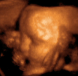
Although three-dimensional images are already obtained, the use of such technology has been wasted as it has been limited to purely "aesthetic" to encourage mothers to see their children in a third dimension, but not to improve diagnosis.
In 2017, Jan Tesarik introduced “virtual ultrasonographic hysteroscopy” to detect, in 3 dimensions, anomalies of the uterine cavity without entering the womb, just as precise and less invasive than conventional hysteroscopy and later applied the same technique to the study of the fallopian tubes (virtual hysterosalpingoscopy), the ovarian follicle cavity (virtual folliculoscopy) and gestational sacs (virtual embryoscopy).
Basic operating principles
Sound is a mechanical wave that requires a medium to propagate. The human ear can perceive it with a frequency between 20 and 20,000 Hz and ultrasound is any sound that exceeds this number. The ultrasound machine works through a device that generates ultrasound, taking advantage of the physical phenomenon called the piezoelectro effect, which consists in the fact that when some materials are compressed, they can generate a difference in electrical potential on their surface and therefore an electrical current. This effect also happens in reverse, so that when electricity is applied to them in the form of alternating current, they generate vibrations that produce ultrasound. In the ultrasound machine, the piezoelectric material is found in the head, which performs both the functions of generating ultrasonic waves and receiving them by bouncing them off tissues that have different acoustic impedances, and then converting them back into electric current, which the device transforms. in pictures. Acoustic impedance is the resistance offered by the tissue to the passage of sound, which as it progresses suffers a loss of energy due to the following three phenomena:
- Absorption: It is one of the main mechanisms that cause the attenuation of the sonic wave. A part of the energy becomes heat. This property is given minimally in the water and very high in the bone.
- Dispersion: It occurs when the sound against certain obstacles instead of developing a propagation in a normal linear direction is dispersed.
- Refrection:When a sonic wave passes from one medium to another, it experiences a change of speed accompanied by a variation in the direction of the same, so this effect causes a change in the angular direction of the sound regarding the incidence focus.
- Reflection: When passing the sound from one area to another with different acoustic impedance two new waves are produced, one of which rebounds towards the source of origin giving rise to the effect called reflection. The greater the difference of impedances will be this.
All these phenomena make it possible to generate the ultrasound image by bouncing the sound off the tissues and being received by the receptor crystals, which transform them into electrical current to be sent to the CPU that processes them.
Ultrasound machines used in clinical practice usually have frequencies ranging from 3 to 18 MHz.
Benefits and risks
The ultrasound exam can cause in some cases and depends on the exam, discomfort, but rarely it will cause pain.
- It is the cheapest imaging method of all and is easy to use.
- It does not use any type of radiation so it is a very safe method.
- This technique is able to differentiate certain organs that normally with other methods not.
- It is an ideal technique for those patients with claustrophobia and therefore gives the opportunity to those people to generate images without the need to use magnetic resonance or CT scan.
As for risks, today there is no possible risk generated by an ultrasound examination, but this does not mean that it will not exist in the long term, therefore a series of useful recommendations are established to avoid this, such as For example, control the acquisition time, the frequency and intensity used, use ultrasound only when necessary, use a probe less frequently, etc. Although it has been detected that contrast materials could, in a few cases, have a small percentage of risk of generating an allergic reaction on the patient.
By systems
Ocular and orbital ultrasound
An ocular and orbital ultrasound is an exam to look at the eye area specifically to see the size and structure of the eye.
The eye is numbed with specific anesthetic drops. The ultrasound transducer is placed against the front surface of the eye.
In this test, sound waves travel from the surface of the eye to the computer where it processes the data and generates the image.
Abdominal ultrasound
An abdominal ultrasound is performed to visualize the internal organs of the abdomen such as the liver, gallbladder, pancreas, kidneys, and spleen. It can also be used to examine the blood vessels that go to these organs, such as the inferior vena cava and aorta. It can be used to diagnose tumors and many other diseases.
Vaginal ultrasound
A vaginal ultrasound is used to examine a woman's uterus, ovaries, fallopian tubes, cervix, and pelvic area. It is used to assess the position, size, or presence of fibroids or polyps in the uterus. The endometrium can be studied, knowing the phase of the menstrual cycle. Likewise, it can detect possible ovarian cysts, ectopic pregnancies or perform a follicular count. The test uses an ultrasound probe that is placed inside the patient's vagina. This test can be done annually as part of the normal gynecological check-up. It is also used when tumors, infertility, ectopic pregnancy are suspected, when the patient presents abnormal bleeding and during pregnancy.
Breast ultrasound
The ultrasound of the breast is used to differentiate lumps or tumors that may be palpable or appear on mammography. Its main objective is to detect whether the tumor is solid or liquid to determine its benignity. Mammary ultrasounds are recommended when the breasts are dense or it is necessary to differentiate the benignity of the tumor. The BI-RADS system establishes three types of breast density:
1.- Fat breast.
2. -Medium density.
3.- Heterogeneous density.
4.- Very dense breast.
In fatty breasts, tumors are easy to detect in mammograms, but in dense (3-4) (fibrous) breasts, additional tests are needed. The density of the breast varies with age, generally, the older the breast is, the fatter it is.
Ultrasound scan of the thyroid gland
By means of this diagnostic method it is possible to detect different health problems that affect the thyroid gland, among which cysts can be mentioned, as well as nodules, defining the possibility that one of these may be benign or malignant by means of the characterization through the TI-RADS system. Another application is the detection and monitoring of diseases that diffusely affect the thyroid parenchyma, such as multinodular goiter and thyroiditis. For fine needle biopsy (FNAB), ultrasound/ultrasound is used as a guide to accurately perform the procedure.
Transrectal Ultrasound
The medical ultrasound for the diagnosis of prostate cancer consists of the introduction of a probe through the rectum that emits ultrasound waves that produce echoes when hitting the prostate. These echoes are picked up again by the probe and processed by a computer to reproduce the image of the prostate on a video screen. The patient may feel some pressure with this test when the probe is inserted into the rectum. This procedure takes only a few minutes and is done on an outpatient basis. Transrectal ultrasound is the most widely used method for performing a biopsy. Prostate tumors and normal prostate tissue often reflect different sound waves, so transrectal ultrasound is used to guide the biopsy needle into the exact area of the prostate where the tumor is located. Transrectal ultrasound is not routinely recommended as a test for early detection of prostate cancer. Transrectal ultrasound is also essential in the staging of colorectal cancer.
Scrotal Ultrasound
Scrotal ultrasound is an imaging procedure to examine the scrotum, this is a set of envelopes that cover and house the testicles and excretory pathways.
The procedure consists of the following, the patient lies on his back on the table with his legs apart, the doctor will proceed to place a cloth under the scrotum and the doctor will place some strips of adhesive tape to lift the scrotum and be able to thus perform the test. The scrotal sac is raised slightly with the testicles located on each side, a transparent gel will be applied on the scrotal sac to help the transmission of sound waves using the transducer connected to the computer to which they arrive the data and generates the image.
Ultrasound of the Penis
Ultrasound of the penis is the most used and least invasive method to carry out the initial assessment of the penis and its functions to see if it presents certain diseases or not. To study its functionality, Doppler ultrasound will be used, which can be said test of two types at rest or dynamic, the latter explained is to see why erectile dysfunction problems occur. Its performance of the test is very similar to that of the scrotum but in this case the transducer is placed on top of the penis and the waves are taken.
Vascular ultrasound
It is used to evaluate the circulatory system and help identify blood clots, blockages in veins and arteries, or other pathologies. This exam usually includes a Doppler ultrasound study, to assess blood flow through veins and arteries.
Echocardiography
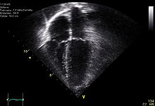
It is used to create images of the heart that are more detailed than images obtained with a plain x-ray. Images can be obtained in real time in two or three dimensions. It allows to evaluate the functioning of the cardiac valves, for the diagnosis of stenosis or insufficiency, and to evaluate the contraction of the cardiac muscle, for the diagnosis of hypertrophy or dilation of the ventricles and atria.
A stress echocardiogram may be done to check how well the heart muscle is working to pump blood to the body. It is used to detect a decrease in blood flow to the heart, caused by a narrowing of the coronary arteries.
Fetal Ultrasound
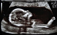
Used to produce images of the fetus while it is in the womb. to assess the growth and development of the baby and to monitor the pregnancy. Through this test, many conditions can be identified that can be dangerous for both mother and child. It is used for:
- confirm pregnancy and location (some fetuses develop outside the uterus)
- determine the baby's gestation age
- confirm the number of babies
- evaluate baby growth
- study placenta and amniotic fluid levels
- identify birth defects
- Research complications
- other prenatal studies
- determine the position of the fetus before delivery.
Kidney ultrasound
Used to obtain images of the kidneys, ureters, and bladder. This test is done when doctors suspect a kidney problem and it allows them to identify:
- the size of the kidneys
- injuries
- anomalies present from birth
- obstructions or stones in the kidneys
- complications from urinary tract infection
- cysts or tumors
Skin ultrasound
This diagnostic technique allows the detection of skin tumors, inflammatory processes, nail disorders, hair diseases and is also applicable to dermo-aesthetics. First used in Chile by Dr. Wartsman, in Spain the technique has been introduced by Dr. Fernando Alfageme Roldán.
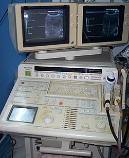
Ultrasound machine components
The processing system
It is the ultrasound machine itself where the processing of the information obtained by ultrasound takes place, it consists of three parts:
- CPU or central processing unit: It is basically a computer with processor and base plate.
- Storage system: Echographs generate images and data which require storage for your study, for this reason they have important amounts of memory.
- Console: To manage the different parameters of the ultrasound and enter data, make measurements and other aspects the device consists of a keyboard, mouse wheels and levers.
- Monitor: It is the screen where echographic images, patient data and the rest of the information are displayed.
The transducer or probe
The transducer is the most important piece of the device, which is responsible for transforming electrical impulses into ultrasonic waves as well as directing them to a specific point, using the physical property called piezoelectric effect. It also performs the same procedure in reverse, so that it transforms the sound impulses that it receives into electrical current that it transmits to the processing system and with which it creates the images that are displayed on the monitor.
To fulfill its function, it must be made up of a casing that is hermetic to the passage of sound, crystals with piezoelectric properties, and a plastic membrane that remains in contact with the skin and over the area that is being explored.
There are different types of transducers depending on their frequency and shape. Although the most used type of them can change their amount of frequency, there are some that have low, medium or high frequency. High frequency is usually used for superficial tissues and low for deep tissues. Usually the ultrasound machine operates at frequencies between 7 and 13 MHz.
As for its shape, it can be linear, convex, microconvex, endocavitary, sectorial and 3D.
Picture modes
The medical equipment can be used in different configurations, depending on the purpose of the study and the target organ.
Still Image Modes
- Mode A (amplitude): This mode is the simplest and is based on the echo technique, where an ultrasound pulse is emitted from a transducer to the region you want to study and the reflections are received by the same transducer. The time from the pulse to the receive is proportional to the depth of the interface. A single ultrasound beam is used and the information collected is represented in graphs representing the widths of the echo received. The advantage is that it provides information quickly with minimal equipment, but only provides one-dimensional information.
- B mode (brillo): In this way, the echo captured is recorded on the screen as a point, whose size and brightness depend on the intensity of the echo. With the movement of the transducer in a single plane you get another series of points, which when added configure a two-dimensional image.
- Mode C: This method does not use the echoes of the waves reflected in the interfaces of the tissues, but takes advantage of the transmitted waves. The transmitter is placed on the object to study and the receiver at the opposite end. With this way you can achieve a total attenuation measure along the route by comparing the pulse width received with the pulse width emitted. Also, comparing the time between emission and pulse reception, the data can be obtained to calculate the acoustic spread of the tissue.
Dynamic Image Modes
- M mode (movement): This configuration is used to analyze the movement of the body structures qualitatively and quantitatively, as in the case of the heart valves. It is a hybrid mode between A and B, since the brightness of each line is modulated to the breadth of the echoes as in mode B, but the echoes are collected in one direction, along the path of lightning as in mode A.
Location mode
- Doppler mode: This way makes use of the Doppler effect, to visualize the blood flow.
- Doppler color: Information about the speed and direction of the flow in real time is represented with color units.
- Spectral doppler: Instead of using colors, it shows the blood circulation on a chart. It can be used to show the level of blockage of a blood vessel.
- Doppler duplex: Uses conventional ultrasound to form images of the blood vessels and organs, which will later be converted into a graph similar to the spectral Doppler.
- Continuous wave Doppler: In this test, the sound waves are sent and received continuously. It allows to measure more accurately the blood flowing faster.
Extensions
Doppler Ultrasound
The doppler ultrasound or simply echo-Doppler is a variety of traditional ultrasound, therefore based on the use of ultrasound, in which taking advantage of the Doppler effect, it is possible to visualize the velocity waves of the flow passing through certain structures in the body, usually blood vessels, that are inaccessible to direct vision. The technique makes it possible to determine if the flow is directed toward the probe or away from it, as well as the speed of said flow. By calculating the variation in frequency of the volume of a particular sample, for example, that of blood flow in a heart valve, its speed and direction can be determined and displayed. The impression of a traditional ultrasound combined with a Doppler ultrasound is known as a duplex ultrasound.
Doppler information is represented graphically with spectral Doppler, or as an image using directional Doppler or power Doppler (non-directional Doppler). The Doppler frequency falls in the audible range and can be heard using stereo speakers, producing a distinctive pulsating sound.
3D and 4D ultrasound
Recently, a revolution has been seen in the field of maternal-fetal medicine. This revolution, moreover, has not only affected medicine itself, but has also provided society with the possibility of establishing a much deeper emotional bond with neonates than was previously believed possible, thanks to a quality of image that allows you to see the appearance of the future baby in photography (3D) or in a moving image (4D).
To achieve this, using the ultrasound machine, ultrasound is emitted in four angles and directions, gently passing the emitter over the patient's belly, to which a gel has previously been applied to improve the efficiency of the process. The ultrasound bounces off and is captured by the computer, which automatically processes the information to reproduce the image of the baby on the screen in real time.
Technological Advances in Ultrasound
Since ultrasound was invented, some of the technological improvements have been introduced in the new devices, which allow better quality images to be obtained, as well as being more efficient in their diagnostic use. They are the following:
- Armonic tissues: Ultrasounds through tissues produce harmonic sounds that can be recognized and separated from the rest of the signal. As a result of this, better quality images are obtained.
- Directional ultrasonic beams: Changing the ultrasonic beam direction can achieve better display of the needle, for example in interventionist procedures.
- Composite image: The device emits different ultrasound beams in different directions, allowing the CPU to add them, produce better images.
- Doppler: It is based on the physical phenomenon called Doppler effect. It allows the ecographer to visualize the movement of fluids like blood passing through blood vessels. Often when activating this option on the device, the moving fluids of another color are marked on the screen.
- Elastography: Quantitatively measure the deformation capacity of a tissue by applying an external force.
- 3D Images: Ecographic 3D images with conventional probes can be obtained using a specialized computer program.
- Expanded field of vision: New ecographers can generate a wider and panoramic image from successive images obtained after a manual sweep with the probe.
- ECO-RM Fusion: Merges the images obtained by magnetic resonance with those of ultrasound through a GPS system that allows synchronization of the two devices.
Clinical uses
Initially, ultrasound was a diagnostic technique developed and used by radiologists, however, today it is increasingly used in other medical specialties as a diagnostic tool: cardiology, gynecology, obstetrics, emergency medicine, intensive care, general medicine, family, urology or pediatrics.
Contenido relacionado
Metaplasia
Quick BASIC
Air safety


