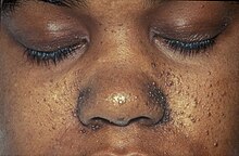
Tuberous sclerosis
tuberous sclerosis is a rare, autosomal dominant hereditary disease with complete penetrance, which causes the formation of abnormal masses (non-cancerous tumors) in some organs of the body, such as: the retina, skin, lungs, kidneys and heart. Generally, it also usually affects the Central Nervous System (the spinal cord and the brain).
This disease is in a group of diseases called neurocutaneous syndromes. The brain lesions of the disease were first described in 1862 by Heinrich von Recklinghausen. Bourneville, years later, made public the anatomo-clinical manifestations. The name tuberous sclerosis is due to the growths produced in the brain, in the form of roots, which calcify with age and become hard.
It has a genetic appearance similar to lymphangioleiomyomatosis that almost exclusively affects fertile women. That is why this female disease has been divided into two types: one with sporadic disease and another with disease associated with tuberous sclerosis.
Epidemiology
Tuberous sclerosis occurs in people of different ethnic groups and of both sexes. Worldwide, it is claimed that there are about 1 to 2 million individuals, and it is believed to affect 1 in 6,000 newborns. In the US, there would be between 25,000 and 40,000 cases. The incidence has been estimated at less than 1 case per 100,000 person/year.
Etiology
Tuberous sclerosis is caused by mutations in two genes (TSC1 and TSC2). If one of the genes is affected, the disease can occur. The TSC1 gene is located on chromosome 9 and gives rise to a protein called hamartin. Unlike the TSC2 gene, which is located on chromosome 16 and causes the protein called tuberin. The TSC1 gene was found in 1997 and the TSC2 gene was discovered in 1993.
These proteins hamartin and tuberin are considered to be involved as tumor growth suppressors, regulating cell differentiation and proliferation in which nerve cells divide to give rise to new generations of cells with individual characteristics.
In recent years, it has been detected that the tuberous sclerosis gene is linked to the ABO blood group locus and to the c-abl oncogene (both located on the long arm of chromosome 9 (9 q 34).). More studies are needed to confirm more genetic loci, some already evidenced: 9q34, 16q13.3.
Clinical picture
Symptoms of tuberous sclerosis can present at birth. Although in some people the advancement of symptoms may occur later. There is variability in the degree of the disease, that is, some patients present a mild form of the disease, others may present severe disabilities. In rare cases, abnormal masses can be life-threatening. Both parents are not needed to transmit the mutation, with only one member it is enough for the child to suffer from the disease. Even so, in most patients Tuberous Sclerosis is caused by new mutations, so there is generally no family history of the disease, so the disease is obtained through a process called gonadal mosaicism (the mutation affects to a part of the gametes: ovules or spermatozoa).
Skin and mucous manifestations
- Hypochromatic or acronomic handles
These are spots that appear on the skin, in the shape of a lanceolate leaf or an ash leaf.
- Angiofibromas
They are reddish round tumors that arise from the dermis protruding on the epidermis, with a size ranging from a pinhead to a pea, they usually appear around the chin, cheeks and nose in the shape of butterfly wings (nasolabial folds). Another characteristic location of angiofibromas is subungual or periungual on the hands and feet.
- Orange skin
With an irregular orange-like texture, usually in the dorsal or lumbar region.
Neurologic Manifestations
- Epileptic crisis
Around the second year of life in 80-90% of cases. If they appear early, they usually take the form of West or Lennox-Gastaut syndrome.
- Psychological alterations
Mental disorders, behavioral disorders or psychotic disorders may be associated with tuberous sclerosis.
- Astrocytomas of intraventricular giant cells adjacent to Monro's foramen
Tumors located in Monro tend to grow and annual controls are needed to evaluate surgery, the rate of growth varies even in the same patient. The chances of recurrence are present, but it decreases towards adolescence until it calcifies. The symptoms may be early or late, depending on the patient's heredity, among these may present with seizures, dizziness, sleep disorders, intracranial pressure due to obstruction, eye pressure.
- Qualifications
That they do not grow, but play a fundamental role in seizures by functioning as batteries, (which when discharged cause seizures or be a factor that worsens a pre-existing epilepsy) and in the behavior of the patient depending on the area of the brain where it is located most.
- Tuberos
They are tubercle-shaped lesions found in the brain (hence the name) made up of different tissue, which, depending on its location, can cause mental retardation, autism, differential perception of the environment and everyday life, stereotypes, speech delay, lack of self-recognition of the body, hyperactivity (misdiagnosed as attention deficit) among others.
Ophthalmic Manifestations
- Retinian Hamartomas.
Visceral injuries
Kidneys
- Angiomiolipomas: They are benign tumors composed of vascular, muscle and fatty tissue. In individuals with CET usually appear in both kidneys multiplely. Its growth and alteration of kidney function are asymptomatic if the picture is unknown. Colic, hematuria, sudden low blood pressure and fatigue may occur when it already represents a surgical problem, requiring total or partial nephrectomy. If it maintains good renal functionality, when angiomiolipomas are larger than 6 cm, you can opt for selective embolization. The sudden rupture of an angiomiolipoma can happen at any time (called Wünderlich Syndrome) causing a renal decomposation, as the blood inside the angiomiolipoma floods the rest of the kidney, requiring emergency medical assistance.
- Unique or multiple cysts: They may appear next to angiomiolipomas and are likely to be malignant.
Cardiac
- Rabdomioma or cardiac hamartoma: Unique or multiple are at birth, can cause ventricular obstruction, of mostly benign origin, they are reabsorbing with the passage of time without leaving usually sequelae, the size of these may vary.
Lungs
- Polichistosis: it is usually the most common.
- Pulmonary Linfangioleiomatosis: occurs in ET with a higher incidence in women than in men after 30 years. A chronic involvement of hormonal origin (hyperparathyroidism) is studied. In this disease, lymphatic tissue is generated in the lung alveoli, obstructing them, causing a decrease in progressive respiratory capacity, with transplant surgery or fatal outcome in some cases. Among the symptoms there may be: tiredness, decreased oxygen in blood, among others.
Vascular
- Displasias: Some of these can be brain aneurysms.
Skeletal Injuries
Bone lesions are usually 1–3 mm geodes, pseudocystic, metacarpal and metatarsal or phalangeal, or areas of hyperostosis.
Oral manifestations
- Fibrous plates: Located in the area of gums, lips and tongue.
- Skeletal lesions: Sunken palate, fissured lip and hyperostosis.
- Idiopathic gingival enlargement (clinical dentistry Mac Graw Hill, carranza, pag 380)
- Other: Calcified odontogenic tumor, demoplastic fibroma, mucous and/or intraosic hemangiomas, odontogenic mixedoma, facial asymmetry, bifida ovula, eruption delay and diastemas, among others.
| Features | Percentage of apparition | Feature | Percentage of apparition |
|---|---|---|---|
| Skin injuries | 95% | Kidney tumors | 70% |
| Development challenges | 95% | Heart tumors | 50% |
| Convulsions | 85% | Autism | ~50% |
| Ophthalmological injuries | 80% | Pulmonary disease | ≤40% |
Diagnosis
The diagnosis of this disease is based on clinical manifestations. In those individuals who meet the clinical diagnosis, it is possible to identify the existing mutation in around 85% of cases. Of these patients in whom the mutation can be identified, in 31% of cases the mutation occurs in the TSC1 gene, while in the remaining 69% said mutation is present in the TSC2 gene. The TSC1 variant presents mutations on chromosome 9 (9q34) in the gene that codes for the hamartin protein. The TSC2 variant presents mutations on chromosome 16 (16p13) in the gene that codes for the tuberin protein
Approximately two-thirds of affected individuals are affected as a consequence of a "de novo" (a new mutation arises that was not present in the patient's parents). There are also cases of genetic mosaicism, in which the disease has not been diagnosed in a family until an affected individual appears. In one of the parents of the affected individual and therefore their ancestry (grandparents, great-grandparents, etc.), healthy cells and mutated cells that possess the allele that cause tuberous sclerosis will coexist, but their phenotype will be healthy. When the proportions of these cells change in the offspring, along with many other still unknown factors, they cause the disease to be expressed in their children.
Treatment
Due to the great variety of symptoms, and the wide spectrum that they can present, there is no specific treatment for this disease. So the treatment is based on treating each symptom that the affected person presents.
In the US, the Food and Drug Administration (FDA) has approved several drugs to treat some of the major manifestations of the disease. The antiepileptic drug vigabatrin was approved in 2009 to treat infantile spasms and was recommended as first-line therapy for infantile spasms in children with tuberous sclerosis by the 2012 International TSC Consensus Conference. The adrenocorticotropic hormone was approved in 2010 to treat infantile spasms. infantile spasms, too. Everolimus was approved for treatment of tuberous sclerosis-related tumors in the brain (subependymal giant cell astrocytoma) in 2010 and in the kidneys (renal angiomyolipoma) in 2012. Everolimus has also shown evidence of efficacy in treating epilepsy in some people with tuberous sclerosis. In 2017, the European Commission approved everolimus to treat refractory partial seizures. According to a study conducted in Mexico, the use of cannabidiol—a cannabinoid found in the cannabis plant—is has been shown to be effective for the treatment of seizures.
Neurosurgical intervention may reduce the severity and frequency of seizures in patients with tuberous sclerosis. Embolization and other surgical interventions can be used to treat renal angiomyolipoma with acute bleeding. Surgical treatments for symptoms of lymphangioleiomyomatosis in adult patients include pleurodesis to prevent pneumothorax and lung transplantation in cases of irreversible lung failure.
Other treatments include the ketogenic diet for intractable epilepsy and pulmonary rehabilitation for lymphangioleiomyomatosis.
In Spain, the National Health System has a network of Centers, Services and Reference Units of the National Health System (CSUR) which were created in response to the need to ensure territorial cohesion between autonomous communities and gather knowledge and experiences that help to minimize the inequality of access to health services that treat rare pathologies. The center qualified to care for child patients with neurocutaneous syndromes related to Tuberous Sclerosis pathologies, designated by Resolution of the Minister of Health, Social Services and Equality, corresponds to the Hospital Sant Joan de Déu in Barcelona, a team coordinated by Dr. Héctor Salvador, and the researcher Dr. Federico J. Ramos. The reference CSUR for adults falls on Hospital U. Germans Trias I Pujol, also in Barcelona.
Evolution
The evolution of tuberous sclerosis has a tendency of progress as the affected person advances in age, just as the existing alterations increase. Depending on the affected organ of the individual, it will have one evolution or another and the age of death is related to the size of the tumors. The lung, kidney and central nervous system are the ones that determine the prognosis. From the onset of the disease, a series of early stimulation treatments must be taken (psychomotor skills, physiotherapy, speech therapy...) highlighting that throughout his life he will need support in reading and writing and swimming to strengthen the muscles due to hypotonia that produce epileptic seizures. In the various pathologies, apart from epileptic seizures, the most serious is the behavioral problem, for this reason psychological support is also necessary for the orientation of their relatives.
Throughout the evolution, a series of interventions will be carried out, electroencephalogram if there are epileptic seizures, their periodicity will depend on the degree of the seizures. Cranial CT scan every five years, for correct control of subependymal nodules and their location in relation to Monro's foramen. Brain MRI, in the event that surgical excision of a cortical cerebral tuber is considered, since this examination defines the brain structures better than the CT scan. And finally, psychometry and quantification of the intelligence quotient will be done, especially in children with school problems or at the time of starting school, to place them at the appropriate educational level.
It is necessary to carry out surveillance with the practice of periodic controls, to detect early the appearance of tumor complications. It has been pointed out that the average age of death of these patients was around 24 years (Webb et al., 1996) but other studies (Jancar, 1996) indicate that their longevity has increased in recent times.
Prevention
The most effective treatment for this entity is its prevention.
In the case of a family history, genetic counseling is recommended. If one of the parents is affected, the possibility of transmitting the disease is estimated at 50%. the couple should be warned of the risk involved in procreation but should not be discouraged, since this is only the couple's decision.
There is also the availability of prenatal diagnosis of known DNA mutations, but since the disease frequently appears as new mutations, it can rarely be prevented.
Genetic counseling
This disease follows an autosomal dominant inheritance. The offspring of an individual carrying the mutation will therefore have a 50% chance of inheriting the mutation. In those pregnancies in which there is a high risk of the future baby suffering from the disease, it is possible to resort to prenatal diagnosis. However, this is only possible in those cases in which the causative mutation of the gene has been previously identified in some member. Mosaicism also causes complications when making a prenatal diagnosis in amniocentesis, due to possible differences between the cells that have been sampled, and the number of mutated cells from the fetus. In addition, the healthy or diseased phenotype that the new individual can express cannot be determined with certainty even knowing the proportion of mutated cells.
Contenido relacionado
Psittacidae
Orbicularis oculi muscle
Apnea


