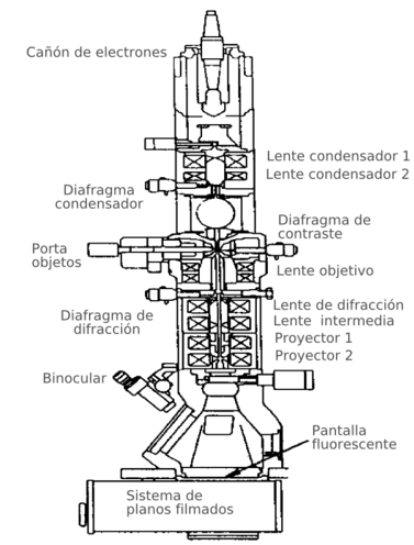
Transmission electron microscope
A transmission electron microscope (TEM for its acronym in English, or MET in Spanish) is a microscope that uses a beam of electrons to visualize an object, because the amplifying power of a light microscope is limited by the wavelength of visible light. The characteristic of this microscope is the use of an ultrafine sample and that the image is obtained from the electrons that pass through the sample.
Transmission electron microscopes can shrink an object up to a million times.
History
The first transmission electron microscope was developed between 1931 and 1933 by Ernst Ruska and his collaborators. The basic optics of that first electron microscope remain to this day; The changes in modern microscopes consist of adding more lenses to increase the scope of magnification and give it greater versatility. The first commercial transmission electron microscope was built by Siemens in 1939. 123
Structure
Because electrons have a much shorter wavelength than visible light, they can show much smaller structures.
The main parts of a transmission electron microscope are:
- Electron Canyon, which emits the electrons that crash or cross the specimen (depending on which type of electronic microscope is), creating an increased image.
- Magnetic lenses to create fields that direct and focus the electron beam, as conventional lenses used in optical microscopes do not work with electrons.
- Vacuum system is a very important part of the electronic microscope. Because electrons can be diverted by air molecules, an almost total vacuum must be made inside a microscope of these characteristics.
- Photo or fluorescent screen which is placed behind the object to visualize to record the increased image.
- Registration system which shows the image produced by electrons, which is usually a computer.
The transmission electron microscope emits an beam of electrons directed towards the object to be magnified. A part of the electrons bounce or are absorbed by the object and others pass through it, forming an enlarged image of the sample.
Electron Gun
From top to bottom, the TEM consists of an emission source, which can be a tungsten filament or a lanthanum hexaboride source (LaB6). In the case of tungsten, the filament can be either in the shape of a hairpin or small and spike-shaped. LaB6 sources use a small single crystal. Connecting said gun to a high voltage source (~120kV for many applications) it will begin to emit electrons into a vacuum. This electron extraction is usually enhanced with the aid of a Wehnelt cylinder. Once extracted, the lenses at the top of the TEM manipulate the electron beams, allowing them to be focused to the desired size and located on the sample.
The manipulation of electrons is achieved by combining two physical effects. The interaction of electrons with a magnetic field causes them to move according to the vector formula F= (q.v) x B (being v and B, the velocity vector of the electron, B the magnetic field vector and "x&# 34; the vector product). This effect allows the emitted electrons to be manipulated by means of electromagnets. This technique allows the formation of a magnetic lens of variable focal length, depending on the distribution of the magnetic flux. In addition, an electric field can deflect the trajectory of electrons at a fixed angle. By means of two consecutive deflections, the trajectories of the electrons can be displaced laterally. This technique allows the lateral displacement of the electron beams in the TEM, this operation being especially important for the scanning of the beams in the STEM variant. From the combination of these two effects as well as the use of a display system (such as a phosphor screen) the level of control of the beams required for the operation of the TEM is obtained.
The TEM lenses allow the convergence of the beams and the control of its angle. This control is exercised by modifying the amount of current that flows through the quadrupole and hexapolar lenses and allows the magnifications of the TEM to be modified. The quadrupole lens consists of a set of four coils located at the vertices of a square. The hexapolar lens simply increases the degree of symmetry of the resulting field.
Typically a TEM contains three lens assemblies with many possible variants in lens configuration, particularly the energy filtered TEM or EFTEM. The sets are respectively called condensing lenses or condenser, objective lenses or simply objective, and spotting lenses. projection or projector. The condenser lenses are responsible for the initial formation of the beam after the emission of the electrons. The objective lenses focus the beam on the sample and finally the projection lenses are responsible for expanding the reflected beam towards the phosphor screen or other display device such as film. The magnifications of the TEM are given by the ratio of the distances between the sample and the image plane of the objective.
It is appreciated that the configuration of a TEM varies significantly depending on its implementation. Thus some manufacturers use special lens configurations, such as in spherical aberration corrected instruments, particularly in high voltage field emission TEM applications.
The display system in a TEM can consist of a phosphor screen for direct observation by the operator and optionally an image recording system such as film or a CCD retina combined with a phosphor screen. Normally these display systems can be interchanged at the convenience of the operator.
Vacuum system
To achieve the uninterrupted flow of electrons, the TEM must operate at low pressures, typically in the order of 10− − 4{displaystyle 10^{-4} a 10− − 8{displaystyle 10^{-8} Pa. The need for this is due to two reasons: first, allow a voltage difference between the cathode and earth without a voltaic arch. Second, reduce the frequency of electron collisions with air atoms to despicable levels. Since the TEM, contrary to a CRT, is a system that must allow the replacement of components, the insertion of samples and, in particular in old models, the change of film reel makes it essential to reproduce the vacuum regularly. Therefore the TEMs are equipped with complete pumping systems and their vacuum sealing is not permanent.
The display system in a TEM can consist of a phosphor screen for direct observation by the operator and optionally an image recording system such as film or a CCD retina combined with a phosphor screen. Normally these display systems can be interchanged at the convenience of the operator.
Procedure for its use
To use a transmission electron microscope, the sample must be cut into thin layers, no larger than a couple thousand angstroms. Transmission electron microscopes can magnify an object up to a million times.
Theoretical basis
Theoretically the maximum resolution d{displaystyle d} achievable with an optical microscope is in principle limited by the wavelength λ λ {displaystyle lambda } of the light used to examine the sample, and by the numerical opening NA{displaystyle {textrm {NA}}} of the system.
d=λ λ 2nwithout α α ≈ ≈ λ λ 2NA{displaystyle d={frac {lambda }{2,n,sin alpha }}}}approx {frac {lambda }{2,{textrm {NA}}}}}}}}
The physicists of the early centuryXX. they theorized on possible ways to overcome the limitations imposed by the relatively large wavelength of visible light (from 400 to 700 nm) by using electrons. Like all the matter, electrons exhibit both wave and particle properties (as Louis-Victor of Broglie already proposed). As a result, an electron beam can be made to behave as an electromagnetic beam. The wavelength of the electron is obtained by matching the Equation of De Broglie to the kinetic energy of an electron. Additional relativistic correction should be introduced, as electrons in a TEM team reach speeds close to that of light c{displaystyle {textrm {c}}}.
λ λ e≈ ≈ h2m0E(1+E2m0c2){displaystyle lambda _{e}approx {frac {h}{sqrt {2m_{0}Eleft(1+{frac {E}{2m_{0}c^{2}}}}}}}}}}}}
In an electron microscope, electrons are generally produced in a filament, usually tungsten, similar to that of a light bulb, by a process known as thermionic emission or by field emission. The emitted electrons are then accelerated with the help of an electrical potential (measured in V, or volts) and focused by electrostatic or electromagnetic lenses.
Applications
The main function of the transmission electron microscope is the study of metals, minerals and the study of cells at the supramolecular level.
It has a very important role in the metallurgy industry.
It is used in microbiology, to observe the structure of viruses.
It is also used in pathological anatomy, to diagnose based on the cellular ultrastructure.
Contenido relacionado
Triode
Nuclear energy
Large Electron-Positron Collider
Photoelectric cell
William Bradford Shockley


