Tibia
The tibia (shin) is a long, paired, triangular prism-shaped bone located in the anterior and internal part of the leg; It presents two curvatures in the opposite direction: the upper one, concave outwards; another inferior, concave inward (in the form of an italic S). Like all long bones, it has two epiphyses, two metaphyses and one diaphysis. The proximal epiphysis participates in the knee joint, relating to the femur, while the distal epiphysis shares the ankle joint with the distal epiphysis of the fibula.
In humans
The tibia lies medial to the fibula with which it articulates at its proximal and distal ends. Likewise, between both bones there is a fibrous membrane called "interosseous membrane" which provides stability to both joints by forming a syndesmosis.
Etymology
The Spanish word “tibia” comes from the Latin “tībia”, which means 'flute'.
Development
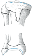
The tibia begins to ossify from three centers; one for the body and two others for the extremities. Ossification begins in the center of the body, around the seventh week of fetal life, and gradually extends to the extremities.
The center of the upper epiphysis appears before or shortly after birth at about 34 weeks' gestation; takes a flattened shape, and has a thin tongue-like process in front, which forms the tibial tuberosity; which appears through the lower epiphysis in the second year.
The lower epiphysis fuses with the tibial shaft at approximately 18 years of age, and the upper epiphysis fuses with the tibial shaft by about the second year of life.
Occasionally there are two additional centers, one for the tab-like process of the upper epiphysis, which forms the tuberosity, and one for the medial malleolus.
Upper extremity; tibial plateau
The end that articulates with the femur is broad and has two internal and external glenoid cavities (facies articularis superior) that articulate with the condyles of the femur. It has a flat upper face called the "tibial plateau", from which emerges an eminence between the glenoid cavities called the tibial spine or intercondylar eminence (eminentia intercondylaris). This eminence fits into the intercondylar fossa of the femur, it is divided by a notch in two: internal tubercle (tuberculum intercondylare mediale) and external tubercle (tuberculum intercondylare laterale).
There are two rough triangular surfaces called the prespinal (area intercondylaris anterior) and retrospinal (area intercondylaris posterior) surfaces. The two glenoial cavities rest between two bulky masses called the tibial tuberosities; the internal tuberosity or medial condyles (condylus medialis) present a rough impression behind for the direct tendon of the semimembranosus, and in front a channel for the horizontal tendon of the semimembranosus. The external tuberosity or lateral condyle (condylus lateralis) has an articular facet called the fibular facet of the tibia bone (facies articularis fibularis).
The two tuberosities are separated by a vertical notch in front of this is a triangular surface, rough and full of holes below this is the anterior tubercle or anterior tuberosity (tuberositas tibiae) of which splits a ridge ending in Gerdy's tubercle for attachment of the tibialis anterior muscle.
Body of tibia (corpus tibiae)
It consists of three edges and three faces:
Borders
- Previous era (previous March): also called the earlier crest of the tibia.
- External (margo medialis): is located on the opposite side to the internal mallo. At the bottom it has a joint surface for the Peron.
- Internal branch (margo interosseus): has the insertion of aponeurosis palmar.
Faces
- Internal face (facies medialis): It's smooth, it's in contact with the skin. It presents roars for the goose leg.
- External face (side phases): is the tube for the previous tibial.
- Later face (later phases): finds the oblique crest of the tibia (sulcus malleolaris).
Lower extremity
It is shaped like a pyramid, in its lower part it has the medial malleolus (malleolus medialis), which is the widened part that can also be palpated and is the site of union with the talus (facies articularis inferior). Between the tibia and fibula is the interosseous membrane.
On the posterior aspect of the tibia is the soleus line (linea musculi solei), which is the insertion site for the soleus muscle.
The notch on the lateral surface of the lower end of the tibia (incisura fibularis), for the fibula, is located in the anterior and internal part of the leg, parallel and to one side of the fibula. With the talus below and the fibula outside and above (facies articularis malleoli medialis).
Vascularization
The tibia draws its arterial blood supply from two sources: a feeding artery, as the main source, and periosteal vessels derived from the anterior tibial artery.
Articulation
- With the femur forming the joint of the knee.
- With the butne formed the thybioperonea joints, superior and lower.
- With the astrágalo, forming along with the perone the joint of the ankle.
Muscle Insertions
- Previous Tibial
- posterior tibial muscle
- Poplíteo
- Soleo
- Semitendinos
- Semimembranous
- Sartorio
- Grácil
- Rotulian
Function
- Get the body weight.
- It serves as support for locomotive.
In other species
The structure of the tibia in most other tetrapods is essentially similar to that of humans. The tibial tuberosity, a ridge to which the patellar ligament attaches in mammals, is instead the point for the tendon of the quadriceps muscle in reptiles, birds, and amphibians, which do not have a patella.
Contenido relacionado
Kalanchoe
Dieffenbachia
Sageraea


