Synapse
The synapsis (from the Greek σύναψις [sýnapsis], 'union', 'link') is a specialized approximation between neurons, either between two association neurons, one neuron and a receptor cell, or between a neuron and an effector cell (almost always glandular or muscular). In these contacts the transmission of the nervous impulse is carried out. This begins with a chemical discharge that causes an electrical current in the membrane of the emitting cell (called pre-synaptic); once this nerve impulse reaches the end of the axon, the connection is responsible for exciting or inhibiting the action of another cell called the receptor cell (called post-synaptic).
This article covers the general properties of synapses. There are specific articles for Chemical Synapse and Electrical Synapse.
Word origin
The word synapse comes from synaptein, which C.S. Sherrington and his collaborators formed with the Greek words sin-, meaning & # 34; together & # 34;, and hapteina , meaning & # 34; with firmness & # 34;.
Activity Framework
These chemical-electrical bonds are specialized in sending certain types of survival signals, which affect other neurons, or non-neuronal cells such as muscle or glandular cells.
There are two different types of base activity, survival activity and survival activity.
Synaptic activity of survival develops in these contexts:
- Between two neurons: the stimulus is carried by amino acid neurotransmitters.
- Between a neuron and a muscle cell: the stimulus is transported by the neurotransmitters of an ester type.
- Between a neuron and a secret cell: the neurotransmitters of a neuropeptide type are transported to the stimulus.
Synaptic survival activity develops in these contexts:
- In neuroprocessing activity.
- In food consumption activity.
- In extreme homeostatic conservation activity.
The synapse occurs when presynaptic and other postsynaptic chemical-electric activity is recorded. If this condition does not occur, one cannot speak of a synapse. In this action, chemically-based ionized neurotransmitters are released, whose charge cancellation causes the activation of specific receptors that, in turn, generate other types of chemical-electrical responses.
Each neuron communicates with at least a thousand other neurons and can simultaneously receive up to ten times as many connections from others. It is estimated that in the adult human brain there are at least 1014 synaptic connections (between 100 and 500 trillion). In children the number would reach 1000 billion, but this number decreases over the years, stabilizing in adulthood.
Synapses allow neurons in the central nervous system to form a network of neural circuits. They are crucial to the biological processes that underlie perception and thought. They are also the system by which the nervous system connects and controls all the systems of the body.
Histology
| Axon terminal |
|---|
| Structure of a classic neuron. |
From the histological and functional point of view, a neuron has three main areas: the body or soma, the dendrites and the axon. These last two elements are in charge of establishing synaptic relationships: the dendrites are like "antennas" that receive most of the information that comes from other cells; the axon, for its part, is like the "cable" with which one neuron connects to others.
Connections can be made at very short ranges, a few hundred micrometers away, or much greater distances. Neurons in the spinal cord, for example, communicate directly with organs such as muscles to cause movement (neuromuscular synapses).
The classic model of the synapse had its center in the neuron, and included the presynaptic and postsynaptic terminals. This bipartite model only partially explained the fine "plastic modifications" of the synapse seen in some physiological situations.
Ultrastructure
A prototypical synapse consists of "mushroom"-shaped cytoplasmic dilations (synaptic button) from each neuron that approach one against the other. In this zone, the cell membranes of both cells determine a narrow space. The zone of approach of both neurons is approximately 20-30 nanometers (nm) thick, and is called the synaptic gap (or cleft).
These synapses are asymmetric in both their structure and function. Only the presynaptic neuron secretes the neurotransmitters, which bind to the transmembrane receptors that the postsynaptic cell has in the cleft.
The presynaptic nerve terminal (also called the synaptic button) normally emerges from the end of an axon, while the postsynaptic zone normally corresponds to dendrites, the cell body, or other cellular zones.
The area of the synapse where the neurotransmitter is released is called the active area.
In the active zones, the membranes of the two adjacent cells are closely brought together by cell adhesion proteins.[citation needed]
Just behind the membrane of the postsynaptic cell is a complex of interlocking proteins called Postsynaptic density. Postsynaptic density proteins perform numerous functions, ranging from the anchoring and movement of neurotransmitter receptors in the plasma membrane, to the anchoring of various proteins that regulate the activity of these receptors.
Types of synapses
According to what is transmitted
The transmission can be electrical or chemical.
Electrical Synapse
An electrical synapse is one in which the transmission between the first neuron and the second is not produced by the secretion of a neurotransmitter, as in chemical synapses, but by the passage of ions from one cell to another through gap junctions, small channels formed by the coupling of protein complexes, based on connections, in closely adherent cells.
The electrical synapse is the most common in less complex vertebrates and in some places in the mammalian brain. The cell membranes of presynaptic and postsynaptic neurons are closely in contact, through gap junctions or nexuses which have molecular channels through which ions pass. Thus the nerve impulse is transmitted directly from one cell to another. They are faster than chemical synapses but less plastic; they are less prone to alteration or modulation because they facilitate the exchange between cytoplasms of ions and other chemicals. In vertebrates they are common in the heart and liver.
Electrical synapses have three very important advantages:
- Electrical synapses have a bidirectional transmission of action potentials,
- In electrical synapses there is a synchronization in neuronal activity, which makes possible coordinated action among them.
- Communication is faster in electrical synapses because the action potentials pass through an ionic protein channel directly without the need for release molecules.
Chemical Synapse
The chemical synapse is established between cells that are separated from each other by a space of about 20-30 nanometers (nm), called the synaptic cleft or gap.
The release of neurotransmitters is initiated by the arrival of a nerve impulse (or action potential), and is produced by a very rapid process of cellular secretion: in the presynaptic nerve terminal, the vesicles containing the neurotransmitters remain anchored and prepared next to the synaptic membrane. When an action potential arrives, an influx of calcium ions occurs through voltage-gated calcium channels. Calcium ions initiate a cascade of reactions that ultimately cause the vesicular membranes to fuse with the presynaptic membrane, releasing their contents into the synaptic cleft.
Receptors on the opposite side of the cleft bind neurotransmitters and force open ion channels in the postsynaptic membrane, causing ions to flow in or out, changing the local membrane potential.
The result is excitatory in the case of depolarization flows, or inhibitory in the case of hyperpolarization flows. Whether a synapse is excitatory or inhibitory depends on the type or types of ions that are channeled in postsynaptic flows, which in turn is a function of the type of receptors and neurotransmitters involved in that synapse.
The sum of excitatory and inhibitory impulses arriving at all the synapses that are associated with each neuron (1,000 to 200,000) determines whether or not the discharge of the action potential occurs along the axon of that neuron.
According to the location where the synapse is made
In the nervous system, synapses can occur in various sectors of the body of the neuron:
- Axodendrytic syndrome
Union of the terminal branches of the axon of the presynaptic neuron, with the dendrites of the postsynaptic cell, in which they intertwine or end in the dendrites directly.
- Axosomal synapsis
The union of the terminal branches of the axon of the presynaptic neuron that form a basket or network around the body (soma (neurology) of the postsynaptic cell.
- Axoaxon sinapsis
The junction where some terminals (axons) of the presynaptic neuron terminate in the axons of the postsynaptic neurons.
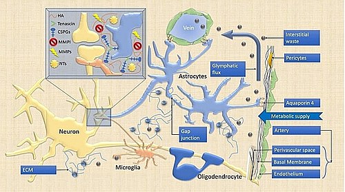
Bipartite, tripartite and tetrapartite synapses
The classic model of the synapse had its center in the neuron, and included the pre-synaptic and post-synaptic terminals that corresponded to two contiguous neurons, this two-part model was called bipartite synapse.
Subsequently, other cellular elements present in the neuropil were added to the synapse: oligodendrocytes, astrocytes and microglia as fundamental elements for the formation and remodeling of the tripartite synapse
According to research related to the extracellular matrix, synapses would consist of up to four elements (tetrapartite):
- neuronal pre-synaptics
- neuronal post-septics,
- the nearby neuroglia and
- the extracellular matrix
Classes of synaptic transmission
There are three main types of synaptic transmission; The first two mechanisms constitute the main forces that govern neural circuits.
- Exciting transmission
one that increases the possibility of producing an action potential.
- Inhibitive transmission
one that reduces the possibility of producing an action potential.
- Modular transmission
one that changes the pattern and/or frequency of the activity produced by the cells involved.
Synaptic Strength
The strength of a synapse is determined by the change in membrane potential that occurs when postsynaptic neurotransmitter receptors are activated. This voltage change is called the postsynaptic potential, and it is a direct result of ionic flows through postsynaptic receptor channels. Changes in synaptic strength can be short-term and without permanent changes in neuronal structures, lasting seconds or minutes, or long-lasting (long-term potentiation or LTP), in which continued or repeated activation of synaptic strength synapse implies that second messengers induce protein synthesis in the nucleus of the neuron, altering the structure of the neuron itself. Learning and memory could be the result of long-term changes in synaptic strength, through a mechanism of synaptic plasticity.
Integration of synaptic signals
Generally, if an excitatory synapse is strong, an action potential in the presynaptic neuron will initiate another potential in the postsynaptic cell. In a weak synapse, the postsynaptic excitatory potential ('PEPS') will not reach the threshold for action potential initiation. In the brain, each neuron maintains connections or synapses with many others, each of which can receive multiple signals. When action potentials fire simultaneously in several neurons that are attached at weak synapses to another neuron, they can force the initiation of an impulse in that cell even though the synapses are weak.
On the other hand, a presynaptic neuron that releases inhibitory neurotransmitters, such as GABA, can generate a postsynaptic inhibitory potential ("PIPS") in the postsynaptic neuron, lowering its sensitivity and the likelihood that it will be generated. an action potential in it. Thus the response of a neuron depends on the signals it receives from others, with which it can have different degrees of influence, depending on the strength of the synapse with that neuron. John Carew Eccles performed some important experiments in the early days of synaptic research, for which he received the Nobel Prize in Physiology or Medicine in 1963. Complex input/output relationships form the foundation of transistor-based computing, and it is believed that they function in a similar way in neural circuitry.
Properties and regulation
After the fusion of the synaptic vesicles and the release of the transmitter molecules in the synaptic cleft, the neurotransmitter is rapidly removed from the space by specialized recycling proteins located in both the presynaptic and postsynaptic membranes. This reuptake prevents desensitization of postsynaptic receptors and ensures that subsequent action potentials generate a PEP of the same intensity. The need for reuptake and the phenomenon of desensitization at receptors and ion channels means that the strength of the synapse can be diminished if a train of action potentials arrives in rapid succession, a phenomenon that leads to a dependence frequency at synapses.[citation needed]
The nervous system takes advantage of this property for computations, and can adjust synapses by phosphorylating the proteins involved.
The size, number, and replacement rate of the vesicles is also subject to regulation, as are many other aspects of synaptic transmission. For example, a class of drugs known as selective serotonin reuptake inhibitors or SSRIs affect certain synapses by inhibiting the reuptake of the neurotransmitter serotonin. In contrast, a very important excitatory neurotransmitter, acetylcholine, is not reuptaken, but is removed by the enzyme acetylcholinesterase.
Synapses in plastic phenomena
Modification of synaptic parameters can modify the behavior of neural circuits and the interaction between the different modules that make up the nervous system (modal). These changes are encompassed in a phenomenon known as neuroplasticity or neuronal plasticity.
Pathologies that affect the synapse
- Parkinson's Disease
It is a neuronal degenerative disorder located in the substantia nigra, these are in charge of producing dopamine (neurotransmitter) essential for the movement of the body to be carried out correctly. When not enough dopamine is available, the symptoms that characterize this disease occur.
- Epilepsy
They are recurrent discharge crises between inhibitory and excitatory impulses. Recurrent inhibition can occur when a principal neuron synapses with an inhibitory neuron. The hyperexcitable state results from the increase in synaptic excitatory neurotransmitter.
- Alzheimer's Disease
It is a degenerative process of the neurons of the cerebral cortex that is irreversible up to now.
Immune Synapses
By analogy with the synapses described, the encounter between an antigenic cell and a lymphocyte is called an immune synapse.
Contenido relacionado
Systematic botany
Asteraceae
Anacardiaceae


