Sydenham's Chorea
Sydenham's chorea —also known as "minor chorea", "St. Vitus disease" or "dance of Saint Vitus", chorea sancti viti, "corea aguda" o "rheumatic chorea"— is an infectious disease of the central nervous system, due to rheumatic fever after pharyngitis caused by the bacterium Streptococcus pyogenes. Some rare cases may be associated with pregnancy, and in this case are called "chorea gravidarum" or chorea gravidarum.
Not to be confused with Huntington's chorea, which is a degenerative, non-infectious disease. It is also easy to confuse it with other unrelated diseases, such as lupus erythematosus. This increases the importance of a correct differential diagnosis.
Eponymy
The disease is named after the physician Thomas Sydenham (1624-1689), known as 'the English Hippocrates'. However, it had been described before him by the German Gregor Horstius.
Etymology
The Latin term chorea, in turn from the Greek khoreia (χορεία), comes from the same root as chorus (khoros, χορός), showgirl, choreuter and choreography, and refers to the involuntary movements of the disease, which simulate a violent dance.
History
The expression chorea sancti viti referred to a dancing mania, a hysterical disorder that seems to have been very common in the 15th and 16th centuries. These psychiatric episodes were called chorea magna ("corea major"), reserving the name chorea minor (minor chorea) for what we know today as Sydenham's chorea. As the dance of San Vito has disappeared as a clinical entity, today the name "corea mayor" as a synonym for Huntington's chorea.
Minor chorea may have been observed by Theophrastus Philippus Aureolus Bombastus von Hohenheim, known as Paracelsus (1493-1541), who named it chorea naturalis ("natural chorea"), although it may have referred to mental illness.
Chorea minor was typified in 1625 by Horstius, but the most complete and precise description was published in 1686 by Sydenham, as a result of which the disease now bears his name.
It is highly unlikely that Horstius or Sydenham ever saw a true case of Saint Vitus dancing (as a hysterical symptom) since he himself had disappeared by the time they lived. Therefore, it is assumed that their descriptions refer to chorea minor.
Classification
The disease is part of the pathological group called "pediatric neuropsychiatric autoimmune disorders associated with streptococcal infection" (PANDAS for its acronym in English), and is classified as follows:
- Neurological disorders (nervous system diseases).
- Diskinesias (abnormal involuntary movements).
- Koreas (arrhythmic irregular contractions).
- Secondary Koreas (due to another disorder).
- Infectious secondary Koreas (produced by microorganisms).
- Rheumatic infectious secondary Koreas (because of rheumatic fever) Streptococcus).
Prevalence
It has been officially classified as a rare disease because it affects fewer than 200,000 people in the United States.
It was previously believed that this type of chorea affected 50% of rheumatic fever patients. The most recent figures reduce this percentage to 26%.As streptococcal infections are declining in developed countries,they only represent a serious problem in emerging countries.
Of patients with strep, only between 1 and 3% develop rheumatic fever, so the incidence is 0.5 per 100,000 population between 5 and 17 years of age. The disease only has a predilection for this age range; it does not discriminate based on other parameters such as ethnicity or social class. Characteristic nodules or rashes appear in only 10% of those affected.
20% of diagnosed patients experience a recurrence.
Epidemiology
As a disease associated with acute rheumatic fever, Sydenham's chorea has declined dramatically since its origins in the 15th century. With the invention of antibiotics and their use to treat streptococcosis, its incidence in industrialized countries has been greatly reduced. Despite this, there have been two major epidemics in the United States in recent years. The cause of this is unknown.
However, it is still a problem in developing countries. In Brazil, for example, more than 50% of cardiac surgeries are performed to correct sequelae left by rheumatic fever. 80% of patients in this Korea worldwide suffer from cardiac lesions.
This chorea is seen in children and adolescents between the ages of 5 and 15. The average age of symptom onset is 9.7 years. 20% of patients experience a second attack of the disease, within the two years following the resolution of the first. It attacks twice as many women as men, and it has therefore been suggested that sex hormones (particularly estrogen) may play a role in the development of the syndrome.
The gravid form is common in women who have suffered rheumatic fever in childhood, or who have been treated with estrogen therapy, estrogen-containing oral contraceptives, or phenytoin-type anticonvulsants.
Causes
The intimate etiology of Sydenham's chorea is not known. This is apparently an autoimmune or antibody-mediated inflammatory response involving certain regions of the basal ganglia.
Pathology
Patients have been shown to have abnormally elevated metabolism in certain areas of the brain, possibly reflecting hyperactivity of the autoimmune process. Positron emission tomography shows a high metabolic level of glucose in the striatum and other basal structures, which is reversed when the patient's clinical condition improves. Nuclear magnetic resonance evidenced enlargement of the caudate nucleus, globus pallidus, and putamen.
Pathogenesis
Patients presenting with Sydenham have previously suffered from rheumatic fever, a consequence of adenitis, pharyngitis or adenopharyngitis caused by Streptococcus pyogenes, a common bacterium. S. pyogenes belongs to group A β-hemolytic streptococci.
Rheumatic fever is a late inflammatory reaction to this type of cocci, which apparently requires a genetic predisposition, as certain families have a higher risk factor than the general population.
In addition to hereditary factors, it is believed that there are predisposing environmental factors, such as overcrowding and malnutrition. The estrogenic factor is very suspicious, since most of those affected are women around puberty.
The specific mechanisms by which the bacterium causes rheumatic fever and chorea is unknown, but evidence suggests that antibodies produced to fight the infection attack certain areas of the nervous system. This can occur if one of the proteins present in the coconut is also found in certain cells of its own, making the human immune system unable to distinguish between the two and attacking them equally. This theory is supported by the fact that—in affected patients—streptococcal M proteins trigger the production of antineuronal antibodies, and that both react with each other in the brain. The M protein from group A of β-hemolytic streptococci contains numerous amino acid sequences that are also present in human tissues. Furthermore, antibodies such as immunoglobulin G interact with neuronal antigens at the level of the caudate nucleus and the subthalamic nucleus in these patients.
Clinical picture
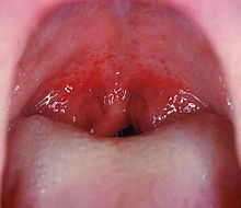
Throat pain and irritation often precede the clinical manifestations of chorea itself. This is due to oropharyngeal infection with S. pyogenes. After 1 to 5 weeks, sudden, acute symptoms of rheumatic fever develop.
Usually, the first and main concrete symptom of Sydenham's chorea usually refers to unexplained changes in the stroke of the handwritten letter.
The choreic signs consist of uncontrollable and spasmodic contractions of various muscle groups, ineffective and similar to fasciculations (the latter do not produce joint displacement). Most of the time, the patient has lost fine motor skills in the fingers and hands, which explains the pathological changes in handwriting.
The choreic movements are continuous, incessant, repetitive and, although they are usually located in the limbs, it is not uncommon to observe them also on the face and neck. This phenomenon is more common in the gravid form. Although they do not normally seriously affect the life of the individual, in severe cases they can cause speech disorders, locomotion, the use of the arms, and, in general, impair the ability physical of the patient.
Clinical manifestations may also include emotional or mental disturbances: loss of emotional control, unjustified crying or laughing attacks, as well as obsessive-compulsive disorders.
Diagnosis
It's not always easy to diagnose Sydenham's chorea. Because the original infection may precede the onset of choreic symptoms by up to 6 months or more, the patient or parent rarely recalls previous tonsillitis or pharyngitis. Since there are usually no clinically visible sequelae of streptococcosis of this date, the following diagnostic tests should be performed:
Diagnosis of S. pyogenes infection
- Analyze blood levels of antibodies for S. pyogenes, for example, anti-streptolysis 0 (AS0). Although 80% of patients with rheumatic fevers show high levels of AS0, there are cases where this test is negative.
- To rule out streptocococcosis with negative AS0, it is necessary to practice a culture of exudado de fauces. Oropharyngeal material is obtained through a hypnotated and is cultivated in a laboratory to verify the growth of betahemolytic streptococci in group A. This trial is also not definitive, because although most patients with pharyngitis, rheumatic fever or chore give positive results, it is often negative with the disease clearly established. Symmetrically, from 60 to 80% of healthy and asymptomatic teenagers—without chore, rheumatic fever or pharyngitis—show positive results.
- Test eritrosedimentation. Measure the deposit of red blood cell sedimentation in a sample of centrifugal blood. A high result in this trial tests a present inflammatory process. Although the results are high in patients with rheumatic fever of months of evolution, erythrosedimentation can also negatively affect the appearance of choreic signs.
Infection with streptococci is also considered confirmed in those patients with rheumatic fever who are undergoing the second round of the disease, or who have a history of rheumatic heart valve disease. If they test positive for one major or two minor criteria (of those explained below), the infection is considered proven.
Diagnosis of rheumatic fever
There are no single diagnostic tests that can accurately diagnose rheumatic fever, but every effort is needed to do so because it is clearly associated with this type of chorea. To achieve this, a list of symptoms and signs called the "Jones Criterion" is used. The list is made up as follows:
Major criteria
- Carditis: All layers of the heart tissue are inflamed (pericardial, epicardial, myocardial and endocardial. The patient has heart murmurs, mitral regurgitation and aortic insufficiency.
- Polyarthritis: This type of migratory arthritis typically affects the joints of the elbow, knees, ankles and wrists. It's very painful, but it responds well to anti-inflammatory.
- Korea: Korean symptoms are well established.
- Erythema: Unwise rash that affects the trunk and the proximal part of the extremities and radiates to the face. It has well defined edges and usually migrates from the central areas to the periphery.
- Subcutaneous nodules: Firms and indolores, are located on bones or tendons.
Minor criteria
- Fever.
- Artralgia.
- Rheumatic fever or rheumatic heart disease.
- Acute phase reactions: leucocytosis, elevated erythro, dramatic increase of C-reactive protein.
- An abnormally prolonged P-R interval in the electrocardiogram (the pause between the contraction of the atriums and that of the ventricles is too long).
Evidence of strep infection
- Any of the diagnostic criteria explained in the previous section.
- Recent scarlet.
To confirm the diagnosis of rheumatic fever, the patient must test positive for:
- Two of the major criteria and show evidence of recent streptococci; or
- A higher and lower criterion, showing evidence of recent streptococci.
In spite of everything, some patients can be diagnosed with rheumatic fever without meeting the Jones Criteria. These are:
- Some Sydenham choreics, when other possible chore causes have been ruled out, or
- Patients with late-appearing carditis or insidious symptomatology without another proven cause than rheumatic fever.
Diagnosis of Sydenham's chorea
Therefore, the correct diagnosis of this disease is based on the presence of coccus infection and rheumatic fever, all accompanied by evaluation of other characteristic signs and symptoms, as well as a complete study of the medical history. of the individual.
There are specific tests for Sydenham's chorea:
- The patient is asked to take his tongue out. If the tongue, instead of staying out of the mouth, goes in and out quickly like the snakes, it is a clear sign of chore.
- Another typical symptom is the "sign of the milker". The patient is ordered to take the hands of the examiner and to exert constant pressure. If you are unable to do so, and instead your hands open and close as if you were milking a cow, it is considered positive.
- He will be asked to extend his arm in front of him with his palm forward, as a policeman stops the transit. If there is a chore, it will be extremely visible in that position.
- Signs of atetosis, stiffness or tremors will be sought.
- The patient's march will be analyzed. When you ask him to walk, the choreic will abruptly change the position of the trunk and lay his head at every step. The movement of the legs will be slow, clumsy and labory, because of the involuntary contractions and postures that overlap the movements that he consciously initiates to move. The legs stop briefly in the air at every step (because of an involuntary contraction in the middle of the movement), conferring on the Korean march a strange and distinctive "dancerous" quality.
The defining studies —as explained— are diagnostic imaging, since certain basal structures are inflamed or enlarged. Additionally, the choreic electroencephalogram is abnormal, showing abnormal slowness and irregularity in the brain waves.
Once a positive diagnosis has been reached for Sydenham's chorea, it is necessary to carry out a complete cardiac evaluation, since it is highly probable that the previous rheumatic fever has left sequelae. Heart and lung sounds and blood flow through the valves will be studied. An x-ray will look for cardiomegaly, a very common cardiac hypertrophy in rheumatic carditis. An echocardiogram will verify possible structural alterations as a result of rheumatism.
Differential diagnosis
Differential diagnosis should rule out choreas associated with systemic lupus erythematosus and those subsequent to non-streptococcal infections, particularly viral ones.
The existence of a family history should be verified to differentiate the condition from hereditary chorea, as well as to define other probable causes: exposure to different toxins, unwanted drug effects, brain lesions, etc.
Other types of pathologies capable of producing chorea are included below, along with the features that differentiate them from Sydenham:
- Korea of Huntington: is dominant autosomal hereditary, is associated with psychiatric symptoms and progressive dementia, has atrophy of the enclosed core and abnormalities in chromosome 4.
- Systemic erythematous lupus: It does not have basal ganglia enlargement or strep infection.
- Sida: It is reactive to antibodies for HIV.
- Hyperthyroidism: Abnormal values of thyroid hormones.
- toxic Korea: Follow the administration of levodopa, stimulants, antidepressants, neuroleptics or estrogens. In children, phenothiacin or other tranquilizers can cause chore-like symptoms, often leading to diagnostic errors.
- ACV: Evidence of cerebral hemorrhage.
- Wilson's disease: Degenerative evil of the liver tissue that leads to insufficiency, abnormalities in the metabolism of copper, dystony and dysarria. It's recessive autosomal.
- Neuroacantocytosis: In addition to the chore, it carries deformities in the erythrocytes.
- Atrophy dentatorrubral-palidoluisiana: Attacks the Japanese population. Alongside the chore produces ataxia, epilepsy and dementia.
Treatment
For strep
Since the S. pyogenes is highly sensitive to penicillin, early treatment with this drug is imperative.
The administration of the antibiotic will be oral or intramuscular, and it will be done simultaneously with a total restriction of the normal activities of the child. The difficulty of diagnosing streptococcus is so great that penicillin is recommended for all patients suspected of having rheumatic fever.
For Polyarthritis
If the arthritis is severe, she will be treated with codeine or salicylates such as aspirin, or other non-steroidal anti-inflammatory drugs. However, too early administration of salicylates can complicate the diagnosis, since it can prevent the appearance of the classic migratory arthritic symptom. For this reason, anti-inflammatory therapy should be postponed as long as possible. As a side note, it should be known that the administration of high doses of aspirin or other salicylates in young children can cause Reye's syndrome, with a fatal prognosis.
Salicylic therapy should be closely monitored, as it may affect the blood or liver. The response of these tissues to salicylates will be closely monitored by blood and urine tests for possible toxic effects.
For Carditis
Those patients who show heart failure or inflammation of the heart need to receive a corticosteroid such as prednisone. An attempt will be made to avoid the numerous undesirable effects of this therapy by limiting it in duration and dose as much as possible. It is harmful to stop steroid therapy abruptly; it is necessary to gradually reduce the doses, compensating for their absence with an increase in salicylates. The administration of aspirin should continue for 2 to 4 weeks after stopping the steroids.
Cardiac patients may also require diuretics to eliminate excess fluid, medication to strengthen cardiac contraction (glycosides such as digitalis), and rest if necessary.
In a few cases it will be appropriate to perform surgery or valvuloplasty, aimed at correcting structural damage to the valves.
For Korea
If the chorea is not causing significant functional disabilities, it is recommended not to proceed with neurotransmitter blocking therapies. It must be remembered that most of Sydenham's patients see their symptoms resolve spontaneously.
If the spasmodic contractions are so intense that the patient is at risk of self-harm or trauma, dopamine antagonists should be used, including antipsychotics such as haloperidol or pimozide. These drugs have potentially adverse effects, such as the development of tardive dyskinesias.
Some patients react favorably to anticonvulsants such as sodium valproate. Pimocide is reserved for cases that do not respond to valproate, or for severe forms of chorea (paralytic chorea). If valproate and pimocide fail, immunomodulatory therapies, immunoglobulin G IV, or plasmapheresis will be attempted.
Prophylaxis
Primary prophylaxis
Antibiotics in children with this oropharyngeal infection are very effective. It is a primary prophylaxis, which, if applied within the first week of inflammation, helps prevent the initial attack of acute rheumatic fever.
Secondary prophylaxis
Patients with rheumatic fever or Sydenham's chorea should receive secondary therapy to prevent disease recurrence, such as injectable penicillin, oral every 21 days, or daily oral therapy with the chemotherapy drugs sulfadiazine or the antibiotic erythromycin. The therapy will continue for life or at least for a few years (until the child reaches adulthood).
Forecast
Most children with Sydenham's chorea make a full recovery. A small percentage may be permanently disabled if the chorea does not respond to treatment and persists.
The duration of the most severe symptoms is usually between 3 and 6 weeks, but some children drag it out for months.
Complications
As has been said, most of the complications affect the heart, usually in the form of rheumatic endocarditis or valvular damage. However, associations have been observed between Sydenham's streptocosis-rheumatic fever-chorea triad and neurological pathologies such as tics, obsessive-compulsive disorder, attention deficit syndrome or autism.
Research
Since 2007, research is being carried out to demonstrate the intimate mechanisms of action of this group of diseases, namely how streptococcus triggers rheumatism, rheumatism triggers rheumatism, and how chorea can lead to the others indicated disorders.
Additionally, studies are being carried out to discover the interactions between environmental, genetic and developmental factors, which can determine the vulnerability of certain children to the bacteria or define the immunity of others. Patients who develop fevers and chorea from pharyngitis are suspected to have an abnormal set of molecular features, and attempts are being made to identify them by various assays.
Contenido relacionado
Fertilization
Lung cancer
Goodpasture syndrome


