Sweat gland
The sweat gland is a gland that is located in the reticular dermis and hypodermis and consists of long, thin tubes, closed at the lower end, where they come together, forming a ball. For the pores that open to the outside, secrete sweat, liquid sebaceous fat, with a salty taste, and a texture similar to urine.
In dermatology, the sweat glands form together with the sebaceous glands, the hair follicles and the nails, the so-called phanera or "skin appendages".
Sweat glands are divided into two groups: the eccrine sweat glands and the apocrine sweat glands.
Eccrine sweat glands
Eccrine sweat glands are the largest sweat glands in the human body, they are found on virtually all skin, with the highest density on the palms of the hands and soles of the feet, then on the head, but most except in the torso and extremities. In other mammals, they are relatively rare and are found mainly in hairless areas such as the footpads. They reach their peak of development in humans, where there can be between 200 and 400/cm² of skin surface.
They consist of a secretory glomerulus and a long excretory duct that opens directly into a hole in the skin surface.
Anatomy
There are about 600 sweat glands per square centimeter of skin, with the highest concentration on the palms of the hands, soles of the feet, and the front region of the face.
The glomerulus lies deep in the skin, close to the dermis.
The secretory portion or adenomere is of the acinar type, with a narrow lumen, it is formed by secretory cuboidal epithelium.
The basal zone of the acinus consists of contractile myoepithelial cells, a basement membrane, and nerve endings.
The duct is long and consists of two cell layers: luminal ductal and
basal ductal, not secretory.
They secrete one liter a day in basal conditions and can lose up to 10 liters in extreme conditions.
Sweat glands play important roles in hydrochloric metabolism, in thermoregulation by evaporation of sweat and moisture from the skin surface, which is also related to grasping objects with the hands.
Analysis of the three-dimensional distribution of nerve fibers showed that they wrap around the layers of myoepithelial cells that surround the secretory portions. In the exocrine salivary glands, the nerve fibers extend to the star-shaped myoepithelial cells, but do not wrap around the secretory acini. The spatial arrangement of the nerve fibers in relation to the myoepithelial cells suggests that the multiple myoepithelial cells of the sweat glands synchronously contract their secretory portions.
The control of sweat production by the eccrine sweat glands is carried out by the sympathetic vegetative nervous system; with increased activity of the sympathetic system, the amount of sweat secretion increases.
Apocrine sweat glands
Apocrine sweat glands are made up of a large secretory lobe and a dermal excretory duct that opens into the pilosebaceous follicle, excreting its contents along with sebum. These glands are in involution and are not very important in humans, they are responsible for the secretion of pheromones and are only located in the armpit, perineum, pubis and areolas of the nipples in the mammary glands. The glands that excrete wax in the ear canal and in the eyelid are modified apocrine sweat glands.
The apocrine sweat glands produce substances that, when broken down by bacteria, are responsible for the characteristic odor of areas such as the armpits and sexual organs. Children before puberty have a different odor from adults since they do not produce apocrine sweat and their sebaceous secretion is less.
Replacement
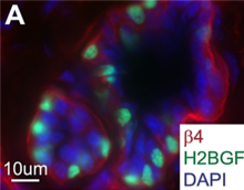
Sweat glands contain stem cells with regenerative potential. Mouse sweat glands were shown to harbor stem cells in sweat ducts and secretory glomeruli that respond differentially to injury and exhibit distinct regenerative potentials. Parasympathetic innervation maintains the progenitor population of sweat glands in an undifferentiated state, which is necessary for gland organogenesis.
Pathology
Inflammation of a sweat gland is called hidradenitis.
Contenido relacionado
Dynamoebales
Aorta
Biceps femoris muscle


