Stomach
The stomach (from the Latin stomăchus, derived from the Greek στόμαχος [stomachos], from the prefix στόμα [stoma], «mouth») is the portion of the digestive tube located between the esophagus and the intestine. In the human species it is located in the upper left region of the abdomen, below the diaphragm. It is a chamber in which the ingested food is mixed and stored, which is emptied in small intervals towards the duodenum thanks to peristaltic movements. Complete gastric emptying takes several hours after a heavy meal. The stomach is highly distensible, so it varies considerably in size depending on whether it is full or empty. It is divided into four main regions, which are called: cardia that joins it to the esophagus, fundus, body, and pylorus that communicates it with the intestine. It secretes several substances that together receive the name of gastric juice, formed mainly by hydrochloric acid and pepsin, a proteolytic enzyme that breaks proteins into smaller fragments to facilitate their absorption. In the stomach, food is transformed into a pasty mush called chyme.
Human Anatomy
The size of the stomach is variable depending on its degree of distension, when it is half full it is on average 25 cm high, 12 wide and 8 anteroposteriorly. The average capacity is around 1200 ml. It is located in the upper left region of the abdomen, below the diaphragm, its position is variable depending on whether the person is standing or lying down. Anatomically it can be divided into several areas:
- Cardias. It is an orifice that communicates the stomach with the lower portion of the esophagus. It has muscle fibers that help prevent gastric acid content from flowing to the esophagus.
- Fundus. It is the upper portion of the stomach, close to the cardia.
- Body. It is the central or main portion of the stomach.
- Inside. Receive this name the lower region of the stomach next to the plumber.
- Pylorus. It is located between the stomach and the duodenum. It is a thickening of the muscle fibers of the wall of the digestive tract that forms a sphincter in order to control the gastric emptied. It is usually in a state of contraction, but it relaxes regularly in coordination with the peristal movements.
The flattened shape of the stomach at rest determines the presence of an anterior and a posterior face. It also delimits a lesser curvature that extends from the cardia to the pylorus forming the concave edge of the stomach and a greater curvature that forms the convex side of the stomach, its length is four times greater than that of the lesser curvature.[citation required]
The stomach wall is made up of the characteristic layers of the entire digestive tract: mucosa, submucosa, muscularis, and serosa. The mucosa contains cells that produce mucus, hydrochloric acid, and digestive enzymes. The muscular layer consists of longitudinal, circular and oblique fibers. The serosa corresponds to the outermost envelope of the organ.[citation needed]
The stomach has fixation systems at its two ends, which are joined by the lesser curvature through the lesser omentum (omentum). At the level of the cardia, there is the posterior gastrophrenic ligament, which joins it to the diaphragm. On the pyloric side it is attached to the underside of the liver by the gastrohepatic ligament. These fixation systems determine their relationships with other abdominal organs.
Gastric folds
The mucosa that covers the internal layer of the stomach is not smooth, it presents a set of large and elongated longitudinal folds that have the function of multiplying the surface of the mucosa. When the stomach is full, the folds disappear and reappear again when the emptying process takes place. On the inner surface of the folds, there is a set of holes called crypts, gastric pits or foveolae where the gastric glands flow.
Arterial supply
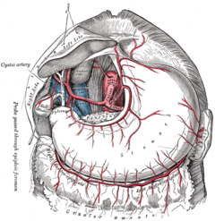
Irrigation is provided by branches of the abdominal aorta. The celiac trunk gives rise to the left gastric artery, which runs along the lesser curvature until it anastomoses with the right gastric artery, a branch of the proper hepatic artery (which leaves the common hepatic artery, a branch of the celiac trunk); these two arteries come to form what is the superior gastric coronary artery. The gastroduodenal artery also arises from the common hepatic artery, which gives rise to the right gastroepiploic artery that runs along the greater curvature until it anastomoses with the left gastroepiploic artery, a branch of the splenic artery (which comes from the celiac trunk); these form what is the inferior gastric coronary artery. This irrigation is complemented by the short gastric arteries which, coming from the splenic artery, reach the fundus of the stomach.
Venous return
The venous return is very parallel to the arterial, with right and left gastric veins, in addition to the prepyloric vein, which drain into the portal vein; short gastric and left gastroepiploic veins draining into the splenic vein; right gastroepiploic vein terminating in the superior mesenteric. Through the short gastric veins, a union (anastomosis) is established between the portal vein system and the inferior vena cava through the veins of the submucosa of the esophagus.[citation needed]
Lymphatic drainage of the stomach
Lymphatic drainage is provided by lymph node chains that run along the greater curvature (right and left gastroepiploic nodes and right and left gastric nodes). They are supplemented by the celiac and pyloric lymph nodes. These nodes are of great importance in gastric cancer, and must be removed in case of extension of the cancer. The removal is done according to the ganglion barriers, there are sixteen ganglion groups that correspond to three sections: perigastric nodes such as those of lesser and greater curvature, located in the arterial trunks such as the celiac trunk and those far from the stomach such as the retropancreatic and paraaortic. However, it has been proven that the lymphatic drainage of the stomach does not have a fixed pattern and any lymph node may be the first to be affected by the spread of a neoplastic process.
Histology
The stomach wall is made up of the characteristic layers of the entire digestive tract: mucosa, submucosa, muscularis, and serosa.
Mucous membranes
The tunica mucosa of the stomach presents multiple folds, ridges, and crypts. It is divided into three layers: epithelium, lamina propria, and lamina muscularis mucosa.
- Epithelium. On the surface of the mucosa there are simple epithelial cells that receive the name of superficial mucous cells. Epithelial cells form columns of secret cells that are called gastric glands that lead to foveolas or gastric fosites that end in the light of the stomach. In the gastric glands there are different types of cells, each of which produce a different substance:
- Mucous cells of the neck: They produce mucous.
- Parietal cells: They produce chloric acid and intrinsic factor.
- Main cells: produce pepsinogen and gastric lipasa.
- Endocrine cells
- Cells G. They are located in the pilric antro and produce gastrin, a substance that passes directly into the blood and stimulates the secretion of chloric acid and pepsinogen.
- Cells D that segregate somatostatin. However most of this hormone is produced by delta cells located in the Langerhans islets of the pancreas.
- enterochromafine cells (EC cells) that segregate serotonin.
- Histamine-releasing ECL cells, an essential substance to stimulate the secretion of chloric acid by the pacifier cells.
- Same size as mucosa: formed by laxo connective tissue.
- Muscose muscular tissue: also called mucosae muscleis, has two layers, little differentiated between.
Tunica submucosa
Formed by moderately dense connective tissue (support tissue that connects or unites the various parts of the body), in which there are numerous blood vessels, lymphatics, and nerve endings. It lies beneath the mucosa and forms the Meissner plexus.[citation needed]
Muscle Tunic
Within it are three layers of smooth muscle: internal or oblique, median or circular, and external or longitudinal. The muscular tunic is formed from the inside out by oblique fibers, the circular stratum and the longitudinal stratum. The gastric muscle tunic can be considered as the gastric muscle because thanks to its contractions, the food bolus is mixed with gastric juices and moves towards the pylorus with peristaltic movements.[citation needed]
The muscular tunic has its fibers in different directions, from more internal to more external, having oblique fibers, a circular layer and a longitudinal layer. In a cross section, this difference in the arrangement of muscle fibers can be clearly distinguished. It can be seen that the circular layer is thickened in some places, forming the sphincters that regulate the passage of food.
Serous tunica
The tunica serosa, made up of loose connective tissue covered by an epithelial layer called the mesothelium, surrounds the stomach in its entirety, expanding in its curvatures to form the lesser omentum, the greater omentum, and the gastrophrenic ligament.
Embryology
The stomach is formed by a process of dilation of the primitive digestive tract, specifically the foregut. It begins to be recognized visually from four weeks of gestation. During the dilation process, the dorsal portion grows more rapidly and gives rise to the greater curvature, while the ventral surface gives rise to the lesser curvature. As it grows, there is a 90º turn in the longitudinal axis, in such a way that the greater curvature is oriented to the left, while the lesser one is to the right.
Physiology of the stomach
The stomach receives crushed food from the esophagus, has a great capacity for distention and can hold up to 1.9 liters of food and liquids. The cells that make up the stomach wall produce different substances that help digestion and are collectively called gastric juices, its main components are hydrochloric acid and pepsin. Hydrochloric acid has the function of digesting food proteins and destroys most microorganisms, while pepsin is a protease enzyme that breaks down proteins into smaller peptides and amino acids.
The mixture of food with gastric juices produces a highly acidic semi-liquid substance called chyme. The chyme leaves the stomach through the pylorus and passes into the small intestine where most of the absorption process of nutritive substances takes place.
The function of the stomach is controlled by the autonomic nervous system, with the vagus nerve being the main component of the parasympathetic nervous system. Stomach acidity is controlled by several molecules including acetylcholine, histamine, gastrin, secretin, and prostaglandin E2.
Gastric emptying and mixing
After ingestion, food is mixed with gastric juice, forming chyme that passes in small amounts towards the duodenum, in order not to saturate the absorption and digestion mechanisms of the intestine.
Gastric emptying consists of removing food, previously fragmented and mixed, from the stomach into the duodenum. This process occurs thanks to the peristaltic waves caused by the contraction of the fibers of the muscular layer of the gastric wall.
- Gastric emptied. The peristaltic contraction that originates at the top of the gastric mollus, spreads downwards, in the direction of the pilric sphincter, being increasingly vigorous. As the strong antral peristaltic contraction propels the quimo forward, a small portion of this escapes through the open pylore until reaching the duodenum. The stronger the antral contraction, the more quimo is emptied with each contractable wave.
- Mixed. During the mixing, when the peristaltic contraction reaches the pylore, the sphincter is completely closed, so the emptied does not take place. When the quimo that is being driven forward reaches the closed sphincter, it returns back. As the quimo is driven forward and back into the gastric atro, the mixture is produced.
Mucous discharge
A constantly renewing layer of mucus covers the stomach wall. It is produced by two types of cells: the superficial mucous cells and the neck mucous cells, each of which produces a different mucin. Gastric mucus is made up of mucins, glycoproteins, and water. Among other functions, it protects the mucosa from the corrosive acidic environment that fills the gastric cavity.
Gastrin secretion
Gastrin is a hormone released by G cells located in the antrum of the stomach. It passes into the blood and stimulates gastric emptying and the production of hydrochloric acid by parietal cells. It also contracts the lower esophageal sphincter, relaxes the pyloric sphincter, and stimulates ECL cells to produce histamine. Gastrin is one of the most important substances in the regulation of gastric activity. It is secreted in response to distension of the stomach and elevation of gastric pH that occurs after food intake.
Histamine secretion
Histamine is a molecule that is of great importance in gastric physiology. It is synthesized by ECL cells located in the stomach glands, in response to gastrin. Histamine, after its release, stimulates the H2 receptors located in the parietal cells, causing the secretion of hydrochloric acid. In medicine, H2 antagonist drugs, such as ranitidine, are used in order to decrease acid production and improve the symptoms of various diseases. gastric diseases.
Proton Pump
It is a mechanism of active transport of the cell membrane by which H+ is secreted, which is exchanged for K+ ions. This process is carried out in the parietal cells of the stomach and is the basis for the formation of hydrochloric acid in the gastric cavity. Some medications, such as omeprazole, are capable of inhibiting the proton pump and decrease gastric acidity.
Diseases
- Gastritis.
- Peptic ulcer.
- Gastric cancer.
- Menetrier's disease.
- Pyric stenosis.
- Gastroesophageal reflux.
- Hernia de hiato. It consists in the protrusion of the stomach through the esophageal hiatus, thus penetrating the thorax. Many patients with hiatal hernia do not have just symptoms and do not require treatment. In the most serious cases surgery is required.
- Helicobacter pylori. It is estimated that more than two thirds of the world's population is infected by this bacteria that lives in the gastric epithelium. CR Historically, it was believed that the extremely acidic atmosphere of the stomach would maintain this immune organ of the infection. However, several studies have shown that helicobacter pylori bacteria can colonize the stomach and contribute to the appearance of stomach ulcers, gastritis and gastric cancer. This microorganism is able to survive in the stomach because it produces an enzyme called ureasa that metabolizes ammonia and carbon dioxide to neutralize chloric acid.
In other species
Although the precise shape and size of the stomach varies widely among different vertebrate species, the relative positions of the esophageal and duodenal openings remain relatively constant. Lampreys, hagfish, chimaeras, lungfish, and some teleost fish have no stomach and the esophagus opens directly into the anus. These animals follow diets that either require little food storage, or no predigestion with gastric juices, or both.
The gastric lining is usually divided into two regions, an anterior portion bordered by fundic glands and a posterior portion lined with pyloric glands. The cardia glands are exclusive to mammals, although they are absent in several species, the distribution of these glands varies between species, and they do not always correspond to the same regions as in man. Furthermore, in many non-human mammals, a portion of the stomach anterior to the cardia glands is lined with epithelium essentially identical to that of the esophagus. Ruminants have a complex stomach made up of several chambers.
In birds and crocodiles, the stomach is divided into two regions. Anteriorly it is a narrow tubular region, the proventriculus, bordered by the fundic glands, and connecting the true stomach for the crop. Beyond lies the powerfully muscular gizzard, fringed by pyloric glands, and, in some species, containing stones that the animal swallows to help break up food.
Second brain
The stomach and the entire digestive system itself, is home to the enteric nervous system, made up of around 100 million neurons, which makes it a key point of sensitivity, since they maintain bidirectional communication with the nervous system central, which is transformed into signals that are sent directly to the brain.
Additional images
|
Contenido relacionado
Mimosoideae
Lythraceae
Jet lagged


