Spermatozoon
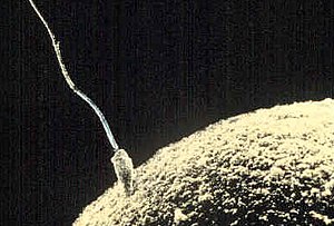
A spermatozoon is a haploid cell that makes up the male gamete. It is one of the most differentiated cells and its function is to form a totipotent zygote by fusing its nucleus with that of the female gamete, phenomenon that will give rise, later, to the embryo and the fetus. In the fertilization of mammals, the spermatozoa are the ones that determine the sex of the new diploid cell (embryo), since they contain an X sex chromosome or a Y chromosome.
Etymology
It comes from the French spermatozoïde. In turn, it was composed of the French words spérma (from the Greek σπέρμα, properly 'seed'), zôion (from the Greek ζῷον 'animal') and the suffix -oïde ('-oid', that is, 'in the form of' or 'similar to').
History
Sperm was first described in 1677 by the scientist Anton van Leeuwenhoek, recognized as the "father of microbiology". However, the first person to visualize them was a medical student named Johan Ham, who told him that he had seen some small "animalcules" on the surface. in the semen. Ham thought that these little animals were the result of the putrefaction of the seminal fluid. Leeuwenhoek, on the contrary, assumed that it was a common component of semen and made the first detailed description of spermatozoa. In addition, he was also the first person to propose that fertilization occurred through the entry of the sperm into the ovum, since at that time it was believed that fertilization took place by vapors emanating from the sperm.
Later, in 1697, Nicholas Hartsocker proposed the homunculus theory. Hartsocker was a Dutch scientist who dedicated himself to investigating the origin of life. Observing the sperm under a microscope led him to think that inside each of them was a homunculus, a kind of miniature human being. In conclusion, his theory states that in each of the spermatozoa the human being is already potentially found, which will later develop in the female womb.
Lastly, it is worth noting the figure of Lazzaro Spallanzani, an Italian physiologist and priest who investigated the unknown that was still fertilization and the role that spermatozoa played in the process. In one of his experiments he took virgin eggs and seminal fluid from frogs and put them in contact, achieving the fertilization of the former. This work could be considered as the first work on artificial fertilization (or insemination) carried out using the experimental method. Later, around 1790, he dedicated himself to investigating artificial insemination in dogs: he injected sperm into a female dog with a syringe and she became pregnant. Thanks to these experiments, the importance of sperm in the fertilization process was demonstrated. In addition, these discoveries served as a basis for the English surgeon Hunter to attempt their application to the human species.
Spermatogenesis
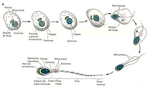
Spermatogenesis is the process in which spermatozoa are produced from the primordial germ cells of man (spermatogonia) through mechanisms of mitosis and meiosis. It is the mechanism of gametogenesis in men and it develops in the testicles (male gonads), although the final maturation of the spermatozoa takes place in the epididymis. This process of spermatogenesis in humans lasts approximately 64-74 days, in this way, any mutation, radiation exposure or other factors affect the semen secreted 74 days after exposure. Spermatozoa are male reproductive cells, destined for the fertilization of the egg; They measure from ten to sixty microns in length and are composed of a head that contains the chromosomal material and a tail or flagellum that acts as a propeller.
Structure of the human spermatozoon
Spermatozoa in humans are pyriform in shape, they only survive in a warm environment, although between 1 and 3 °C below body temperature, and they are the only human cells to have a flagellum; this helps it to be a cell with high mobility, capable of swimming freely.
They are mainly composed of two parts: a head and its flagellum, but within them we can distinguish several structures, which, in cephalic-caudal order (from head to tail, that is, from top to bottom), are: acrosome, nucleus, membrane, neck, midpiece, tail and terminal piece.
They live for an average of 24 hours, although it is possible that they fertilize the egg after three days.[citation required]
Head: acrosome, membrane, and nucleus
The head contains two main parts: the acrosome, which covers the anterior two-thirds of the head; and the nucleus, which contains the genetic load of the spermatozoon (23 chromosomes, in the pronucleus, which, together with the 23 of the ovum, give rise to the mother cell, when adding a total of 46 chromosomes, grouped in pairs). In humans, the sperm head is 5 µm (micrometers) in length. Both the pronucleus and the acrosome are surrounded by a small amount of cytoplasm and covered by a plasma membrane that joins the head to the body of the sperm. It is the most important part attached to the body. This membrane has high levels of polyunsaturated fatty acids, which are primarily responsible for sperm motility.
The acrosome is a layer formed by the enzymes hyaluronidase, acrosin and neuraminidase that will favor the rupture of the zona pellucida for penetration, which surrounds the oocyte. Also, hyaluronidase has been shown to be (together with the movement produced by the tail, the flagellum) responsible for swimming through the cluster of cells (cumulus oophorus) that surrounds the female gamete, separating them and allowing them to reach the oocyte. The function of the hyaluronidase enzyme is to break down the matrix in which cells are attached.
The nucleus, after the acrosome opens the oocyte's zona pellucida, is the only part that enters its cytoplasm,[citation needed] leaving behind the membrane already empty, to later fuse with the nucleus of the ovule, complete itself as a diploid cell and begin cell division (mitosis).
Therefore, since the mitochondria and everything else in the male gamete do not attach to the zygote, all the mitochondria in the new cell come from the maternal side.
The chromatin of a mature spermatozoon is highly condensed due to the replacement of histones with protamines during spermatogenesis.
Flagellum: neck, middle part, tail, end part
The neck is very short, so it is not visible under the light microscope. It is slightly thicker than the other parts of the flagellum and contains cytoplasmic residues of the spermatid. After these elements it contains two centrioles: the distal one, which originates the middle piece, and the other, the proximal one, disappears after having given rise to the flagellum. It contains a basal plate of dense material that separates it from the head and is where it is attached. they anchor 9 protein columns, which are modified centrioles, continuing throughout the tail. The middle piece originates from one of them (the distal).
- The middle part (about 4 or 5 μm in length) has a large number of mitochondria concentrated in a helical pod, which provide energy to the sperm, producing ATP. The sperm needs this energy to make its journey through the cervix, uterus, and female fallopian tubes to reach the egg to fertilize it.
- The tail (35 μm) provides mobility (functional flagelic area covered by membrane only).
- The tail provides mobility, and this can be type A, B, C or D; as seen in the seminogram. Type A would correspond to the sperm with rectilinear movement at a speed greater than 25 microns, compared to the 5-24 microns/s of type B which have a definite pathless movement, a speed less than 5 microns/s for type C, which are barely moved even if movement is detected in them, and a null movement for type D. Therefore, they are grouped into progressive movements (type A and B) and not progressive (C).
Abnormal motility corresponds to percentages less than 50% of A+B or 25% of A —Note that type A motility is rare in sperm in the population (around 1%)—. These abnormalities are called asthenozoospermia or asthenospermia; distinguishing between mild, moderate and severe.
Exclusive characteristics according to species
There is an indirect relationship between ejaculate volume and sperm concentration in different species:
- In humans, sperm have a head of 5 to 8 μm and a tail of 50 μm in length. They have a speed of 3 millimeters per minute. The normal human ejaculate is 2 to 6 ml (mililiters), and carries between 60 to 300 million sperm (according to the duration of the previous abstinence). To fertilize the egg there must be more than 20 million sperm per ml.
- In pigs, ejaculation is about 100 to 600 ml, with a concentration of 300 000 to 1 000 000 sperm/mm3. The length of the sperm is about 90 μm.
In some mammals, including humans, spermatozoa must be produced at a lower temperature than the body's average (2 °C less than normal in humans), so the male gonads are outside the body.
Epigenetics in the spermatozoon
The development of primordial germ cells into mature spermatozoa is a key stage for epigenetic reprogramming. DNA methylation and histone modification produce changes in gametogenesis; and alterations at any level of the epigenome of the spermatozoon can affect fertility and the correct development of the embryo.
Patterns of Methylation in Male Germ Cells
Recent studies in mice and humans show that male germ cells possess a unique methylation pattern compared to somatic tissues. Promoter methylation patterns in sperm, such as hypomethylation, would allow the expression of specific germ cell genes involved in spermatogenesis; while hypermethylation would lead to the repression of pluripotency and specific genes of somatic tissues. Many of these differently methylated sites in sperm and somatic tissues lie outside of gene regions and CpG islands, so they appear to play roles other than controlling gene expression. Methylation patterns in centromeric and intergenic sequences may be necessary for the formation of the specialized chromatin structure found in germ cells.
Somatic cell methylation patterns are established early during embryonic life and are maintained throughout development and adulthood. Germ cells, however, will undergo two waves of demethylation in order to establish sex-specific patterns that give rise to imprinted genes. Unlike in the egg, the epigenetic patterns of sperm begin to be acquired prenatally. The initial acquisition is related to the expression of Dnmt3a and Dnmt3L, which is consistent with the role of DNMT3 enzymes as de novo methyltransferases. These patterns are completed after birth in the pachynemal phase of meiosis.
Chromatin remodeling
Mammalian sperm chromatin is unique in that it is highly organized, condensed, and compacted. Chromatin remodeling is facilitated by histone hyperacetylation and by DNA topoisomerase II, which produces temporary nicks in the DNA to relieve torsional stress due to supercoiling.
The protamines condense the DNA strands and form a basic packaging unit of chromatin called a toroid. They confer a higher level of DNA packaging than that of somatic cells. All of this protects the chromatin during transport through the male and female reproductive tracts. In addition, protamines are necessary for the silencing of the paternal genome and the reprogramming of the imprinting pattern of the gamete. However, 15% of the histones are not replaced in human sperm chromatin, causing it to be less compacted.
In spermatogenesis, protamines progressively replace histones in a stepwise fashion. First, somatic histones are replaced by testis-specific histone variants. In spermiogenesis, tissue-specific histone variants are exchanged for transition proteins (TP1 and TP2) in a process that requires DNA remodeling. Transition proteins are necessary for normal chromatin condensation, to reduce the number of DNA breaks, and to prevent the formation of secondary defects in sperm and eventual loss of genomic integrity. Finally, in the elongation of the spermatids, the transition proteins are replaced by protamines. This sequential process facilitates molecular remodeling of the male genome in spermatid differentiation.
In humans, the P1/P2 ratio is approximately 1.0, and changes in this ratio are associated with infertility. Protamines are about half the size of histones. They are basic nuclear proteins characterized by a nucleus rich in arginines and cysteine residues. High levels of arginine cause a net positive charge, thus facilitating its binding to DNA. Likewise, cysteine residues facilitate the formation of multiple disulfide bridges between and between protamines, which are essential for higher-order packing of chromatin. P2 protamines contain fewer cysteine groups, making DNA more susceptible to damage.
Contenido relacionado
Dactylis
Cathestechum
Alnus


