Small intestine
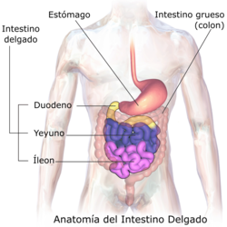
The small intestine is the section of the digestive system that connects the stomach to the large intestine. They are divided into three parts: duodenum, jejunum and ileum.
It fulfills the functions of digestion, absorption, barrier and also immunity.
It is one of the organs with the highest number of cell turnover in the entire organism, since its entire internal surface is renewed every five days.
Function of the small intestine
The small intestine absorbs the necessary nutrients for the body with the help of symbiotic bacteria or intestinal flora. It is located between two sphincters: the pyloric, and the ileocecal, which connects it with the large intestine. It constitutes the largest mucosal surface of the organism. Its length ranges between 3 and 7 meters (m), depending on numerous variables such as the size of the individual. In the cadaver, as a consequence of smooth muscle hypotonia, its length increases.
It has a layer of cells inside that form a barrier. Its mission is, in addition to digesting substances, to act by defending the body from the external enemy of the environment (substances that we ingest and microorganisms present in the intestine). It achieves this by keeping the intercellular tight junctions closed, to prevent the uncontrolled access of substances, toxins, chemicals, microorganisms and macromolecules, which could otherwise enter the bloodstream. It is now known that tight junctions, previously thought of as static structures, are actually dynamic and readily adapt to various circumstances, both physiological and pathological. There is a complex regulatory system that orchestrates the state of assembly of the protein network of intercellular tight junctions.

When the gates between cells (the tight junctions between cells) malfunction and instead of being closed or nearly closed, as they should be, they are open without control, an increase in intestinal permeability occurs. This opening causes substances to enter the body and, depending on the person's genetic predisposition, autoimmune and inflammatory diseases, infections, allergies or cancers, both intestinal and in other organs, may develop.
The chyme that is created in the stomach, formed by the food bolus mixed with hydrochloric acid, pepsinogen and other substances from peristaltic movements, is mixed in turn with biliary and pancreatic secretions (in addition to the duodenal itself) so as not to break the layers of the small intestine (since it has a highly acidic pH) and is carried to the duodenum. Food transit continues through this tube along which the digestion process is completed, the chyme is transformed into chyle and the absorption of useful substances is effected.
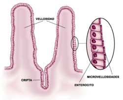
The phenomenon of digestion and absorption depends to a large extent on the contact of the food with the intestinal walls, so the greater this is and on a larger surface area, the better the digestion and absorption of food will be. This gives us one of the most important morphological characteristics of the small intestine, which is the presence of numerous folds that amplify the absorption surface, such as:
- Circular folds.
- Intestinal velocity (0.5 mm high and a nucleus of its own foil).
- Microvellosities of the small intestine: Microvellosities are extensions of the plasma membrane in cylindrical-shaped enterocytes, which serve to increase the contact of the plasma membrane with an internal surface. If the epithelium is of absorption, the microvellosities have in the central axis filaments of actin, if not out of absorption this axis would not appear. Covering the surface is a glycolix cover. Its function is to increase the absortive surface of the cells, and it is estimated that it allows an approximate increase of 20 times.
The small intestine absorbs every day several hundred grams (g) of carbohydrates, 100 g of fat, 50-100 g of amino acids, 50-100 g of ions and 7 l of water. The absorption capacity of the normal small intestine is much higher than these figures and can reach 500-700 g of protein and 20 liters (l) of water per day.
Shape and relationships of the small intestine
The duodenum is characterized by its relationship with the stomach, it is the main portion where pancreatic and hepatic juice arrives, but the jejunum and ileum are more difficult to distinguish and there is no separation between them.
In general, they can be distinguished because:
- The yeyune has a greater diameter than the ileon (3 cm the yeyun, 2 cm the ileum).
- The yeyune has more circular folds, more intestinal hairs and finer, while the ileum has less.
- In the ileum the lymphoid follicles (peyer plates) and vascular irrigation in the form of arches is greater than in the yeyuno. In addition, its walls are thinner and less vascularized.
Topographically, both the jejunum and the ileum occupy the infracolic space, although:
- The yeyuno stands a little higher and left (umbilical region) than the ileon (low and right).
- In general, the yeyunal handles are of more horizontal direction, while the ileals are of vertical direction.
The end of the small intestine is the terminal ileum which opens into the cecum via the ileocecal valve.
In the constitution of the intestinal wall, in addition to the usual layers of mucosa, submucosa, muscle and serosa, the presence of accumulations of lymphoid tissue that reaches up to the submucosa stands out. They are located in the antimesenteric border and their number is 30 or 40, and they measure up to 2.5 cm in diameter. As previously mentioned, they are more numerous in the ileum.
The entire length of the small intestine is attached to the posterior wall via the root of the mesentery. This attachment of the mesentery to the posterior wall begins at the level of the L2 vertebra, crosses the hook of the pancreas (where the superior mesenteric artery enters), crosses in front of the inferior vena cava, externally follows the common and external iliac vessels to terminate at the right iliac fossa, at the level of the promontory, lateral to the right sacroiliac joint, about 6 cm from the midline of the intestine.
Arterial supply
Irrigation comes from the superior mesenteric artery, a branch of the aorta, which walks inside the mesentery and from which the arteries arise:
- Lower pancreaticduodenals.
- Yeyunal branches and ileal branches: these Yeyunal and ileal branches have the particularity of forming artery arches that anastomosan one with others. First order arches are formed, new arches from these (second order) and even third order in the ileum. Finally, it originates the
- ileocholic artery, which ends up giving four branches: a) ascending clic that goes up by the ascending colon, b) anterior cecal, c) posterior cecal, and d) apendicular artery for the appendix. Other branches of the upper mesenteric artery go to the right angle of colon:
- Right and finally for the proximal part of the transverse colon
- The average wind artery, which is anastomated with the previous one. Therefore, the upper mesenteric artery irrigates the whole yeyun, the ileon and the right half of the large intestine including the appendix.
Venous drainage
Venous drainage is quite similar, being carried out by the superior mesenteric vein, the main constituent of the portal vein, along with the inferior mesenteric vein and the splenic vein.
Enteric Nervous System
It is in charge through both afferent and efferent nerves, respectively, of the motility and sensitivity of the intestine.
Histology of the small intestine
The intestinal mucosa is specialized in the digestion and absorption of nutrients and for this it has to increase its surface that gives light, in three ways:
- Circular folds, Kerckring valves or plications, which are visible at the naked eye and are permanent folds formed by mucosa and submucosa.
- Intestinal or villi speeds, which have a size of 0.5 to 1 millimeter and give the velvety texture of the interior of the intestine.
- Lieberkühn's necks, which are tubular glands located between the hairs. The Mother Cells appear at the bottom of these crypts.
The intestinal mucosal epithelium is made up of different cell types, which are:
- Absorbent or integer cells: The plasma membrane of these cells presents in its luminal pole multiple microvellosities that confer the appearance of a brush rib on an optical microscope.
- Caliciform cells: they are mucin or muco secretaries.
- Endocrine cells: are Algerian cells, also called basal granule cells. They belong to the APUD system. (also called SNED: diffuse neuroendocrine system)
- Paneth Cells: that produce lisozymes when bacterial infections (are defensive).
- Undifferentiated Mother Cells: responsible for the renewal of all cell types.
The lamina propria has loose connective tissue, with vessels and nerves. It is invaded by a lymphocytic population and by smooth muscle fibers from the muscular layer of the mucosa. It is called the Brucke muscle [citation needed] and it is the motor muscle of the villi [citation needed].
The central lacteal or milk duct is a central lymphatic vessel of the villus. It is found in every cross section of the villus. The lining of the quifer is discontinuous.
The glycocalyx is essential in the completion of the digestive process, in that it is the last link in degradation. Of the absorbed elements, the fats go to the central quiferous, and the rest to the blood.
If there are mucous glands in the submucosa, we are in a duodenum, and if not in a jejunum, an ileum. The duodenum presents these glands that secrete a mucin that neutralizes the acidic pH of the chyme.
In the digestive tract, the presence of MALT, mucosa-associated lymphoid tissue, is characteristic. This lymphoid tissue is found in the chorion or lamina propria of the mucosa. It is usually a diffuse or nodular lymphoid tissue. Plasmocytes are usually found alongside this lymphoid tissue. In the ileum, the lymphoid tissue is especially notorious for its arrangement in plaques, called Peyer's patches. The lymph node produces a change in the lining epithelium.
The Brunner's glands are the glands of the duodenal submucosa, which produce mucus in order to protect the mucosa from stomach acids. They are characteristic glands of the duodenum.
The number of goblet cells increases from the duodenum to the rectum, the absorptive cells decrease from the duodenum to the rectum. In the stomach there are no goblet cells, since the epithelium itself is mucigenic.
Diseases
Vascular diseases
Vascular diseases of the small intestine correspond to different etiologies. For this reason there is no universally accepted classification system. However, all these etiologies can manifest in the form of haemorrhage.
The most frequently observed vascular anomaly is angiodysplasia, which is defined as a dilated vascular complex located on the surface of the gastrointestinal tract, being present in 40% of hemorrhages of undetermined origin.
Tumors
In the small intestine, carcinogenesis occurs at a significantly slower rate than in other areas of the gastrointestinal tract, thus, despite being the largest area of the gastrointestinal tract, primary tumors of the small intestine represent only 2% of the total. total of gastrointestinal tumors. This fact is due to several factors, such as the rapid passage of a mainly liquid content and few bacteria. However, up to 40 different types of small bowel tumors have been identified, 75% of which are benign. The diagnosis of these, however, is complicated because the initial symptoms can be confused with other diseases.
Celiac disease
Celiac disease or celiac disease is an autoimmune disease caused by a permanent intolerance to gluten, a group of proteins present in wheat, oats, barley and rye (TACC) and derivatives, in people with a genetic predisposition. Traditionally considered a digestive disorder only, it is now known to be a systemic disease, since the abnormal immune response caused by gluten can lead to the production of different autoantibodies that can attack various organs and systems.
Lesions caused by celiac disease in the small intestine are not limited to the presence of intestinal villous atrophy, but often consist of minimal changes without villous atrophy, with mild to moderate inflammation, especially in older children two-year-olds and adults. Cases that include severe malabsorption and signs of malnutrition are practically exceptional, especially in children older than two years and adults. The symptoms that can appear are very varied, there is no common pattern, with mild, intermittent or even completely absent digestive symptoms, and all kinds of non-digestive symptoms.
Diagnosis is complicated, especially in children older than two years and adults, For this reason, currently approximately 83% of cases remain undiagnosed. A strict gluten-free diet for life improves symptoms and prevents the numerous associated complications, including all kinds of cancers.
Crohn's Disease
It is an inflammatory bowel disease that can affect the entire thickness of the intestinal wall, which is called transmural involvement, and that can appear simultaneously in several segments of the digestive tube or tract.
The terminal ileum (the last portion of the small intestine) is the most common site of involvement (up to 40-50% of all people with Crohn's disease), followed by the colon.
Other affectations
- Meckel Diverticle: is a small blind sac (5 or 6 cm long), present in the small intestine. It is a vestigeal organ of the onphalomesentric duct. It is the most common malformation of the gastrointestinal tract, being present in 2 % of the population. It can be asymptomatic all life or swell, ulcer or bleeding.
- Short intestine syndrome or small intestine failure. It causes malabsorption of food and related to a disease or surgical removal of a large portion of the small intestine.
- Irritable bowel syndrome: Functional disorder that affects 10% of the population. It is characterized by abdominal pain and alterations in the rhythm of depositions with the absence of organic pathologies.
- Intestinal motility disorders: usually grouped into two groups:
- Rapid Transit with the appearance of diarrhea, as in infectious gastroenteritis conditions.
- Slowed Transit, which usually occurs with constipation, as in the case of paralytic ileum, functional dyspepsia and intestinal pseudo-obstruction.
- Inguinal hernias: The direct inguinal hernias of the small intestine are produced by a weakness of the abdominal musculature and protruyen in the area of the triangle of Hasselbach.
- Cholera: is an infectious disease caused by bacteria Vibrio choleraewhich directly affects the small intestine and is often presented in the form of an epidemic. The bacteria causes alteration of permeability to the small intestine that ends up leading to: brusque diarrhea, vomiting, which lead to dehydration with the possibility of a fatal circulatory shock in a few hours.
- Theniasis: Disease caused by Had the cow (Taenia saginataor Lonely worm, silver parasite of the Cestoda class, the adult form that lives in the first portions of the small intestine of the human being. They usually measure between 2 and 5 meters in length (can reach up to 10 meters). Normally only one worm is found in the intestine of the infected person. The disease is relatively common in Africa, parts of Eastern Europe, the Philippines and Latin America.
Contenido relacionado
Fagaceae
Erythroxylaceae
Cynodon


