
Skull
The skull (from the Greek: κρανίον, kranion and from the Latin: cranium) is part of the system bone or skeletal system, is a bony box that protects from blows and contains the brain mainly. The human skull is made up of the articulation of 8 bones, which form an open and ovoid cavity of variable thickness, with an approximate capacity of 294,594 ml (in adults). In zoology, the ossified skull is also called the osteocranium.
Semantic clarification

The skeleton of the head, or neofacial skeletal massif, is the set of the bones of the skull (ossa cranii PNA) and the bones of the face (ossa faciei PNA), known as skull in colloquial terms, although anatomically it is the bony head, the skull being a part of the head. It is common for skull to designate the entire bony head, which is improper in the study of anatomy. However, in other areas (embryology, biology, etc.) the skull is considered a synonym for head skeleton.
The distinction between skull and face is very clear: the skull houses the brain, fundamentally the -neurocranium-, while the face inserts the muscles of mimicry and chewing and houses some of the sense organs. The skull fulfills a very important function, since it is concerned with containing the entire central nervous system, with the exception of the spinal cord.
Bones of the skull
The skull is the skeleton of the head and various bones make up its two parts: the neurocranium and the splanchnocranium. The neurocranium is the bony case of the brain and its membranous coverings. In an adult, it is made up of a series of eight bones: four unpaired, centered in the midline (frontal, ethmoid, sphenoid, and occipital) and two paired bilateral series (temporal and parietal). The bones of the so-called neurocranium together make up two other anatomical structures: The frontal, parietal and occipital bones usually make up a dome-like roof structure, called the calvaria or cranial vault, while the sphenoid and temporal bones form part of the base of the skull. skull.
The splanchnocranium or viscerocranium, also called the facial skeleton, makes up the anterior part of the skull and is made up of the bones that surround the mouth (maxillae and mandible), the nose/nasal cavity and most of the orbital cavities. This consists of 14 irregular bones: two centered unpaired bones (mandible and vomer) and six bilateral paired bones (maxilla, inferior nasal turbinate, zygomatic, palatine, nasal, and lacrimal). The jaws and mandible house the teeth; In other words, they provide the cavities and supporting bone for the maxillary and mandibular teeth. The jaws form the majority of the upper facial skeleton, attached to the base of the skull. The mandible forms the lower facial skeleton, being this mobile in nature as it articulates with the base of the skull at the temporomandibular joints.
Cranial Regions


The skull, as a cavity, can be considered from the inside of that cavity as endocranium, or from the outside as exocranium. In turn, as a whole, it can be divided by means of a horizontal section that passes through the median frontal eminence and through the external occipital protuberance, into two portions:
- a top, cranial or calota vault (PNA Calvary);
- a lower part, the base of the skull (basis cranii NAPA).
This division is not so arbitrary. Part of the different embryological origin of bone structures: endochondral ossification for the bones of the cranial base, and intramembranous ossification for the bones of the skull.
The vault is made up of the frontal (vertical part), the parietals, the temporal scales, and the occipital (upper part). It is covered by the scalp; The bones are joined by joints called sutures: coronal or frontoparietal suture, between the frontal and parietal bones, sagittal or interparietal suture, between the two parietal bones, and lambdoid or parieto-occipital suture, between the occipital bones and the parietal bones.
Origin, development and growth
Cranial cephalic structures originate from the mesenchyme derived from the neural crest cells and the paraxial mesoderm. The bones that make up the skull do not have the same origin, which is why the difference is made between the regions of the vault and the cranial base.
- Membrane neurocráneo (dermatocráneo) - cranial vault
Calotate bones are flat lining bones. They are generated by the process of intramembranous ossification from fibrous connective tissue plates (mesenchyme) that surround the brain. In this way, centrifugally, flattened membranous bones develop (ossify). At birth, the bones of the calvarium are not fused or fully ossified, leaving interosseous spaces covered by fibrous tissue (sutures and fontanels).
- Cartylaginous neurocráneo (condrocráneo) - base of the skull
The bones of the skull base develop by the process of endochondral ossification from the chondrocranium, a structure made up of several separate osteogenic cartilaginous nuclei that extend throughout the entire skull. region (prechordal chondrocranium originating from neural crest, and chordal chondrocranium originating from paraxial mesoderm)
- Fontanelas and sutures - Newborn Cráneo

At the time of birth, the flat bones of the skull are not completely ossified and are separated from each other by spaces occupied by fibrous connective tissue derived from the neural crest that will contribute in the future to the definitive formation of bones and their articulation (symphibrosis). These spaces are the metopic, coronal, sagittal, and lamdoid sutures. In those places where more than two bones articulate, the sutures are wide and form the six fontanelles:
- two odds and stocks: previous and later fontanelas;
- and two sides and pairs: posterolateral (mastoid) and anterolateral (esphenoidal).
Sutures and fontanelles are of paramount importance during childbirth, since they allow a superimposition mechanism between the bony plates of the skull (modelling) that enables the passage of the fetal head through the birth canal. Birth. During the puerperium, the bones return to their original position. During childhood, palpation of the anterior fontanelle allows verification of normal development and ossification of the skull, as well as intracranial pressure.
- Growth and consolidation
The sutures and fontanelles take years to ossify completely and achieve total coaptation between the bony parts of the skull. The growth of the vault bones that continues into adulthood is at the expense of the fibrous material of the sutures and fontanels. This mechanism admits a certain compliance of the cranial box for the growth of the brain and an adaptation according to the development and growth of the facial mass. Full cranial capacity is reached around 6 years of age.
Joints
The joints of the cranial bones are synarthrosis, immobile joints that fix the bone pieces together by means of cartilage (synchondrosis) or fibrous connective tissue (symphibrosis).
Those bones that form part of the cranial base, developed by endochondral ossification, are joined together through synchondrosis. And the bones from the vault of the skull (and the bones of the face as well), developed from connective tissue stubs, are joined together through symfibrosis or sutures (suturae PNA).
Depending on the configuration of the articular surfaces involved in bony union, there are four types of sutures (symphibroses) in the skull:
- the dented sutures arise from the occipital union "by gear" of the joint surfaces. It is the joint that binds to the frontal, occipital, parietal, sphenoids and ethmoid bones (fronto-parietal articulations; parieto-parietal; parieto-occipital; fronto-etmoidal; fronto-esfenoidal) and joints with the facial massif (fronto-malar; fronto-nasal, etc.).
- The squamous sutures arise from the binding of rough surfaces and "sized to bezel". It's the temporo-parietal joint.
- schindilesis is the articulation of the vomit (cara) with the sphenoids (craneous), formed by the union of a surface with a crest shape (lower sphenoidal crest) that fits with a complementary surface in the form of a slot (between the wings of the vomer).
- the harmonic suture contact flat and linear surfaces (between the viscerocraneous bones (face)).
Sexual dimorphism in the human skull
In the mid-19th century, anthropologists found it crucial to distinguish between male and female skulls. An anthropologist at the time, James McGrigor Allan, argued that the female brain was similar to that of an animal. This allowed anthropologists to declare that women were in fact more emotional and less rational than men. McGrigor then concluded that women's brains were more analogous to those of babies, which is why he considered them inferior at the time. To further these claims of female inferiority and silence feminists of the time, other anthropologists joined in. to studies of the female skull. These cranial measurements are the basis of what is known as craniometry. These cranial measurements were also used to establish a connection between women and blacks. Research has shown that although in the first years of life there is little difference between male and female skulls, in adulthood male skulls tend to be larger and more robust than female skulls, which are lighter and smaller, with a cranial capacity approximately 10 percent less than that of men. However, subsequent studies show that women's skulls are slightly thicker and therefore men may be more susceptible to head injuries than women. However, other studies show that men's skulls are slightly thicker in certain areas. As well as some studies that show that women are more susceptible to head trauma (concussion) than men. Men's skulls have also been shown to maintain their density with age, which may help prevent head injuries, while women's The density of the skull of women decreases slightly with age.
Male skulls may have more prominent supraorbital ridges, a more prominent glabella, and more prominent parietal bones. Female skulls tend to have rounder orbits and narrower jaws. Male skulls have on average a larger and broader palate, squarer orbit], larger mastoid processes, larger sinuses, and larger occipital condyles than those of females. Male human jaws tend to have squarer chins and thicker, rougher muscle attachments than female jaws.[citation needed]
Skulls of other animals
Fenestra


Fenestrae are anatomical features of the skulls of various types of amniotes, characterized by bilateral symmetrical foraminae (fenestrae) in the temporal bone. Depending on the lineage of a given animal, there may be two, one, or no pair of temporal fenestrae, above or below the postorbital and squamosal bones. Superior temporal fenestrae are also known as supratemporal fenestrae, and inferior temporal fenestrae are also known as infratemporal fenestrae. The presence and morphology of the temporal fenestra are essential for the taxonomic classification of synapsids, of which mammals are a part.
Fenestrae (from the Latin fenestrae, meaning windows) are openings in the skull. There are four big types:
- Fenestra totorbital
- Fenestra mandibular
- Fenestra quadjugal
- Fenestra subscamosa, an opening between two parts of the scaly bone in some rodents
- Temporary Fenestra
Physiological speculations associate it with increased metabolic rates and an increase in jaw musculature. Early Carboniferous amniotes did not have temporal fenestrae, but two more advanced lines did: the synapsids (mammal-like reptiles) and the diapsids (most reptiles and later birds). Over time, the temporal fenestrae of diapsids and synapsids modified and increased in size to allow for stronger, more muscular jaw bites. Dinosaurs, which are diapsids, have large forward openings, and their descendants, the birds, have modified temporal fenestrae. The synapsids have a fenestral opening in the skull, located in the posterior part of the orbit. In their descendants, the cynodonts, the orbit was fused with the fenestral opening after it began to expand in therapsids. Thus, most mammals also have it. Later primates separated their orbit from the temporal fossa by the postorbital bar with haplorhines later evolving the postorbital septum.
Classification
There are four types of amniote skulls, classified by the number and location of their temporal fenestrae. These are:
- Anapsida - no openings
- Synapsida - a low opening (under postorbital and scaly bones)
- Euryapsida - a high opening (over postorbital and scaly bones); euryápsidos actually evolved from a diapsy configuration, losing their lower temporary fenestra.
- Diapsy - two openings
Evolutionarily, they are related as follows:
- Amniota
- Class Synapsida'
- Order Therapsida
- Mammalia Class - Mammals
- Order Therapsida
- (No rating) Sauropsida - reptiles and birds
- Class Reptilia
- Subclase Parareptilia
- Infraclass Anapsida'
- Subclass Eureptilia
- Infraclass Diapsy
- Class Birds
- Infraclass Euryapsida
- Infraclass Diapsy
- Subclase Parareptilia
- Class Reptilia
- Class Synapsida'

A hypopotamus skull.

A tyranosaur skull.

A cat skull

A Brass-baked skull, a typical rodent

Snooze.
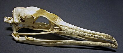
Phalacrocorax carbo.


Contenido relacionado
Plantar muscle
Grelo
Poaceae