Respiratory system
The respiratory system or respiratory system is the set of organs possessed by vertebrates (including the airways and lungs) to exchange gases with the environment atmosphere. Through the airways, air circulates in the direction of the lungs, and gas exchange takes place in these organs. Its structure and function is highly variable depending on the type of organism and its habitat.
The main organ of the human and mammalian respiratory system is the lung. In the pulmonary alveoli, the gas exchange process is produced by passive diffusion, thanks to which the blood captures atmospheric oxygen and eliminates carbon dioxide (CO2), a waste product of metabolism. The apparatus The human respiratory system consists of the nostrils, mouth, pharynx, larynx, trachea, and lungs. The lungs consist of bronchi, bronchioles, and pulmonary alveoli.
The respiratory muscles are the diaphragm and intercostal muscles. On inspiration, the diaphragm contracts and descends, thus the thoracic cavity expands and air enters the lungs. On expiration or exhalation, the diaphragm relaxes and rises, the thoracic cavity decreases in size, causing the vent to escape. air from the lungs to the outside.
In addition to gas exchange, the respiratory system plays an important role in maintaining the balance between acids and bases in the body through the efficient removal of carbon dioxide from the blood.
Human Respiratory System
In humans, the respiratory system consists of the airways, lungs, and respiratory muscles that move air both in and out of the body. In the pulmonary alveoli, oxygen and carbon dioxide molecules are exchanged passively, by diffusion between the gaseous environment and the blood. In this way, the respiratory system makes oxygenation and elimination of carbon dioxide possible, which is a waste substance from cellular metabolism. The system also fulfills the function of maintaining the balance between acids and bases in the body through the efficient removal of carbon dioxide from the blood.
Parts of the respiratory system
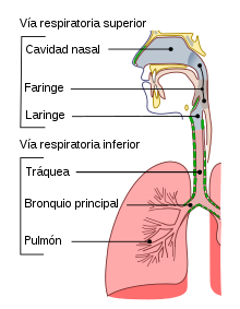
The human respiratory system consists of the following elements:
- Nasal stuff: There are two large cavities whose function is to allow the entry and exit of the air, which is moistened, filtered and heated through structures called cornets.
- Faringe: Tube-shaped structure located on the neck and coated with mucous membrane; connects the mouth cavity and nostrils with the esophagus and the larynx.
- Laringe: It is a duct that allows the passage of air from the pharynx to the trachea and lungs. In the larynx are the vocal cords that leave a space called glotis.
- Vocal chords. They are two folds located in the larynx that vibrate when the air passes through them producing the voice.
- Gloss. It is the narrowest portion of laryngeal light, space that is limited by vocal cords.
- Epiglottis: Epiglottis is a cartilage located on top of the glotis that obstructs the passage of the food bowl at the time of swallowing, preventing it from going to the respiratory system. It marks the limit between oropharynx and larynx.
- Trachea: It is a tube-shaped conduit that has the function of making air passage possible between the larynx and the bronchus. Its wall is reinforced by a set of C-shaped cartilages that make it difficult for the route to collapse by external compression over the neck.
- Pulmons: Organs whose function is to make gaseous exchange with blood. Within each lung, the bronchial tree gradually divides by giving ever smaller ramifications. The trachea gives rise to the two main bronchus that are divided into secondary bronchus or lobars. Each lobar bronchus is divided into tertiary or segmentary bronchus that are divided into bronchioles. The bronchiole continues the process of branching and gives rise to the terminal bronchiole from which the respiratory bronchioles that is where the alveolar sacs are found.
- Bronquio: Fibrocartilaginous tubular conduct leading the air from the trachea to the bronchioles.
- Bronquilo: Conduct that drives the air from the bronchus to the alveoli.
- Alvelo: The alveolos are located at the end of the last ramifications of the bronchioles. They have the form of small bags and are the place where the gas exchange occurs with the blood. Its wall is very thin, because it is formed by a unicelular layer, that is, formed by a single cell. In addition to the two lungs, the human organism has about 600 million alveolos that if deployed in its entirety would occupy a surface of 60 m2, this enormous surface is the one that makes it possible to obtain the amount of oxygen necessary for the vital functions.
- Intercostal muscles: Links located in the existing space between two consecutive ribs. They have an important role in mobilizing the chest during inspiration.
- Diaphragm: Muscle that separates the chest cavity from the abdominal cavity. When contracted lowers and increases the size of the chest cavity by causing inspiration. When relaxed, it decreases the size of the chest cavity and causes spiration.
- Pleura and pleural cavity: Pleura is a serosa membrane covering both lungs. It consists of two layers, the parietal pleura in contact with the chest wall and visceral pleura in contact with the lungs. Between both layers is a space called pleural cavity. Pressure in pleural cavity is less than atmospheric pressure which makes it possible to expand the lungs during inspiration.
Ventilation
The function of the respiratory system is to move volumes of air from the atmosphere to the lungs and vice versa. This is possible thanks to a process known as ventilation.
Ventilation is a cyclical process and consists of two stages: inspiration, which is the entry of air into the lungs, and expiration, which is the exit. Inspiration is an active phenomenon, characterized by the increase in thoracic volume that causes a negative intrapulmonary pressure and determines the displacement of air from the outside to the lungs. The contraction of the main inspiratory muscles, diaphragm and external intercostals, is responsible for this process. Once the intrapulmonary pressure equals atmospheric pressure, inspiration stops and then, thanks to the elastic force of the ribcage, it retracts, generating a positive pressure that exceeds atmospheric pressure and determining the output of air from the lungs..
Under normal conditions, expiration is a passive process, when the diaphragm relaxes it rises and returns to its initial position. However, in forced expiration, the rectus abdominis muscle contracts, which propels the abdominal viscera upwards, this process further decreases the intrathoracic volume and increases the amount of air that is displaced to the outside.
Ventilation control
Ventilation is very carefully controlled to enable arterial PaO2 and PaCO2 levels to remain within narrow limits despite the demands O2 uptake and CO2 removal rates vary widely. The respiratory system has a set of sensors that gather information, which reaches the central controller of the brain, which coordinates the information and sends impulses to the effector respiratory muscles, which cause ventilation.
Sensors (inputs)
The main sensors in the control of breathing are the chemoreceptors, which respond to changes in the chemical composition of the blood or other liquid. They have been classified anatomically as central and peripheral.
- Central smokers. They are close to the ventral surface of the spinal bulb are surrounded by the extracellular fluid of the brain and respond to H+ changes in that fluid. The level of CO2 in the blood regulates ventilation mainly by its effect on the pH of the cerebrospinal fluid.
- Peripheral smokers. They are found inside the carotid bodies, in the fork of the primitive carotid arteries, and in the aortic bodies, above and below the aortic cayado. Respond to the decline of the PO2 arterial and increased pCO2 and H+ concentration. They are responsible for any increase in human ventilation as a response to arterial hypoxemia.
In the lungs there are also sensory receptors that are involved in the control of the caliber of the airways, bronchial secretion, as well as in the release of mediators by mast cells or other inflammatory cells, this information reaches the superior centers through through the sensory fibers of the vagus nerve.
Central brain control
The control of ventilation is possible thanks to a complex interconnection of neurons located in various locations in the brain from where the orders originate that through the nerves reach the different muscles in charge of pulmonary ventilation. The normal automatic process of breathing is involuntary and originates from impulses that come from the brain stem, however, some voluntary control can be had within certain limits, since stimuli from the cerebral cortex can be prioritized with respect to those from the stem. cerebral.
The brain's main respiratory centers are located in the medulla oblongata and automatically establish the rhythmic pattern of breathing. A Dorsal Respiratory Group can be distinguished that can modify the basic rhythm according to the needs of the organism and a Ventral Respiratory Group that contains neurons that are activated when a high level of ventilation is required. There are two other nuclei of neurons located in the pons called the Pneumotaxic Center and the Apneustic Center that also influence the frequency and depth of inspiration.
The cerebral cortex plays a role in the voluntary control of ventilation, since it is possible to perform voluntary hyperventilation or hypoventilation for short periods of time. Other parts of the brain such as the limbic system and the hypothalamus can affect the pattern of breathing, for example in emotional disturbances.
Effectors (outputs)
As actuators of the respiratory system are the diaphragm, intercostal muscles, abdominal muscles and accessory muscles. In the context of ventilation control it is essential that these muscle groups work together in a coordinated manner. There is evidence that in some neonates, particularly premature ones, there is a lack of coordination in the activity of the respiratory muscles, especially during sleep. For example, the thoracic muscles can do the work of inspiratory work while the abdominal muscles do the work of expiratory.
Gas exchange
Once the pulmonary alveoli are filled with air after the inspiration process, oxygen has to diffuse into the blood, while carbon dioxide follows the opposite path, that is, it passes from the blood to the pulmonary alveoli. This process occurs by a simple diffusion mechanism motivated by a random cross-linking of the molecules that pass from where they are in higher concentration to where the concentration is lower. The phenomenon is due to the fact that the molecules are in continuous movement and move in all directions colliding and bouncing between them repeatedly. There is a physical law according to which when a gas is in a closed chamber and its concentration is different at the two extremes, the particles tend to move from where the concentration is high to where it is low, finally reaching an equilibrium situation, a process known as simple diffusion. In the respiratory system, diffusion occurs in the alveolus very rapidly, taking place in the first 0.25 seconds of the 0.75 seconds of blood circulation time through the pulmonary capillaries.
Oxygen transport in the blood
Once oxygen passes into the capillary blood in the pulmonary alveoli, it must be distributed throughout the body to satisfy the requirements of the cells, which need this element as a priority. The partial pressure of oxygen is higher in the pulmonary alveoli than in the capillary blood, which is why the process of simple diffusion between both media occurs. On the other hand, the partial pressure of oxygen is lower in the cells of the tissues than in the blood, so that when oxygenated blood reaches the tissues of the whole body, part of its oxygen is released, which is incorporated by diffusion. through the membrane into the cell to make possible the cellular respiration that takes place in the mitochondria.
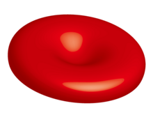
The capacity of the blood to transport dissolved oxygen directly is very low, since this element is poorly soluble in water. For this reason, the body has developed a protein called hemoglobin that has the ability to capture oxygen and transport it with great efficiency. If hemoglobin did not exist, the heart would have to pump about 80 liters of blood per minute, which would be completely impossible. Thanks to hemoglobin, cardiac output is only 5 liters of blood per minute, this figure being enough to keep all cells oxygenated. resting body cells. Each hemoglobin molecule has the capacity to transport four oxygen molecules, a single red blood cell has 250 million hemoglobin molecules and in a milliliter of blood there are around 5 million red blood cells.
- Hb+4O2→ → Hb(O2)4{displaystyle mathrm {Hb+4 o_{2}rightarrow Hb(O_{2})_{4}}} }
Hb= Hemoglobin, O=Oxygen.
Adaptation to height
As altitude increases, atmospheric pressure decreases. For this reason, at high altitudes, the phenomenon of hypoxia can occur, since the organism must produce extra energy expenditure for the same amount of oxygen to enter the body. The consequences of hypoxia are:
- Immediately. Tachycardia, increased cardiac expenditure, increased resistance of the pulmonary artery and hyperventilation occurs which, if excessive, can cause metabolic alkaloses.
- Chronicles. There is an increase in the mass of red blood cells, renal compensation of respiratory alkalosis, increase in muscle capillary density and increase in the number of mitochondria and their oxidative enzymes.
Blood gas values
- PaO2: It is the partial oxygen pressure in the blood, its normal values can vary between certain limits, depending on the individual's age, sex and body weight. Oscila between 66 and 100 mmHg.
- PaCO2: It is the partial pressure of carbon dioxide in the blood. Normal values range from 35 to 45 mmHg. If the figure is higher than 45 mmHg, this indicates that the body is retaining too much carbon dioxide in the blood.
To obtain these parameters, it is necessary to extract blood from an artery, generally the radial artery. Venous blood, which is commonly used to determine other analytical values, is useless, since venous blood contains much less oxygen. When the measured parameters are compared with the reference values, it can be detected if there is any health problem that affects the function of the respiratory system.
Lung volumes
Under normal conditions, a person breathes 15 times per minute and 500 cc of air enters their lungs with each inspiration. During expiration, the same amount that entered the lung leaves. Therefore, in one minute the pulmonary ventilation is 15 x 500 = 7.5 liters, which is what is called minute volume. However, the depth of respirations and their frequency can increase considerably in conditions of physical effort, so the minute volume can reach up to 200 liters per minute, multiplying the value at rest more than 20 times.
- Current volume (VC): It is the amount of air used in each non-forced breathing. It's about 500 ml. This means that in normal conditions during an inspiration 500 cc of air enters in the lungs and during expiration the same amount comes out.
- Spiracy reserve volume. It corresponds to the extra volume of air that can be expelled to the outside when a forced expiration is performed. Its average value is 1000 cc.
- Inspiring reserve volume. It corresponds to the extra volume of air that can be inhaled when a forced inspiration is made. Its average value is 2500 cc.
- Residual volume. It corresponds to the amount of air left inside the lung after a maximum expiration. Its average value is 1200 ml.
Total lung capacity is given by the sum of the 4 aforementioned volumes.
Pressures in the respiratory system
Four different pressures must be considered to understand the functioning of the human respiratory system. These pressures are not constant, as they change throughout the respiratory cycle.
- Atmospheric pressure. It corresponds to air pressure in the atmosphere.
- Alveolar or intrapulmonary pressure. It is the air pressure contained in the alveoli.
- Pleural or intrapleural pressure. It is the pressure existing in the pleural cavity, that is, in the space between the visceral pleura and the parietal pleura. Pleural pressure is negative and therefore less than atmospheric.
- Transpulmonary pressure. It corresponds to the difference between alveolar pressure and pleural pressure.
Concepts
- Hypoxemia: Decrease in the PaO2 80 mmHg.
- Hypoxia: PaO Decrease2 on a cellular level.
- Respiratory insufficiency: Decreasing partial oxygen pressure (OP)2below 60 mmHg at sea level. There are two types:
- Partial: PaO decrease2 60 mmHg with PaCO2 Normal or low.
- Global: PaO decrease2 60 mmHg and increase of PaCO2 45 mmHg (acidosis respiratory).
Atmospheric air composition
| Nitrogen | 78.00 % |
| Oxygen | 21.00 % |
| Argon and helium | 0.92 % |
| Carbon dioxide | 0.04 % |
| Water vapor | 0.04 % |
Alveolar air composition
| Nitrogen | 78 % |
| Oxygen | 15-16 % |
| Water vapor | 6 % |
| Carbon dioxide | 4-5 % |
Most common respiratory diseases
Some respiratory illnesses are caused by viruses and bacteria. If not properly prevented and treated, they can be deadly. Pediatric lung diseases cause 50% of deaths in children under 1 year of age and approximately 20% of all hospitalizations in children under 15 years of age.
- Common cold. It's the most common infectious disease. The incidence is greater in early childhood than in any other period of life. Children under 5 years of age have 6 to 12 episodes of cold per year.
- Rinitis. It is presented as constant nasal colds. It has significant morbidity and can contribute to the development of sinusitis and asthma exacerbations.
- Rinosinusitis. It is defined as successive episodes of bacterial infections of paranasal sinuses, each lasting less than 30 days and separated by periods of at least 10 days, during which the patient is without symptoms.
- Faringitis. More than 90% of cases of sore throat and fever are due to viral infections. Most people develop rinorrhea and mild cough.
- Amigdalitis. It is due to an infectious process that affects the palate amygdala.
- Tracheitis. It is the acute inflammation of the trachea, which is the airway linking the larynx with the bronchus. Bacterial tracheitis most often affects children of school age (about 5 years).
- Bronchitis. It is inflammation of the bronchus, main airways of conduction within the lung. It may be produced by viral or bacterial infections of the lower respiratory system favored by exposure to irritants of the environment including tobacco smoke.
- Chronic obstructive pulmonary disease. The main cause is smoking.
- Enfisema. The main cause is the inhalation of tobacco smoke.
- Asma. Reversible obstruction of minor airways that can progress to respiratory failure if an immediate intervention is not carried out.
- Pulmonary tuberculosis. Infectious disease caused by Koch bacillus.
- Pneumonia. Pneumonia is inflammation of the lung. The most common cause is infective microorganisms, especially bacteria and viruses. It appears more easily when one or more of the defense mechanisms that protects the lung are inadequate.
- Silicosis. Caused by prolonged inhalation of chemical compounds containing crystalline silica, it often occurs in mine workers.
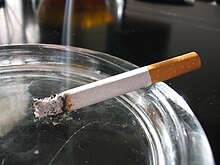
- Lung cancer. Although it can occur in non-smoking or passive smokers, the main cause is the direct aspiration of tobacco smoke.
- Cystic fibrosis. Genetic disease that mainly affects the lungs.
Preventive measures
- Lung cancer. Among the diseases of the respiratory system, lung cancer is highlighted by its frequency and severity. Despite current advances in medical treatments, it continues to cause the death of the affected person on many occasions. Therefore, preventive measures are fundamental, highlighting the fact that they avoid exposure to tobacco smoke, both as an active and passive smoker. The higher the exposure to tobacco the higher the chance of getting this disease. Other substances that have been related to the appearance of lung cancer are asbestos and radon gas.
- Gripe. Influenza is an important cause of mortality when it affects people of advanced age or who have risk factors such as heart disease, immunity deficiency or other lung disorders. This is why annual anti-grip vaccination is recommended as an effective measure to prevent flu in people with some of the above risk factors.
Respiratory system in animals
Living beings have developed several gas exchange systems with the environment in which they live: skin, tracheal, branchial and pulmonary. Through any of these systems they incorporate oxygen from the external environment and eliminate carbon dioxide and water vapor. Man and mammals have only pulmonary respiration, but some organisms, such as amphibians, use several systems simultaneously and have cutaneous and pulmonary respiration.
- Skin breathing. In some animals breathing occurs directly through the skin. For this to be possible, the skin should be very thin and not covered by corneal structures such as scales. Among the animals that have skin breathing are the anelids. Skin breathing is responsible for more than 20% of gas exchange in amphibians.
- Tracheal breathing. Tracheal breathing takes place in many invertebrates, including insects, miriapodes and some arcnides. These animals have a series of holes throughout their body called stigmas by which the air of the atmosphere is introduced.
- Branch breathing. Branch breathing takes place in fish. Branquias are respiratory organs of many aquatic animals. They are formed by a set of very thin sheets surrounded by blood vessels. When the oxygen-filled water passes between the gills, the gaseous exchange with the blood occurs.
- Pulmonary breathing. It takes place in most land vertebrates: amphibians, reptiles, birds and mammals including man. The lung respiratory system consists of a system of ducts that transports the air to the lungs. The lungs consist of a set of alveoles surrounded by blood capillaries. In the alveolos it is where the gas exchange occurs with the blood. The oxygenated blood is distributed throughout the body through the circulatory system.
Simple Organisms
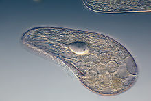
Protozoa are unicellular organisms that do not have a respiratory system, they capture oxygen directly from the environment by diffusion through the cell membrane. Some multicellular organisms such as hydras and jellyfish also do not have a respiratory system.
Insects
Insects breathe through a set of holes open to the outside located on both sides of the thorax and abdomen called spiracles or stigmas. Each spiracle has a small valve that opens when the animal needs to get oxygen. From the spiracles there are ducts called tracheas that have the function of transporting air to the interior of the animal. The tracheae become tracheoles and branch progressively to form small tubes, each of which carries oxygen to a small group of cells. Sometimes the animal performs contractions or dilations of the abdomen to facilitate the movement of gases into the duct system.
Fish
Fish breathe through gills that take in oxygen from the water (gill respiration). The water with dissolved oxygen enters through the fish's mouth and through lateral pharyngeal openings exits through the gill slits, which are covered by protective plates called opercula that function like a lid and can be opened and closed. The blood vessels in the gills take oxygen from the water flowing through them and expel carbon dioxide. The system is very efficient and is produced thanks to a mechanism called the countercurrent exchange system, since the blood circulates in the opposite direction to the flow of water.
Amphibians
Amphibians breathe through gills in their early development phase, when they are tadpoles. In adult life they have cutaneous and pulmonary respiration simultaneously.
Reptiles
Reptiles have pulmonary respiration. The skin is thick and hard, so with few exceptions they do not have cutaneous respiration as in amphibians. Aquatic reptiles must periodically come to the surface to capture atmospheric oxygen. Some sea turtles have developed an amazing adaptation to aquatic life, in such a way that they are able to remain active for 30 minutes underwater without coming to the surface to breathe. This is believed to be possible due to several physiological adaptations, including slow heart rate and the brain's ability to function with reduced oxygen concentrations.
Each group of reptiles has adopted some special characteristics in their respiratory system. Many snakes have only one functional lung, as the elongated shape of their bodies has led to the increase in length of one lung and the atrophy of the second. In turtles, the thorax is not expandable due to its rigid shell, so they use alternative mechanisms to get air into the lungs during inspiration.
Birds
Birds have lungs, but their respiratory system has special organs that do not exist in mammals, the air sacs. Most birds have 8 air sacs, one cervical, one clavicular, two cranial thoracic, two caudal thoracic, and two abdominal. They act as cul-de-sacs where air accumulates, in such a way that it circulates through the bronchi in both directions without the accumulation of residual air in any lung space. Another specific characteristic of the respiratory system of birds is the vocal organ that receives the name of syrinx and is located at the base of the trachea.
Mammals
The respiratory system of mammals is pulmonary and shares very similar characteristics in all species including man. There is a system of ducts that transport air, but they are not involved in the gas exchange that occurs in the pulmonary alveoli located at the end of the airways. The alveoli form a large surface area for gas exchange in which there is a very small gap between blood and air. Inspiration takes place thanks to the contraction of the diaphragm and the intercostal muscles that expand the size of the thorax and produce a negative pressure that acts as a suction mechanism, expiration is passive. During the exercise, forced inspirations and expirations are produced that considerably increase the volume of ventilation. The walls of the alveoli have a substance called pulmonary surfactant composed of phospholipids that reduces surface tension, preventing alveolar collapse.
Contenido relacionado
Castanea seguinii
Aulonemia
Arctagrostis


