
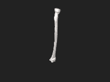
Radius (bone)

The radius (radius) is a bone located on the lateral or external part of the forearm, parallel to the ulna. Due to its morphology, it is a long bone, prism-shaped and slightly curved. Its upper or proximal end, more rounded, connects with the elbow joint and the lower or distal, more flattened, with the wrist joint, on the side closest to the thumb.
Body of radius (corpus radii)
It presents an external curvature that is generally not very pronounced and an internal one of concavity looking at the ulna, much more accentuated. It has a triangular prismatic shape, consisting of three faces and three edges:
Faces
- Previous face (previous facies): it is almost flat, slightly concave on its average part. In it is the otter hole of the radius and the long bending muscle of the thumb is inserted, and in its lower third the square pronador.
- Later face (facies posterior): rounded in its upper third, is covered by the short suppander. In the rest of its extension it is flat or slightly concave and in it the long abductor muscles of the thumb and short extender of the thumb are inserted.
- External face (lateral facies): convex and rounded at the top, in it the short supinator is inserted. Its average part is a rough surface (tuberositas pronatoriadestined to insert the round pronador. Its lower part is smooth and is related to the tendons of external radial muscles.
Borders
- Previous draft (previous margo): part of the bicipital tuberosity (tuberositas radii), forming an outgoing crest that softens when reaching the height of the hole nutritious confusing with the external face of the bone.
- Later draft (later): is obtuso and difuminated, especially at its ends.
- Internal edge (margo interosseus): is thin, cutting, almost always concave as the body of the bone, and provides insertion to the interoseous ligament. In its lower part it is bifurcated, delimiting in the corresponding part of the bone a kind of small triangular face of upper vertex that is part of the lower radicubital joint.
Top end
At the upper end there is a voluminous and rounded portion, called the head of the radius (caput radii), shaped like a cylinder. In the upper part there is a dome-shaped depression, called the glenoid cavity of the radius or articular fossa (fovea articularis capitis radii) which corresponds to the condyle of the humerus and is surrounded by an articular circumference (< i>circumferentia articularis). The head of the radius is supported by a narrow portion of the bone, the neck of the radius (collum radii). Below the neck, in the anterior medial part of the bone, an ovoid eminence rises, into which the inferior biceps tendon is inserted: the biceps radial tuberosity.
Bottom End
The lower end, or carpal, is the largest part of the bone. As a whole, it takes the shape of a truncated quadrangular pyramid and, therefore, has six faces: superior, inferior or carpal, anterior, posterior, internal and posteroexternal.
- Upper face: it is confused without clearly different demarcation line with the body of the bone.
- Lower face: triangular, articular, and presents a carpiana joint that is divided into two ribs for the joints of the semilunar and the scafoid(facies articularis carpalis). On the outside side of this joint surface is a robust apophysis called apophysis styling of the radio (processus styloideus radii). It is in contact with the skin and descends a little more than the styling apophysis of the cubit. The vertice of the radio systeroid apophysis provides insertion to the external side ligament of the wrist joint. On its base and on its side edge (crista suprastyloidea) insert the long suppander tendon.
- Previous face: flat in transverse sense and concave in vertical sense, is in relation to the square pronador muscle.
- Later face: on the back side two channels are observed (sulci tendinum musculorum extensiverum), one for the tendons of the own extender of the index and the common spreader of the fingers; and an external channel in which the tendon of the long extension muscle of the thumb is accommodated separated by a small spinal tuber prominence of the radio (tuberculum dorsale radii).
- Post-external face: it has two tendinous, internal and external channels for the first and second outer radial tendons and for the long abductor muscle tendons of the thumb and short thumb extender, respectively.
- Internal face: in it is the sigmoid cavity of the radio or cubital scotadura (incisura ulnaris), for your joint with the cube.
Muscle Insertions
The radius provides attachment to 9 muscles:
Front face
- Brachial biceps
- Short supenter
- Surface toe flexor
- Long thumb flexor
- Square cleaner
Back side
- Short supenter
- Long thumb reducer
- Short thumb extension
External face
- Short supenter
- Round cleaner
Styloid process of radius
- Long or braquirorradial
Joints
- With the humerus forming the humerradial joint.
- With the cubit forming the upper radio-cubital joint, the lower radio-cubital joint and the intermediate radio-cubital joint through the interoseo ligament.
- With the bones of the carpus (semilunar and scaphoids) forming the radio-carpian joint.
Function
- The radio serves to insert the muscles that allow the arm to move and thus facilitates the pressure.
Gallery


Contenido relacionado
White blood cell
Petiole
Popowia