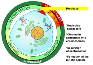
Prophase
The Prophase is the first phase of mitosis and meiosis. In it is it produces the condensation of all the genetic material (DNA), which normally exists in the form of condensed chromatin within a highly ordered structure called the chromosome, and the bipolar development of the spindle.
One of the earliest events of prophase in animal cells is the migration of two pairs of centrioles to opposite ends of the cell. A proper nucleus is needed to perform its condensation function.
Mitosis
Mitosisis the period during which chromosomes can be easily recognized in the nucleus, becoming evident as the nuclear chromatin condenses. Each chromosome consists of two identical rod-shaped bodies, or chromatids, linked together by a structure known as the centromere. In prophase, the pairs of centrioles separate to opposite poles. As they separate, fibers appear that form a spindle-shaped structure. These, collectively known as asters, start from the centrioles and radiate outward. In turn, the nucleolus disappears and the nuclear envelope disintegrates.
Prophase lasts approximately 40% of the total time of mitosis. It involves a series of changes, both morphological and in the physical-chemical state of the cell. This stage of typical mitosis can be observed using immunocytochemical techniques and a series of antibodies, where the following intracellular changes occur:
- A disorganization and reorganization of the skeleton cyto
- Dispersion of the actin and myosin throughout the cell
- Fragmentation and reorganization of micro tubules in order to form the acromatic spine
- The chromosomes appear as delicate extended or rolled filaments within the nuclear sphere, a process known as chromosomal condensation
Meiosis
During meiosis, two successive division processes occur: Meiosis I and Meiosis II. Meiosis I is divided into Prophase I, Metaphase I, Anaphase I and Telophase I. Meiosis II is divided into Prophase II, Metaphase II, Anaphase II Telophase II and Cytokinesis.
Prophase I in turn is subdivided into:
- Reptatenos: at this stage chromosomes are recognizable under the optical microscope, although chromosomes have been replicated at a very early stage there is no indication that they are formed by two sister clemátides, a situation that becomes visible when using an electronic microscope.
- Zigothene: at this point of Profase I occurs the sinapsis (meiosis) of the homologous chromosomes and the synatic quiases are formed
- Packaging: the resulting structure is called tetrada or bivalent because it is formed by the two chromosome chromosome chromosome chromosome chromosome chromosome chromosome chromosome chromosome chromosome chromosome chromosome chromosome chromosome chromosome chromosome chromosome chromosome chromosome chromosome chromosome chromosome chromosome chromosome chromosome chromosome chromosome chromosome chromosome chromosome chromosome chromosome chromosome chromosome chromosome chromosome chromosome chromosome chromosome chromosome chromosome chromosome chromosome chromosome chromosome chromosome chromosome chromosome chromosome chromosome chromosome chromosome chromosome chromosome chromosome chromosome chromosome chromosome chromo At this point, the phenomenon of chromosome cross-cutting or crossing-over can be presented: during cross-cutting, a fragment of a chromosome can be separated and exchanged by another fragment of its corresponding counterpart.
- Diplomen: chromosomes, after "crossing-over", are joined by contact points called quiasmas.
- Diaconies: the quiasmas move towards the ends of the chromosomes.
Prophase II is short-lived. The nuclear envelope and nucleolus disappear. Chromosomes begin to condense. The spindle is formed between the centrioles, which have moved to the poles.
Contenido relacionado
Spinal nerve
Marine biology
Avenula blaui
