Prolactin
Prolactin (PRL), pituitary lactogenic hormone or mammotropin, is a protein hormone secreted mainly by the pituitary gland (adenohypophysis).
It stimulates milk production in the mammary glands, progesterone synthesis in the corpus luteum, cell growth and cell differentiation, fat metabolism and immunomodulation among others.
PRL has many biological functions in different tissues and organs of the body, and is considered as a hormone with different and unrelated effects.
The existence of elevated levels of PRL in the blood (hyperprolactinemia) is a disorder that can be caused by a tumor (adenoma) in the pituitary gland.
Features
Prolactin (PRL) acts on various physiological processes such as: lactation, pregnancy, cell growth and cell differentiation, fluid homeostasis, immunomodulation, regulation of secretory gland differentiation, and formation of the corpus luteum.
PRL is secreted in the pituitary and also in multiple sites outside of it.
The PRL hormone has tropical action (from the Greek root τρόπος tropos, which means 'in the direction', 'heading towards'), and is known as mammotropic hormone pituitary gland, or mammotropin, which stimulates the mammary gland to secrete breast milk.
A complementary concept is that of trophic action (from the Greek τροφικός trophicos 'food from', 'nutrition to'), meaning that mammotrophin as a trophic hormone pituitary lactogen stimulates the function of its target cell (the acinus epithelial cell) of the mammary gland for milk generation.
Structure
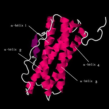
Prolactin is a protein hormone, formed by a chain that in mammals consists of 226-229 amino acids (aa). It has a molecular mass between 25,500-26,140 Daltons.
The 3D structure of prolactin contains the following motifs: beta strand (Beta strand), helix (Helix), turn (Turn).
| Ref. | Structural motivations, location and extension in the amino acid chain (aa) |
|---|---|
| Beta strand. | aa 31-34 |
| Helix . | aa 43-72 |
| Turn . | 73-75 |
| Helix . | aa 78-82 |
| Turn . | 87-90 |
| Helix . | aa 105-118 |
| Helix . | aa 120-131 |
| Beta strand. | 133-135 |
| Helix . | aa 138-165 |
| Beta strand . | 166-168 |
| Helix . | aa 181-185 |
| Helix . | aa 189-223 |
Most PRL from the pituitary has a weight of 23 kiloDaltons (kDa), but other forms of 14, 16, and 24 kDa exist. The 14 kDa PRL is generated in the hypothalamus.
Genetics
The prolactin gene is called PRL and GHA1 is located on chromosome 6 (human) in its short arm or p arm, band 22.2 and 21.3, it has 10 kilobases (kb) in length, it has five exons and four introns. The mRNA results in a polypeptide of 227 amino acids (aa), which has a signal peptide of 28 aa.
Discharge
Prolactin is secreted mainly by lactotroph cells of the
anterior lobe of the pituitary gland during pregnancy and lactation.
PRL secretion from lactotroph cells is modulated by higher level function of the hypothalamus. It makes prolactin-releasing factors (PRFs) and prolactin-inhibiting factors (PIFs). In mammals, the hypothalamus exerts an inhibitory effect on the synthesis and secretion of prolactin, through the physiological dopaminergic tone.
PRL is also secreted in extra-pituitary sites, including the endometrium, myometrium, decidua, immune cells, brain, breast, prostate, skin, and adipose tissue, acting as a signaling molecule at the paracrine level. /autocrine, and locally as a growth promoting factor.
Circadian Rhythm of PRL
Plasma prolactin concentrations are higher during sleep than during wakefulness.
These variations are modulated by the hypothalamus and the VIP peptide seems to be involved in its release during periods of REM sleep.
The hormones that have a synergistic effect are: estrogen, progesterone, and growth hormone (GH).
Nipple sucking during lactation favors the synthesis of a greater amount of this hormone. It is regulated by positive feedback.
Prolactin is a hormone that tends to vary easily given certain factors that increase or decrease stress. Sleep parameters influence secretion
Regulation
The PRL hormone is produced and controlled by the function of the
hypothalamic-pituitary axis.
The hypothalamus exerts an inhibitory effect on the synthesis and secretion of prolactin, which is also influenced by other local autocrine regulatory factors, as well as by other pituitary cells via paracrine regulation.
In the anterior pituitary, the neurotransmitter called dopamine inhibits the synthesis and release of the hormone prolactin, inhibits the proliferation of lactotroph cells and favors their apoptosis.
- Dopamine neurons, which control the secretion of prolactin, are found within the arched nucleus of hypothalamus. These neurons have been subdivided into three subpopulations according to the anatomy of their projections: the tuberoinfuular dopaminergic neurons (TIDA), tuberohipophysaries (THDA) and hypophysial periventricular neurons (PHDA).
Other neurotransmitters modulate prolactin secretion such as: histamine, norepinephrine, adrenaline and acetylcholine.
PRL secretion is stimulated by suckling, thyrotropin-releasing hormone (TRH), estrogens, and vasoactive intestinal polypeptide.
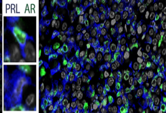
ESR1= Estrogen receptor in green.
Estradiol is an important physiological regulator of pituitary secretion of prolactin, causing increases in its synthesis.
- In the lactotrope cell the estradiol, through the coupling of the estrogen-receptor complex of estrogen (ES-R), in the specific element of response to estrogens in the promoter of the gene that encodes the synthesis of prolactin, increases the synthesis of prolactin through a biphase transcription, which culminates in an increase in prolactin RNAm; this has a direct action on the gene capable of altering transcription.
- In the lactotrope cell the estradiol, through the coupling of the estrogen-receptor complex of estrogen (ES-R), in the specific element of response to estrogens in the promoter of the gene that encodes the synthesis of prolactin, increases the synthesis of prolactin through a biphase transcription, which culminates in an increase in prolactin RNAm; this has a direct action on the gene capable of altering transcription.
Local transformation of androgens to estradiol by the aromatase enzyme maintains the prolactin cell population in the male pituitary gland through the aromatization of testosterone to estradiol.
PRL receiver
Prolactin receptors (PRL-R) are located in cells of various organs. The circulating PRL molecule binds to them after being secreted by lactotroph cells.
The prolactin receptor (PRL-R), is classified as a member of the cytokine receptor family.
The PRL-R receptor can also be activated by growth hormone (GH) and human placental lactogen (hPL).
Prolactin receptor (PRLR) and growth hormone receptor (GHR) are associated with the onset and development of breast cancer. Breast cancer cells endogenously express the GHR, PRLR, and GHR-PRLR heterodimer.
Function
Prolactin (PRL) is vital in several physiological processes such as: lactation, pregnancy, cell growth and differentiation. The PRL is associated with more than 300 functions that can be grouped into five categories:
- reproduction,
- osmorregulation,
- development and growth,
- carbohydrate and lipid metabolism
- immunoregulation.
It is classically called mammotropin or pituitary lactogenic hormone.
In women
Prolactin (PRL) increases the secretion of milk from the mammary gland. Among its effects on the cells of the mammary acini is an increase in the synthesis of lactose and a greater production of milk proteins such as casein and lactalbumin. Although it is true that the PRL concentration is high before childbirth, milk secretion only takes place after childbirth, since the high amount of estrogen and progesterone in pregnant women has an inhibitory effect on milk secretion. When the levels of these hormones drop after pregnancy, lactation occurs.
In primates and humans, the endometrium also generates PRL during the corpus luteum dominance phase and during gestation. In the corpus luteum, PRL stimulates the expression of estrogen receptors and LH.
Prolactin also has an inhibitory effect on the secretion of gonadotropins, so its hypersecretion can cause absence of menstruation in women.
In men
In men, the behavior of prolactin (PRL) can affect adrenal function, electrolyte balance, breast development, sometimes galactorrhea, decreased libido, and impotence, and affects the functions of the prostate, seminal vesicles, and testicles.. Also, normally after intercourse with orgasm, PRL would be one of the main causes of the refractory period (including often drowsiness).
Normal values
- Men: 2-18 nanograms (ng) per milliliter (mL) (ng/mL)
- Women who are not pregnant: 2,3-25 ng/mL
- Pregnant women: 80-400 ng/mL
Aside from pregnancy, the most common cause of elevated prolactin levels in the blood, called hyperprolactinemia, is the presence of a prolactinoma, a prolactin-producing tumor in the pituitary gland. Prolactinomas are the most common pituitary tumors and are generally benign. They are more frequent in women, but can also appear in men. The symptoms they produce, if they do, are related to excess prolactin and therefore milk production in non-pregnant women, which is called galactorrhoea.
Pathology
Hyperprolactinemia
Hyperprolactinemia is increased levels of the hormone prolactin in the blood. Prolactin is a sex hormone that plays a key role during breastfeeding.
Some of the disorders that cause hyperprolactinemia are a dopaminergic deficit in the CNS or, if not, a pituitary tumor.
PRL excess promotes weight gain, obesity, metabolic syndrome, and impaired glucose and insulin profiles and lipid profiles. These are decreased glucose tolerance and an increase in fasting insulin.
Exposure of pancreatic islets to PRL is known to stimulate insulin secretion and β cell proliferation.
Macroprolactinemia: Increased prolactin levels without significant biological effects.
One way to increase the amount of prolactin produced is to follow sleep parameters similar to the time 'before electric light'.
The interaction between PRL and the prolactin receptor (PRL-R) mainly promotes pro-tumoral events, such as migration, invasion, metastasis, chemoresistance, inhibition of apoptosis.
Prolactinoma
Prolactinomas are benign tumors that make up about one-third of all pituitary tumors. One of the main characteristics is that they rarely undergo malignant transformation or local invasiveness. Alterations in the regulation of the cell cycle seem to lead to the expansion of an original mutated cell, forming an adenoma. Alterations in the mechanisms that physiologically regulate anterior pituitary cell turnover may be involved in the pathogenesis.
Contenido relacionado
Atherosclerosis
Phlomis herba-venti
Likelihood level of evidence


