Porifera
The porifera (Porifera), also known as sponges or sea sponges, are a phylum of aquatic animals found within the Parazoa subkingdom. They are filter feeders thanks to a developed aquifer system of pores, channels and chambers that generate water currents caused by the movement of flagellated cells: choanocytes. There are about 9,000 species of sponges in the world, of which only about 150 live in fresh water. Fossils of sponges (a hexactinellid) are known from the Ediacaran Period (Neoproterozoic or Late Precambrian). They were considered plants due to their immobility until the existence of internal water currents was discovered in 1765 and they were recognized as animals. Its digestion is intracellular. Sponges are the sister group to all other animals. Sponges were the first forms of the evolutionary tree to branch from the common ancestor of all animals.
History
Aristotle (IV century BC), who was at the origins of the scientific taxonomy of animals, he divided them in his work "The Parts of Animals" (in ancient Greek, Περὶ ζώων μορίων), into two large groups: "animals with blood" and "bloodless animals," which he in turn classified into smaller units.
Later, in the IV-V centuries AD. C., the Neoplatonists Dexipo and Harmonio de Hermia began to call such organisms "zoophytes"(in ancient Greek, ζωόφυτα), classifying them as intermediate forms between plants and animals. In medieval Europe, this term was hardly used, but it again came into use in the Renaissance, as it was used in their classifications by zoologists such as Edward Wotton, Guillaume Rondelet, Conrad Gesner, and Ulisse Aldrovandi.
Sponges invariably appeared in the 'zoophytes,' although the size of the group changed; thus, in Wotto's "De differentiis animalium" (1552), sea stars, scyphozoans and ctenophores were included in zoophytes. In Carlos Linnaeus's book "Systema Naturae", the class Vermes included the order Zoophyta with an even larger volume than Wotton's: Linnaeus also included marine urns and some molluscs and worms in the number of zoophytes. At the same time, in the 10th edition of the book (1758), Linnaeus defined Zoophyta as "flowering plants living animal life", and in the 12th edition (1766-1768) as "complex animals that bloom like the flowers of plants".
The animal nature of sponges was first corroborated by John Ellis, who described their ability to create water currents and change the diameter of the Osculum and outlined his observations in a 1765 letter to Daniel Solander (in 1766 the The letter was published in Philosophical Transactions of the Royal Society). In 1752, before Ellis' discovery, the French naturalist Jean-André Peysonnel hypothesized that sponges are not living organisms, but structures erected by sea worms.
The systematic position of sponges among "zoophytes" was gradually cleared up. In Système des animaux sans vertèbres (1801) and Philosophie zoologique (1809) by Jean-Baptiste Lamarck, sponges appear as part of the order Polyps with Polypnyak, along with bryozoans, shells and a series of groups of intestinal cavities. Georges Cuvier in his work "Le régne animal distribué d'apres son organization" (1817) included sponges together with anthozoa in the class Polypae of the section Radiata (the latter corresponds roughly Zoophyta in the sense of Wotton or Linnaeus, although Cuvier no longer regarded Radiata as a transition between plant and animal organisms).
The transition to an in-depth study of sponge biology arose in the studies of Robert Edmund Grant, in which he proposed the first precise interpretation of the structure and physiology of these animals. Already in his first paper (1825), Grant described larvae and eggs in sponges of the genus Spongilla, and carried out a comprehensive microscopic study of the pore structure of sponges. In 1836, introduced the name Porophora for sponges, which was replaced by himself in 1841 by Poriphera, and by Rymer Jones in 1847 by Porifera (all the names mean "bearing pores").
For most of the 19th century, sponges were generally associated with centreans and often simply referred to as included in the latter (although naturalists such as Henri Dutrochet, Paul Gervais, and John Hogg classified sponges as plants). However, as early as 1816, Henri de Blainville proposed the idea of a close connection between sponges and protozoa. 1860, Hogg, and in 1866 Ernst Haeckel, proposed assigning a separate kingdom (according to Hogg: Protista, after Haeckel: Protista), which included sponges, unicellular animals, and part of unicellular plants (Thomas Huxley, who considered sponges to be colonies of single-celled organisms, in 1864 he simply included them in the protozoa) This view seemed to have been confirmed by Henry James-Clark's discovery in 1867 of collared flagella (choanoflagellates), remarkably similar to the special cells of all the sponges: the choanocytes.
However, the last third of the XIX century became, in the words of A. V. Ereskovsky, the "golden age" of the embryology of sponges (and the first stage of systematic studies of their development). From 1874 to 1879, the research of II. Mechnikov, Franz Schulze and Oscar Schmidt, engaged in the study of the structure and development of sponges, irrefutably proved their belonging to multicellular animals. At the same time, a great originality of this group of animals was discovered. In particular, Schulze (1878) and Yves Delage (1892) described the so-called phenomenon of "perversion of the germ sheets" in the development of sponges, clearly distinguishing Porifera from all other Metazoans (including coelenterates); however, in the late 20th century and early XXI, the terminology changed: the opinion began to prevail according to which the germ sheets of sponges in the course of embryogenesis are not formed at all, and therefore there is no point in talking of their "perversion". Therefore, William Sollas (1884) contrasted sponges as Parazoa, separating them from all other Metazoans (which would soon be called Eumetazoa). In the late Haeckel system (1894-1895), sponges were already eliminated from Protista and considered in the animal kingdom as an independent type of Spongiae, and in Ray Lankester's system (1900-1909), they are clearly attributed to Metazoa and they appear as a type of Porifera (the only one in the Parazoa section). This latter point of view was absolutely dominant throughout most of the 20th century, although the range of Parazoa varies among the different authors: as "section", "superdivision" and then "subkingdom".
In the 1900s and 1960s (the second stage of sponge development research, according to A. V. Ereskovsky), interest in studying sponge development waned, although important work by Henry Wilson and Claude Levy appeared. Around 1960, the third stage begins, which is characterized by the predominance of ultrastructural studies using electron microscopy. At the end of the XX century, began the study of the cytogenetic and molecular-biological characteristics of sponges.
General characteristics
One of the most surprising characteristics of sponges is that most of the cells that make up their body are totipotent, that is, they can transform into any of the other cell types according to the animal's needs. Therefore, it is considered that sponges have a cellular organization, unlike the rest of metazoans whose organization is tissue (with tissues). They lack true embryonic layers.
The general body shape of these animals is that of a "sack" with a large opening at the top, the osculum, which is where the water comes out, and many more or less small pores on the walls, which is where the water enters. Food filtration occurs in the internal chamber of the animal, and is carried out by a specialized cell type unique to porifera, the choanocytes. These cells bear great similarity to choanoflagellate protozoa, so there is little doubt today that they are phylogenetically related. Sponges, the most primitive metazoans, probably had a common ancestor with colonial choanoflagellates, perhaps similar to today's Proterospongia or Sphaeroeca, which are simple aggregates of unicellular animals.
Sponges are practically unable to move; many lack body symmetry and therefore do not have a definite shape; there are those that grow indefinitely until they run into another growing sponge or other obstacle, others that embed themselves in rocks, boring them, etc. A given species can take on different aspects depending on environmental conditions, such as the nature and inclination of the substrate, availability of space, water currents, etc.
However, according to studies they have shown that some sponges can move over the substrate from one place to another, but given their extreme slowness (about 4 mm per day) the phenomenon had gone unnoticed.
Excretion, basically ammonia, and gas exchange occur by simple diffusion, mainly through the choanoderm.
Sponges lack a mouth and a digestive system and, unlike other metazoans, they depend on intracellular digestion, so phagocytosis and pinocytosis are the mechanisms used for food ingestion. They also do not have nerve cells, they are the only animals that lack a nervous system.

Anatomy
Pinacoderm
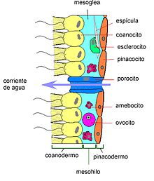
Externally, sponges are covered by a layer of long, wide pseudoepithelial cells called pinacocytes; It is not true epithelium, since it lacks a basal lamina. The set of pinacocytes form the pinacoderm or ectosome, which is analogous to the epidermis of eumetazoans. The pinacoderm is traversed by numerous dermal pores each lined by a coiled cell called a porocyte; the water is attracted towards them and penetrates inside. In some species there is a cuticle, a thick layer of collagen that covers and eventually replaces the pinacoderm.
Choanoderm
The inner surface of a sponge is lined with flagellated cells that together form the choanoderm. The main central cavity is the spongocele or atrium. These flagellated cells, called choanocytes, which are virtually identical to choanoflagellate protozoa, produce the water current and are important in feeding. The choanoderm may be one cell thick (asconoid organization), may be retracted (syconoid organization), or may subdivide to form clusters of independent choanocyte chambers ( organization leuconoid).
Mesowire
Between these two layers is a loosely organized area, the mesohil, in which support fibers, skeletal spicules, and a variety of amoeboid cells of great importance in digestion, can be found. skeletal secretion, gamete production, and the transport of nutrients and wastes. The different elements of the mesowire are immersed in a colloidal mesoglea.
Skeleton
In the mesowire there are numerous elastic collagen fibers (protein part of the skeleton) and siliceous (hydrated silicon dioxide) or calcareous (calcium carbonate) spicules, depending on the class to which it belongs, which are the mineral part of the skeleton and what makes it tough. The rigidity of this body wall will vary depending on whether there is more of the protein part (more flexible) or more of the mineral part (harder and more rigid).
Collagen fibers are of two basic natures; thin and dispersed fibers, and spongin fibers, thicker, that form a reticulum or lattice; they are intertwined with each other and with the spicules, and may include sand grains and spicule debris from the sediment.
The calcareous spicules have little variation in morphology, but the siliceous ones have different shapes and sizes, distinguishing the megascleras (> 100 μm) from the microscleras (< 100µm).
Frequently, spicules and fibers are not arranged randomly but form various ordered structures.
Cell types
Since sponges lack true tissues and organs, the different functions of the animal are carried out by various more or less independent and interchangeable cell types.
- Pinacocitos. Typical pinacocytes form the outer coating of most sponges; it has protective function and also fagocitan.
- Basopinacocytes. They are special pinacocitos located at the base of the sponge that segregate fibers that anchor the sponge to the substrate.
- Porocitos. They are cylindrical cells of the pinacoderm with a central adjustable channel that allows more or less volume of water to pass inside. They are exclusive of calcareous sponges.
- Coanocitos. Coanocytes are the most characteristic cells of sponges. They are provided with a long central flagelo surrounded by a crown or necklace, simple or double, of microvellosities connected with each other by mucous filaments that form a reticle. The flagelos, directed towards vibrational chambers, cause water flows thanks to movements that, although not coordinated in time, are in the direction. The water loaded with particles (bacteria, phytoplankton and organic matter in suspension) crosses the microvellosities, where the food that will be later fagocite is trapped.
- Dudes. and Lofocus. Mesohile cells that secrete scattered collagen fibers that form a brat in the mesohile.
- Spongiocitos. Mesohile cells that secrete thick collagen fibers known as Spongin fibers, which are the main support of the body of many sponges.
- Sclerocytes. They handle production Particlesboth calcareous and siliceous, and disintegrate when the secretion of the spine is completed.
- Myocytes. Fusiform count cells located in the mesohilo, which are available around the circle and the main channels. Your cytoplasm is rich in microfilaments and microtubules. Its response is slow and not conditioned on electrical stimuli, as in sponges there are no nerve cells.
- Archeocites or Amebocitos. Mesohile ameboid cells capable of transforming into any other cell type. They also have great importance in digestion processes, accepting particles made by coanocytes, and are the system of transport and excretion of sponges. Given their totypetence, they are key in asexual reproduction.
- Spherulous cells. They have excretory function; they accumulate refringent granules and release them to the exhalant current.
Levels of organization
Sponges have three levels of organization, each of which considerably increases the surface of the choanoderm with the consequent increase in filtration efficiency; from simplest to most complex:
- Asconoid. Tubular sponges, with radiated symmetry, small (some 10 cm), with a central cavity called spongiocele or atrium. The movement of the scourges of the coanocytes force the entrance of water in the spongiocele through pores that cross the body wall. Coanocytes, which tap the spongiocele, capture the suspended particles in the water.
- Siconoid. They also have radiated symmetry. The body wall is thicker and more complex than asconoids; the coanoderm also covers the atrial cavity. Present radio channels (o) flagged cameras), tapered cameras of coanocytes that open to spongiocele through a pore called apopilo. The water enters the inhaling channels through a large number of pores dermales and then pass to the radial channels by tiny openings called prosopilos. There the food is ingested by the little ones. During their development, siconoid sponges pass through an asconoid stage called olinto.
Only a few species of calcareous sponges have ascon or sicon organization.
- Leuconoide. Most of the demosponges have leucon organization, which is the one that reaches the greatest complexity. Leuconoid sponges lack radiated symmetry, have reduced atrial cavity and have numerous vibratile chambers, globulr cameras upholstered of independent coanocytes one another and sunk in the mesohile and communicated with one another, with the outside and with the bone by a multitude of inhaling and exhalant channels. Apopilos lead to exhalant channels; the various exhalant channels meet to expel water through several circles. A large leuconoid sponge may have several circles, which can be interpreted as a colony of individuals or a single complex individual.
Reproduction and development
All sponges reproduce sexually, but various types of asexual reproduction are very common.
Asexual reproduction
Given the full potential of their cells, all sponges can reproduce asexually from fragments. Many sponges produce yolks, small growths that eventually break off, which in some cases contain stored food. Freshwater species (Spongillidae) produce complex gemules, small well-organized spheres with archaeocytes and several protective layers, including a thick collagen layer supported by amphidisk-like spicules; They are very resistant to environmental inclemencies, such as drying out and freezing (they can withstand -10 °C). Some marine species produce simpler gemmules, called soritos.
Sexual reproduction
Sponges do not have gonads, and gametes and embryos are found in mesohyls. Most are hermaphrodites, but there is great variability, reaching the extreme that in the same species hermaphrodite individuals coexist with dioecious individuals. In any case, fertilization is almost always crossed.
Spermatozoa are formed from choanocytes, when all of them in one chamber undergo spermatogenesis and originate a sperm cyst. The ova originate from choanocytes or archaeocytes and are surrounded by a layer of feeding cells or trophocytes. The sperm and eggs are expelled abroad through the aquifer system; in this case, fertilization occurs in the water and gives rise to planktonic larvae. In some species, spermatozoa enter the aquifer system of other individuals where they are phagocytosed by choanocytes; then these choanocytes detach, transform into amoeboid cells (phorocytes) that carry the sperm to an egg; after fertilization, the larvae are released through the aquifer system.
There are four basic types of larvae in sponges:
- Parenquímula. It is a solid larva, with a layer of monoflagelated cells on the outside and a mass of cells similar to archaeocites in the interior immersed in a matrix.
- Celoblástula. It is a hollow larva composed of a layer of monoflagelated cells that surround an inner cavity.
- Stomach. It is a special type of jealousy, typical of sponges that incubate embryos in their mesohile. It is also hollow, but it has some larger cells (macromers) that leave an opening that communicates with the inner cavity. It suffers from a surprising process of investment in which the flogged cells that were initially internal, end up being external.
- Anfiblástula. It is the result of the investment process of a stump. It consists of a hemisphere formed by large and non-flagged cells (macromers) and another with small and monoflagellate cells (micromers). The amphiblástula is released and ends in the substrate by the micrómers; these are invaded by forming a chamber of flogged cells that will be the future coanoderm; the macromers form the pinacoderm; then a circle is opened originating a small leuconoid sponge called olinto.
Cell aggregation
Sponges have a unique and extraordinary property: when their cells are separated by mechanical means (for example, by sieving them), they immediately come back together and form, in a few weeks, a complete and functional individual; Furthermore, if two sponges of different species are shredded, the cells separate and regroup, reconstructing the separate individuals.
Ecology
Due to their body structure (aquifer filtration system), sponges always inhabit the aquatic environment, whether fresh or marine, and attach to a solid substrate, although some species can attach to soft substrates such as sand or mud. Most sponges are sciophilous (they prefer penumbra). Their main source of food is submicroscopic organic particles in suspension, which are very abundant in the sea, although they also ingest bacteria, dinoflagellates and other small plankton. Its filtering capacity is remarkable; a leuconoid sponge 10 cm tall and 1 cm in diameter contains 2,250,000 flagellate chambers and filters 22.5 liters of water per day.
Despite their simplicity, sponges are highly ecologically successful; they are the dominant animals in many marine benthic habitats and tolerate pollution by hydrocarbons, heavy metals and detergents well, accumulating these pollutants in high concentrations without apparent damage.
Some sponges have photosynthetic symbionts (cyanobacteria, zooxanthellae, diatoms, zoochlorellae) or not (bacteria). They periodically expel symbionts and somatic cells, and regularly secrete mucous substances. In certain sponges, the symbionts come to represent 38% of their body volume.
Few animals eat sponges, due to their spicule skeleton and toxicity. Some opisthobranch mollusks, echinoderms and fish. Often these are very specific species that are exclusively spongiophagous and prey on a specific species of sponge.
Sponges possess a surprising variety of toxins and antibiotics that they use to avoid predation and in competition for substrate. Some of these compounds have been shown to be pharmacologically useful, with anti-inflammatory, cardiovascular, gastrointestinal, antiviral, antitumor properties, etc., and are being intensively investigated. These compounds include arabinosides, terpenoids, halichondrins, etc.
Many invertebrates and various fish use sponges, due to their porous structure, as a place of residence or refuge. Some gastropods and bivalves have encrusting sponges on their shells, and many crabs collect sponges that they place on their shells. These are cases of mutualism, in which said animals achieve camouflage and sponges a method of displacement.
Systematics
The phylum Porifera is divided into three classes:
- Calcarea class. Particles of 1, 3 or 4 radios, of calcium carbonate crystallized in the form of calcite. There are three types of organization. They generally live in shallow coastal waters.
- Class Hexactinellida (virtreal sponges). Syliceous particles (hydrated silicon dioxide) of three or six radios. In general they live in greater depth, between 450 and 900 m.
- Class Demospongiae. Syliceous particles (hydrated silicon dioxide) monoaxone or tetrasxon, which can be replaced by a mesh of spongin fibers. They all have leuconoid organization. They live at any depth.
- Archaeocyatha†. They are an extinct group of uncertain positions related to sponges; they had a short existence, of about 50 million years, during the War.
The Sclerospongiae class was abandoned in the 1990s. It was made up of sponges that produce a solid calcareous matrix, similar to a rock, which is why they are known as coral sponges. The 15 known species were included in the classes Calcarea and Demospongiae.
Phylogeny
Monophyly
Sponges have historically been considered a monophyletic group due to their morphology and common characteristics. The era of molecular study challenged this view by proposing that Porifera was paraphyletic, implying that the ancestral animals were sponges; however, the most in-depth phylogenomic study reconsiders the monophyly of sponges. Sponges are currently considered to have the following relationships:
| Porifera |
| ||||||||||||||||||
Paraphilia
Recent studies on the histology of hexactinellid sponges have revealed that this group has important peculiarities. Based on this, it has been proposed that the porifera phylum be divided into two subphyla, Symplasma (hexactinellids) and Cellularia (calcareous + demosponges); It has even been questioned whether the porifera are a monophyletic group, that is, that they all have a common and exclusive ancestor of sponges (Zrzavý et al.). Simplasmas comprise only the class hexactinellids; they are poriferous with a simple organization, syncytial, although they present cell types such as archaeocytes and spherulose cells; the pinacoderm does not have differentiated pinacocytes and does not show contractility since it lacks myocytes. The cellular ones gather the calcareous and demosponge classes, which present a defined cellular organization, with individualized pinacocytes and choanocytes, and various types of amoeboid cells in the mesohyl.
On the other hand, genetic studies that maintain that sponges form a paraphyletic group gave the following result:
| Animalia |
| ||||||||||||||||||||||||||||||
Porifera as the non-basal phylum of the kingdom Animalia
Other analyzes based on the sequence of genes and amino acids affirm that Ctenophora would be the first group to separate from the kingdom Animalia in contrast to the traditional view that Porifera is the most basal group. This study argues that Porifera is a monophyletic group that diverged as a sister group to the ParaHoxozoa clade. Poriferans have also been shown to share with the latter certain ancient gene duplications and metabolism-regulating genes that are absent in ctenophores. the ctenophores are an important group for understanding the evolution of animals. If the ctenophores really are the sister group of the other animals, it would be difficult to prove how tissues originated in the animals, since sponges do not possess this characteristic. However, it has been shown that the reasons why ctenophores appear as the most basal group of animals under analysis is because they have a deviation in the nucleotide and amino acid sequence, which produces their misplacement, also called long-branch attraction. Studies attempting to avoid systematic error have suggested that sponges are the basal group of animals.
| Apoikozoa |
| ||||||||||||||||||||||||||||||
A diagnostic feature of porifera is the presence of spicules. For this reason, certain extinct groups with current sponge organization were placed outside the Porifera phylum. In particular, groups with a solid calcareous skeleton such as archaeocyats, caetetids, sphinctozoa, stromatoporoids, and receptaculids are problematic. The discovery of some fifteen living species with a solid calcareous skeleton has greatly helped to understand the phylogeny of porifera. These species have various forms and should be classified with caetetids, sphinctozoa, and stromatoporoids if they had been found as fossils. However, with the study of living material, the histological, cytological and larval characteristics clearly show that these fifteen species can be placed among calcareous sponges and others among demosponges. Thus the very abundant corresponding fossil forms would also fit into one of these two classes.
It is widely accepted among specialists that the calcareans and demosponges are more closely related to each other than to the hexactinellids. With the discovery of the aforementioned fifteen living forms, a fourth class was created, the sclerosponges. However, it is a polyphyletic group that should be abandoned, according to Chombard, et al. The archaeocyanates represent a special case; there are no living representatives, although their organization may refer to that of current sponges. Phylogenetic analysis by Reitner & Mehl places them as a sister group to the demosponges. Therefore, the archaeociates would lose their edge category and would become a class within the sponges.
Distribution
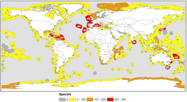
Sponges are common all over the world. Most species inhabit oceanic waters from polar to tropical regions. Even so, sponges have the highest species diversity in the tropical and subtropical parts of the World Ocean. 219 species (according to other sources, about 150) live in bodies of fresh water.
Most species live in calm, clean water, as silt and sand particles suspended in the water or lifted off the bottom by currents can clog pores in sponge bodies, thus hampering the processes of respiration and nutrition. Since sponges lead an attached sedentary lifestyle, they need a solid substrate for their development and growth. In this sense, sponge accumulations occur in places where there are stony materials at the bottom: stones, boulders, pebbles, etc. However, some species can attach themselves to soft bottom sedimentary sediments with the help of a root-like base of their body.
Most sponges live at shallow depths, down to 100-500 m. With increasing depth, the number of sponge species decreases. At depths greater than 1,000–1,500 m, sponges are usually quite rare, such that the number of deep-sea sponge species is small.
In temperate waters, sponges are more numerous, but less diverse, than in the tropics. This is probably due to the fact that there are many sponge-eating organisms in the tropics. Glass sponges are more numerous in polar waters, as well as at great depths in temperate and tropical seas, as their porous body structure allows them to extract food particles from these food-poor waters at minimal cost. Common sponges and calcareous sponges are numerous and varied in calmer, non-polar waters.
Sponges that live in tidal zones are well adapted to a short stay outdoors, when the tide is low they stick out of the water. Their mouths and pores are closed, which prevents excessive moisture loss and drying.
Antiquity
Sponges are the oldest group of living animals. According to the fossil record, they appear approximately 760 million years ago, in the Tonic before the Ediacaran fauna. This early fossil is known as Otavia.
Use
By dolphins
A 1997 report described the use of sponges as tools by dolphins in Shark Bay in Western Australia. A dolphin would attach a sea sponge to its face, which it would presumably use for protection when searching for food on the sandy seafloor. This behavior has only been observed in this bay, and is displayed almost exclusively by females. A 2005 study concluded that mothers teach the behavior to their daughters, and that all sponges are closely related, suggesting that this is a fairly recent innovation.
By humans
Trade and exploitation
The first inhabitants of the Mediterranean already used the well-known bath sponge; its use was probably discovered by the Egyptians. Aristotle knew about sponges and described their great capacity for regeneration. Roman soldiers used sponges instead of metal cups to drink water during military campaigns, and sponge fishing was one of the events in the ancient Olympic games.
In the North Atlantic, sponges washed up on beaches by the sea have traditionally been used as fertilizer for crop fields. However, economic interest lies in bath sponges, especially the genera Spongia and Hippospongia, whose skeleton is exclusively horny and flexible. The sponge trade has focused for years on the Eastern Mediterranean, the American Atlantic coasts, the Gulf of Mexico and the Caribbean to the north, and Japan. In Florida was the most important manufacturing industry in the world. In the middle of the XX century, overfishing and various epidemics drastically reduced the volume of sponges traded. The decline of this trade was accentuated with the appearance of synthetic sponges.
Some demosponges are harmful to humans as they perforate the shells of mollusks, causing damage to bivalve hatcheries (mussels, oysters, etc.)
Medicine
Sponges have medicinal potential due to the presence in the sponges themselves or in their microbial symbionts of chemicals that can be used to control viruses, bacteria, tumors, and fungi.
Contenido relacionado
Achnatherum
Bocagea
Sapranthus


