Pinocytosis
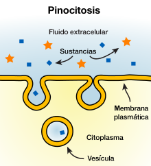
Pinocytosis is a type of endocytosis that consists of the uptake of material from the extracellular space by invagination of the eukaryotic cytoplasmic membrane. This membrane folds up creating an endosome called the pinocytic vesicle or pinozoma that detaches into the cell with the molecules dissolved or suspended inside.
Together with phagocytosis, they constitute the two main types of endocytosis. Pinocytosis differs from phagocytosis mainly in that pinocytosis occurs in all cells and affects molecules (such as most proteins) and phagocytosis only occurs in certain specialized mammalian cells and processes larger particles (such as bacteria). Pinocytic vesicles typically measure between 100 and 200 nanometers in diameter. There are four types of basic pinocytic mechanisms: macropinocytosis, clathrin-mediated endocytosis, caveolae-mediated endocytosis, and clathrin-independent endocytosis and of the caveolae.
In humans, this phenomenon is observed in cells of the intestinal mucosa, when these allow the entry of fat vesicles during the absorption of nutrients. Another type of cell in which it has been frequently observed is the human ovum. When the egg matures in the woman's ovary, it becomes surrounded by "nurse cells." These cells apparently release dissolved food to the ovum, which incorporates it by pinocytosis.[citation needed]
Pinocytosis process
The process of pinocytosis occurs continuously in the membrane of most cells; and the speed at which it occurs is variable, being especially fast, for example, in macrophages since every minute approximately 3% of their membrane is absorbed in the form of a vesicle. The rate at which vesicles are formed can increase when the molecules come into contact with the cell wall. Pinocytosis can be measured through the accumulation of markers present in the intracellular fluid (such as enzymes or other compounds), and the The amount of particles internalized by the cell depends both on the amount of the markers present in the extracellular medium and on the volume of the vesicles. The energy required to perform this process is quite considerable, and is supplied by adenosine triphosphate as well as by the presence of calcium ions.
Vesicles originate from the surface of the cell membrane, usually when a molecule binds to its specific receptor. These receptors are located in areas of the cell membrane called lined depressions. Inside these depressions is the fibrillar protein called clathrin, as well as other contractile filaments such as actin and myosin. Once the molecule binds to receptors, the fibrillar protein-coated vesicle passes into the cytoplasm by invagination, engulfing the molecule and a portion of the extracellular fluid. The walls of the pinocytic vesicle fuse in what is called an early endosome, a process in which the trans-SNARE proteins (v-SNARE and t-SNARE) are involved.
In the intermediate phase the vesicle is called the digestive vesicle. The hydrolysis process begins when one or more lysosomes bind to the vesicle, emptying the enzymes called acid hydrolases inside. These hydrolases digest the substances found inside the vesicle such as proteins, carbohydrates or lipids, and transform them into smaller molecules of amino acids, glucose, or phosphates, among others, which cross the vesicle membrane towards the cytoplasm.
In the last phase, the vesicle is called the residual body, and in most cases it is excreted through the cell membrane because it is made up of indigestible substances, in a process called exocytosis.
In clathrin-mediated pinocytosis, once the vesicle enters the cytoplasm, the clathrin coating is degraded and triskelions (made up of clathrin molecules) are released into the cytoplasm.
Macropinocytosis
Macropinocytosis is a clathrin-independent endocytic mechanism that can be activated in virtually all animal cells. The diameter of a macropinocytic vesicle, or macropinosome, is greater than one micrometer (1 µm). In most cell types it does not occur continuously, but is induced for a limited time in response to cell surface receptor activation by specific cargoes, including growth factors, integrin ligands, apoptotic cell debris and some viruses. Other processes related to macropinocytosis are directed cell migration, activation of antigen-presenting dendritic cells.
These ligands activate complex signaling mechanisms related to GTPase enzymes, resulting in a change in actin dynamics and the formation of protrusions on the cell surface, called frills. When the frills collapse back onto the membrane, large fluid-filled endocytic vesicles, called macropinosomes, are formed that can increase fluid uptake up to ten times momentarily. Macropinosomes acidify and then fuse with late endosomes or endolysosomes, eventually degrading.
Some bacteria induce macropinocytosis by injecting toxins into the cell to increase the production of macropinosomes, which aid their own proliferation.
Clatrin-mediated endocytosis
Clatrin-mediated endocytosis occurs in all mammalian cells, and generates a continuous supply of essential nutrients to the cell such as low-density lipoprotein that binds to the LDL receptor or transferrin that binds to the receptors Tfn. It is named like this because clathrin is the main protein that structures the cavities that in invagination come to form the pinocytic vesicles.
This type of pinocytosis serves several important functions in different cells. One of these is the modulation of signal transduction in developing tissues and organs in the body by controlling the levels of signaling receptors on the cell surface. They also regulate cell and plasma homeostatic levels by controlling the amount of sodium-potassium pumps present in the cell membrane, or by capturing plasma proteins after filtration from the kidneys. They also help regulate membrane potential by modulating the amount of calcium channels present in synapses or involved in the recycling of synaptic vesicle after neurotransmission has taken place.
Clatrin-mediated endocytosis was formerly called receptor-mediated endocytosis, but this was discontinued as it was discovered that most pinocytic channels involve a specific receptor-ligand.
Caveolar-mediated endocytosis
This type of pinocytosis is usually slower than the rest and the vesicles generated are smaller (between 50 and 60 nm in diameter), so it is not generally considered a mechanism that contributes significantly to fluid uptake. Despite this, in certain types of cells such as endothelial cells or adipocytes, the caveolae constitute between 10 and 20% of the cell membrane surface.
Clatrin and caveolae-independent endocytosis
The fact that this mechanism is negatively defined is an indicator that the mechanism of formation is not yet known. Despite this, there are some examples of cavities independent of clathrin and caveolae. These, more generally known as "rafts" (rafts in English), are small lipid structures, 40 to 50 nm in diameter, that diffuse freely on the cell surface; and fulfill specific functions such as the classification of proteins or membrane glycolipids. These rafts can be taken up within other vesicles, and internalized in the endocytic process.
For example, both the shiga toxin and the cholera toxin bind to the glycolipids of this type of raft to be subsequently internalized within vesicles mediated by clathrin.
Etymology
The term pinocytosis was proposed by W. H. Lewis in 1931, and comes from πίνω (pinó-, "to drink") and κύτος (kutos, "container, receptacle") in ancient Greek.
Contenido relacionado
Eragrostis
Cyperaceae
Burramyidae
