
Pancoast-Tobias syndrome
Pancoast syndrome, Pancoast-Tobías syndrome, Ricaldoni syndrome, upper vertex tumor or apico-costo-vertebral syndrome is a set of characteristic symptoms caused by the presence of a tumor in a pulmonary apex.
Etiology
This syndrome is frequently the consequence of the local extension of a tumor of the lung apex, which ends up affecting the chest wall and the base of the neck. Although it can be of any histological variety, the one that predominates is epidermoid despite being preferably centrally located). it generally compresses the roots of C7 and the first 2 dorsal roots, on the other hand, adenocarcinoma is the most frequent cause of malignant pleural effusion, "hemorrhagic". Other less frequent causes are metastases from solid tumors, other intrathoracic tumors, infections (bacterial, fungal, tuberculosis and even hydatidosis), amyloidosis, and haematological lineage neoplasms.
Clinical manifestations
- Pain in the cubital face of the forearm: It is the most early symptom and is due to the involvement of the eighth cervical nerve and the first and second chest (radiocular syndrome due to the involvement of the brachial plexus). It also has pain in the shoulder, brachialgia that characteristically radiates at the cubital edge of the arm. There may even be atrophy of the hand muscles.
- Thoracic pain: Because of the destruction of the first and second ribs.
- Pourfour du petit syndrome.
- Claude-Bernard-Horner Syndrome: By involvement of the starry sympathetic node. It consists of Enophthalmos, palpebral ptosis, myosis and facial anhydrosis.
- Upper vena cava syndrome: Due to difficulty, by compression and invasion, of the drainage of the superior vena cava. Depending on the existing collateral circulation, the speed of installation and the location of obstruction, the clinic will be more or less evident. Cursa with dyspnea, edema of the upper extremity of the body (face, neck and superior members), cyanosis and appearance of colateral circulation cutaneous.
- Poor prognosis factors:
- Disemination to the base of the chest neck and wall.
- Infiltration of mediastinal nodes.
- Appearance of Claude-Bernard-Horner's syndrome.
- Diagnosis: transthoracic percutaneous biopsy (the syndrome is usually present at stages II2b IIIa and IIIb with T3 lung cancer lesion).
- Survival:
Median survival 7 to 31 months
Survival at 5 years of 20 to 25%
- Most common metastases: brain
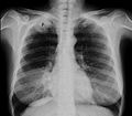
Chest X-ray showing a Pancoast tumor (labeled as P, large cell lung carcinoma, right lung), of a 47-year-old smoking woman.

Computed tomography showing a Pancoast tumor (labeled as P, large cell lung carcinoma, right lung), of a 47-year-old smoking woman.


Contenido relacionado
Aerobic organism
Vertebra
Human eye