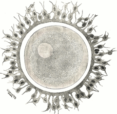
Ovum
The ovules are the mature female sex cells or gametes. They are large, spherical, nonmotile cells and contain half the number of chromosomes (haploid) as a somatic cell.
In women from puberty, and approximately every 28 days, an oocyte matures inside a follicle of an ovary, then it is extruded towards the outside, transforming into an ovum, which usually passes into one of the fallopian tubes.
History
Ova were surely discoveries of the 1660s and 1670s. Then in 1673 the Royal Society decided that neither De Graaf, nor Swammerdam, nor van Horne had been the first to discover that women had eggs. That honor, they said, went to Nicolás Steno.
The ova were described in 1827 in Ovi Mammalium et Hominis genesi ("On the genesis of the ovum of mammals and of men"), a pamphlet presented in 1827 by the biologist Karl Ernst von Baer.
In animals
In animals, the ovum is the female sex cell (female gamete), a mature cell with half the number of chromosomes (haploid) and capable of uniting in fertilization with a sperm, to form a zygote. When the female is born, she does it with all the ovules that she is going to have in her life, and from her birth they will decrease in number.
Graafian follicle
The mature ovum is found floating within a histological anatomical structure in the cortical area of the ovary, called the Graafian follicle, which is also called the mature follicle, late antral follicle or preovulatory follicle.
Among all the antral follicles, the "dominants" will produce mature oocytes ready to ovulate in each mammalian estrous cycle.
Microarchitecture
Using the light microscope, the classic characteristics of the ovule were described:
- large size compared to any somatic cell, 150 micrometers (μm) of diameter on average.
- a small and (central) core
- an abundant cytoplasm and with reserve (of vitelo)
- thick membrane
- periviteline space
- pelvic area /membrana vitelina
- radiata crown
Ultrastructure
With the electron microscope, details of the ovule could be observed:
- ovolema with microvellos
- cortical
- peripheral hue
- pelvic area
| Species | Graaf leaf | Total diameter | Ovocito solo | Hairdrying area |
| Bovina | 14.6 millimeters (mm) | 190 micrometers (μm) | 120 μm | 17,8 μm |
| Ovina | - | 127.5 μm | 104.4 μm | 18.8 μm. |
| Caprina | - | - | 120 μm | - |
| Alpaca | 7-12 millimeters (mm) | 170 micrometers (μm) | - | - |
| Humana | 10-20 mm (mm) 10 000-20 000 micrometers (μm) | 0.15 millimeters (mm) 150 micrometers (μm) | - | 11,6 μm |
Ploidy
The ovules are haploid cells formed in the ovaries by the subdivision by meiosis of a primary oocyte into two secondary oocytes, and these into an ovule and two polar bodies. This process, called oogenesis, in mammals manifests itself macroscopically through the periodic process of ovulation.
The first of the two meiotic divisions—which reduces the number of chromosomes—begins during embryonic development and is interrupted during prophase. It resumes from puberty, when in each cycle an oocyte and the follicle that surrounds it mature, completing the first division —which produces a secondary oocyte— and beginning the second. The second meiotic division —which generates the mature ovule— is in turn interrupted, and is not completed until fertilization occurs. After completion of oogonia meiosis, in addition to an oocyte, two polar bodies will have been formed, the first being the "sister cell" of the secondary oocyte, and the second the sister cell of the ovum.
While polar bodies are small, ova are the largest haploid cells in the human body. The plasmatic membrane of an oocyte is called an ovolem and plays an important role in the fertilization process.
About 400 of the original primary oocytes mature as ova in a woman's lifetime.
Between 24 to 48 hours before ovulation, there is a peak of luteinizing hormone that initiates meiosis II and this stops again in the second meiotic arrest (metaphase II) 3 hours before the ovulation occurs. ovulation.
In plants
In the science that studies plants, the ovule (botany) is called the seminal primordia, which are megasporangia, while the female gametes are called oospheres.
Contenido relacionado
Herman von Helmholtz
Galapagos Islands
Enzyme


