
Optical microscope

An optical microscope is a microscope based on optical lenses. It is also known as a light microscope (using light or "photons") or brightfield microscope. The development of this apparatus is often associated with the work of Anton van Leeuwenhoek. Leeuwenhoek's microscopes consisted of a single small convex lens, mounted on a plate, with a mechanism to hold the material to be examined (the sample or specimen). This use of a single convex lens is known as a simple microscope, which includes a magnifying glass, among other optical devices.
History
- 1590: Zacharias Janssen builds a microscope with two convergent lenses.
- 1611: Johannes Kepler suggests how to manufacture a compound microscope.
- 1665: Robert Hooke uses a composite microscope to study cork cuts and describes small pores in the form of cells he called "cells". Publish your book Micrographia.
- 1674: Leeuwenhoek reports his discovery of protozoa. Nine years later he will be the first to observe bacteria.
- 1828: W. Nicol develops microscopy with polarized light.
- 1838: Schleiden and Schwann propose the cell theory and declare that the nucleated cell is the structural and functional unit in plants and animals.
- 1849: J. Quekett publishes a practical treaty on the use of the microscope.
- 1876: Abbé analyzes the effects of defaction in the formation of the image in the microscope and shows how to perfect the design of the microscope.
- 1881: Retzius describes a large number of animal tissues in a detail that has not been overcome by any other light microscopist. In the next two decades he, Cajal and other histologists develop new methods of staining and put the foundations of microscopic anatomy.
- 1886: Carl Zeiss manufactures a series of lenses, Abbé design that allow the microscopist to solve structures in the theoretical limits of visible light.
- 1908: Köhler and Siedentopf develop the fluorescence microscope.
- 1930: Lebedeff designs and builds the first interference microscope.
- 1932: Zernike invents the phase contrast microscope.
- 1952: Nomarski invents and patents the differential interference contrast system for the light microscope.
Parts of the optical microscope and their functions

Tube, focus zip and macrometric screw.

Interchangeable eyepieces of different increases.

Macro and micrometric screws.
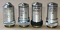
Dismantled goals.

Diaphragm and Condenser.

Platinum and base.
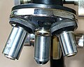
Rev.


- Eye: lens located near the observer's eye. Capture and expand the image formed in the objectives.
- Objective: lens located in the revolver. It magnifies the image, is a vital element that allows you to see through the eyepieces.
- Cabezal: is the head of the microscope that allows you to get better shots of the target.
- Condenser: lens that concentrates the luminous rays on preparation.
- Diaphragm: regulates the amount of light that reaches the capacitor.
- Foco: directs the luminous rays to the capacitor.
- Tube: it is the dark camera that carries the eyepiece and objectives. It can be attached to the arm by means of a zipper to allow the focus.
- Revólver: It is the system that carries the objectives of different increases, and which rotates to be able to use one or another, aligning them with the eyepiece.
- Macro and micrometric screws: They are focus screws, move the platinum or tube up and down. The macrometric allows wide displacements for an initial and micrometric approach, very short displacements, for the most precise approach. They can carry a lock controller that fixes the plate or tube to a certain height.
- Platinum: It is a horizontal platform with a central hole, on which the preparation is placed, which allows the passage of the rays from the light source below.
- Brass clamps: Two clamps, serve to hold the slide on the plate and a zip system that allows to move the preparation. It may be fixed or attached to the arm by a zipper to allow the focus.
- Brazo: It is the structure that holds the tube, the platinum and the focus screws associated with the tube or the platinum. The union with the base can be articulated or fixed.
- Base or foot: It is the bottom of the microscope that allows it to stand.
Lighting system
In transmitted light microscopes, the illumination system provides the light that will pass through the sample. It is composed of different parts: the light source with the help of a lens called collector, is represented in the plane of the aperture iris diaphragm. This diaphragm is installed in the front focal plane of the condenser and can vary its numerical aperture. The iris diaphragm arranged next to the collector is the field diaphragm. The variation of the diameter of the field diaphragm allows to obtain its image equal to the linear visual field of the microscope. The numerical aperture of the condenser generally exceeds that of the aperture of the microscopic objective: it is the illumination that allows us to better see what we want to observe, such as cells or cell membranes, among others.
Compound optical microscope
A compound microscope is a light microscope with more than one lens. They are especially used to examine transparent objects, or objects cut into sheets so thin that they are transparent.
Main elements of a basic microscope

Microscopes of this type are usually more complex, with several lenses in the objective as well as in the eyepiece. The objective of these lenses is to reduce chromatic aberration and spherical aberration. In modern microscopes the mirror is replaced by a lamp that offers stable and controllable illumination.
Compound microscopes are used to study thin specimens, since their depth of field is very narrow. They are generally used to examine cultures, crushed slides, or a very thin slice of whatever material. It usually depends on the light passing through the sample from below and special techniques are usually necessary to increase the contrast of the image.
The resolution of optical microscopes is restricted by a phenomenon called defaction that, depending on the numerical opening (AN or AN{displaystyle A_{N}}) of the optical system and the wavelength of the light used (λ λ {displaystyle lambda }), establishes a defined limit (d{displaystyle d}to optical resolution. Assuming that optical aberrations were despicable, the resolution would be:


- δ δ =λ λ 2↓ ↓ AN{displaystyle delta ={frac {lambda }{2*A_{N}}}}}}}

Normally, it's supposed to be a λ λ {displaystyle lambda } 550 nm, corresponding to the green light. If the middle is the air, AN{displaystyle A_{N}} maximum practice is 0.95, and in the case of oil up to 1.5.
This means that even the best light microscope is limited to a resolution of about 0.2 micrometers.
Spacing power, immersion objectives and useful magnification
- Separate power
From the theory of diffraction on the formation of images through a microscope, it is obtained that the minimum distance between two separately visible points is:
- δ δ =λ λ 2↓ ↓ AN{displaystyle delta ={frac {lambda }{2*A_{N}}}}}}}
Where λ is the wavelength of monochromatic light in which the object is observed and A is the aperture of the microscope.
- Immersion objectives
The liquid optical medium that fills the space between the object and the objective is called the immersion liquid. Its refractive index is close to that of glass (water, glycerin, cedar and juniper oils, monobromonaphthalene, among others are used).
Although all the components that make up a microscope are important, the objectives are of paramount importance, since the image, in the end, depends to a great extent on its quality. The best objectives are those that are corrected for aberrations.
Aberrations
They are optical alterations in the formation of the image due to the objective lenses themselves.
- Geometric saws (Keystone effect).
- Colour saws.
Correction of aberrations
To avoid geometric aberrations, so-called flat or plasmatic objectives are built, which is usually indicated on the objective itself with the inscription PLAN. Objectives that are corrected for chromatic aberrations are called achromatic (corrected for red and blue), semi-apochromatic (corrected for red and blue and have a larger numerical aperture) and finally apochromatic (which are of higher quality and are corrected for red, blue and green).
Optical Microscope Applications
This instrument has been very useful, especially in the fields of science where microscopic structure and organization is important, being successfully incorporated into research within the area of chemistry (in the study of crystals), the physics (in the investigation of the physical properties of materials), geology (in the analysis of the mineralogical and textural composition of rocks) and, of course, in the field of biology (in the study of microscopic structures of the living matter), to cite some disciplines of science.
Until now it has been used in the histology and pathology laboratory, where microscopy allows certain diagnostic applications, including the certainty diagnosis of cancer, numerous crystalline structures, pigments, lipids, proteins, bone deposits, amyloid deposits, etc.
Stereoscopic microscope or Stereomicroscope


The design of this instrument, also called a "binocular magnifying glass", is different from the one in the diagram above, and its utility is different, as it is used to provide a stereoscopic (3D) image of the sample. For this, and as in conventional binocular vision, it is necessary for the two eyes to observe the image with slightly different angles. Obviously all stereoscopic microscopes, by definition, must be binocular (with one eyepiece for each eye), so the two terms are sometimes confused. There are two types of design, called respectively convergent (or Greenough) and common goal (or Galileo).
The convergent design consists of using two identical microscopes inclined at a certain angle with respect to each other and mechanically coupled in such a way that they focus the image at the same point and with the same magnification. Although it is an economical, powerful design and in which aberrations are very easy to correct, it has some limitations in terms of modularity (ability to modify the system to add accessories) and observation for long periods of time is tiring.
The common objective design uses two parallel optical paths (one for each eye) that are converged at the same point and at an angle with an objective common to both microscopes. The design is more sophisticated than the convergent one, with better modularity and does not generate fatigue in long observation times. However, it is more expensive to manufacture and aberrations, as the image is generated through the periphery of the common objective and at an angle that does not coincide with its optical axis, are more difficult to correct.
Stereoscopic microscopes are usually equipped, in any of their variants, with a zoom system or a magnification changer system that allows the sample to be observed in a variable magnification range, always less than that of a compound microscope. The stereoscopic microscope is suitable for observing objects of relatively large sizes, so it is not necessary to modify the objects to be seen, (laminar) nor is it necessary for the light to pass through the sample. This type of microscope allows distances ranging from a couple of centimeters to tens of them from the sample to the objective, which makes it very useful in botany, mineralogy, and in industry (microelectronics, for example) as well as in medicine (microscopes). surgical) and research, mainly in applications that require manipulation of the viewed object (where stereoscopic vision is essential).
We could say that a stereoscopic microscope can be used, among many other things, to carry out dissections of animals and plants.
Connecting a digital camera to an optical microscope

A mechanical optical adapter is important in digital photography. This adapter serves as a link between the camera and the microscope. It is especially important that the mechanical connection is firm, as any slight movement, ie camera vibrations, will reduce the image quality significantly. Additionally, an optical adapter is required for the light path with which the CCD/CMOS sensor of the camera will project an image of total sharpness and illumination.
Photomicrography (photograph taken with the aid of a compound microscope) is a very specialized field of photography, for which very expensive equipment is available, and not just studio equipment.
With a suitable quality microscope, such as those found in most scientific laboratories, photomicrographs of reasonable quality can be made using a general purpose fixed or interchangeable objective camera.
Basic Methods
There are two basic methods of taking pictures through a microscope. In the first method, the camera objective performs a function similar to that of the lens of the eye and projects a real image of the virtual image seen through the microscope eyepiece onto the sensor. This method is the only one suitable for using cameras with a fixed lens, that is, not interchangeable.
The second method, suitable for cameras with an interchangeable objective, involves removing the objective from the camera and adjusting the microscope so that the eyepiece forms an image directly on the sensor.
The quality of a microscope's optics (objective and eyepiece) is essential in determining the quality of a photographic image. Microscope objectives and eyepieces come in different qualities, determined by the precision with which they have been corrected for aberrations. The cheapest objectives are corrected for spherical aberration for a single color, generally greenish yellow, but not for chromatic aberration for the entire visible spectrum, but only for two or three primary colors. These objectives are called achromats, and they also show some amount of field curvature; that is, the entire field of view of the objective cannot be simultaneously brought into fine focus.
There are flat-field achromats, in which the field curvature has been almost completely corrected, they are called planachromats.
The apochromats are corrected for spherical aberration for two colors and for chromatic aberration for the three primary colors. Even so, they will show field curvature unless they are planapochromats, the best objectives available. The eyepieces also have different qualities. The simplest are the wide field ones.
Compensating eyepieces are designed to compensate for certain residual chromatic aberrations in the objective, and are best used with apochromatic objectives, although they can also be used successfully with higher power achromats. There are photo eyepieces, special for photomicrography, and when used with planapochromatic objectives they give the best possible quality of photography.
Contenido relacionado
ARM architecture
Forth
IP adress