Ontogeny
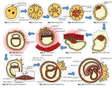
Ontogeny (also called morphogenesis or «ontogenesis») is a branch of biology that describes the development of an organism, from fertilization by the fusion of the male and female gametes to the conformation from a zygote during sexual reproduction to its senescence, passing through the adult form. Ontogeny is studied in developmental biology. «Ontogeny is the history of the structural change of a unit without it losing its organization. This continuous structural change occurs in the unit, at each moment, or as a change triggered by interactions coming from the environment where it is found or as a result of its internal dynamics.
Animal development or ontogeny fulfills two main functions:
- It generates cell diversity (differentiation) from the fertilized egg (cigot) and organizes the various cell types in tissues and organs (morphogenesis and growth).
- Ensures the continuity of life from one generation to the next (reproduction).
Stages of animal development
Fecundation
Fertilization is the union of two gametes with the consequent formation of a zygote. The gametes can be the same (isogametes) or different (anisogametes: sperm and egg). The central process of fertilization is karyogamy, that is, the fusion of the nuclei of the gametes (pronuclei).
Activation
Activation is the set of events that take place in the zygote and that determine that it begins to segment (divide by mitosis).
Embryogenesis
Embryogenesis is the set of ontogenetic processes that spans from the moment the zygote begins to segment until organogenesis is completed. Embryogenesis includes the following phases: cleavage, blastulation, gastrulation, and organogenesis.
- Segmentation is a very fast set of cell mitosis, thanks to which the zygote is divided into multiple smaller cells called blastomers. The first two divisions undergoing the zygote determine the animal's polarity: animal and vegetative poles. The solid spherical body, equal to the cigot, formed by a small number of blastomers, which results from the segmentation is known as the mórula.
- The blasting is the process of formation of the blassula, which is often presented as a hollow spherical body formed by blastomers located on the periphery (blastodermo). The central cavity of the blastula is called blastocele; it is filled with blastocelic fluid and constitutes the primary general cavity (interna) of the animal.
- Gastrulation is the set of morphogenetic processes that are produced from the blond state and which lead to the formation of blastodermal or germinative leaves: ectoderm (outdoor leaf) and endoderm (inner leaf). In some primitive metazoos, the ectoderm and endoderm are separated by a more or less cellularized gelatinose layer, which is called mesoglea or mesohile (diabiotic animals). However, in most of the metazoos a third blastodermal leaf is formed, between the ectoderm and the endoderm, called mesoderm (triblastic animals). In general, it can be said that the ectoderm produces the cells of the epidermis and the nervous system, the endoderm originates both the cells that tap the digestive tract and the organs associated with that system (panges, liver, etc.). Mesoderm leads to various organs (heart, kidneys, gonads), connective tissues and bras (bones, muscles) and blood cells.
- Organogenesis is the set of interactions and cellular displacements leading to organ formation. Many organs are formed by cells originated from different blastodermal leaves. During organogenesis, certain cells carry out long migrations from their places of origin to their final destinations. Among the migratory cells it is worth noting those that are precursors of the blood cells, lymphatic and pigmentary, as well as gametes.
In the zygote there is a region of the cytoplasm (germ plasm) that gives rise to the precursor cells of the gametes. Such precursor cells are called germ cells. All other cells are known as somatic cells. Germ cells migrate to the gonads, where they eventually differentiate into gametes. This differentiation, known as gametogenesis, generally occurs when the animal is physically mature.
Embryogenesis culminates in the formation of an embryo that can then go through various ontogenetic phases (fetus, larva, pupa, juvenile) until it reaches an adult.
The life cycle of animal species is completed with reproduction, aging (senescence) and death.
Segmentation
Zygote segmentation depends largely on the distribution of yolk (reserve material) in the ovule (egg) and, therefore, in the zygote itself (fertilized ovum or egg). Apart from this, the amount of yolk is correlated with the duration of embryogenesis. If there is little yolk, the embryo either soon becomes a larva capable of feeding itself, or it must receive a supply of nutrients from the mother. If there is a large amount of yolk, the embryo becomes a juvenile by feeding exclusively on yolk.
There are numerous terms that describe the condition of the egg based on the amount and arrangement of the yolk:
- Egg: egg without or with very little vite it.
- Oligolicytic egg: egg with relatively little vitela.
- Mesolecytic egg: egg with a moderate amount of vitela.
- Telolecytic egg: egg with lots of vitela.
- Isolectic egg: egg with vite distributed evenly.
- Heterolecytic egg: egg with vitela distributed irregularly.
- Centrallecytic egg: egg with the concentrated vitela in the center of cytoplasm.
- Endoletic egg: egg that contains vitelo inside.
- Egg: Egg with external vitela, contained in viteleogenic cells.
Types of segmentation
Depending on affecting all or only part of the zygote, a distinction is made between total cleavage and partial cleavage.
Total (holoblastic) cleavage occurs in zygotes formed from alecyte, oligolecyte, and mesolecyte eggs. The zygote divides completely into two blastomeres, which then give rise to more blastomeres. Partial cleavage (meroblastic) occurs in zygotes formed from centrolecitos and telolecitos eggs. The zygote does not divide completely; only the cell nucleus divides. The resulting nuclei are surrounded by membranes, forming blastomeres. The yolk remains undivided.
Holoblastic segmentation
Different modalities are distinguished in total segmentation depending on the size and arrangement of the blastomeres.
- Total segmentation modality depending on the size of blastomers: equal and unequal.
- Total equal segmentation: all blastomers that form are of equal size.
- Uneven total segmentation: Relative small-size blastomers are formed, called micromers, and relative-sized blastomers, called macromers. Mycromers occupy the animal pole of the developing organism and are those who form the embryo. The macromers, which contain the reserve material (vitelo), occupy the vegetative pole. The total segmentation equals the eggs that present the vitelo distributed regularly (isolecite eggs). The eggs that are totally segmented, but that have the vitela distributed irregularly (heterolecite eggs) suffer from total unequal sgmentation.
- Total segmentation patterns based on the spatial distribution of blastomers: radial and spiral.
- Total radial segmentation: blastomers are arranged following an imaginary design of parallels and meridians. Depending on the distribution of the vitelo, cigotes that experience total radial segmentation can result in equal blastomers (total equal radial selection) or micromers and macromers (total uneven radial selection). Radial segmentation is said to be undetermined or regulatory because blastomers set their fate relatively late.
- Total spiral segmentation: the various blastomers are placed forming a certain angle with respect to the polar axis, so that, together, they are arranged in a spiral form. The zygotes that experience the spiral segmentation do so by following the unequal pattern (total spiral segmentation). The spiral segmentation is undetermined or in mosaic because the fate of blastomers is fixed at a very early stage of development.
Meroblast segmentation
In partial segmentation, different modalities are distinguished depending on the distribution of the yolk in the ovule from which the zygote is formed.
Partial cleavage of zygotes formed from telolecyte eggs: discoidal cleavage. In discoidal cleavage, the zygote does not divide completely; only the cell nucleus divides. The two resulting nuclei are surrounded by respective cell membranes. Two small blastomeres are thus formed which continue to divide, while the part containing the yolk remains undivided. The blastomeres occupy the animal pole, forming a disc (blastoderm or blastodisc) that is located on the large non-segmented mass of yolk. This mass of yolk forms the yolk sac.
Partial cleavage of zygotes formed from centrolecitos eggs: peripheral cleavage. In peripheral cleavage, the cell nucleus divides several times. The resulting nuclei migrate to the periphery, where each is surrounded by a membrane, forming a blastomere. Thus, the blastomeres occupy the periphery (peripheral blastoderm) and surround the undivided yolk, which occupies a central position. One or several nuclei migrate towards one of the poles to form a set of blastomeres known as polar plasma. These polar plasma blastomeres are the precursors of germ cells.
Different segmentation modalities are not necessarily correlated with the phylogenetic position of the animals. It is frequent that the species corresponding to a certain zoological group present the same segmentation modality. However, different modalities can also occur within the same group. Apart from this, there are variants typical of certain animal groups that do not correspond to any of the segmentation models exposed.
Blastulation
The cleavage of the zygote concludes with the formation of the blastula. The process of formation of the blastula is called blastulation. The blastula is often described as an embryonic state consisting of a sphere of blastomeres (blastoderm) surrounding a central cavity (blastocoel), filled with blastocoel fluid. However, this description corresponds only to a certain type of blastula. Other blastulas differ markedly from this model.
Main types of blastula:
- Celebrities: The gallblasts are spherical blastules formed by a peripheral layer of blastomers (blastoderm) that surrounds a central cavity called blastocele. The gallblasts are formed as a result of the total segmentation of the zygote. When segmentation is total equal (or almost equal, blastomers are similar in size and blastocele occupies a central position. If the segmentation is total unequal, micromers and macromers are formed and the blastocele adopts an eccentric position, towards the animal pole. In this pole the micromers are located, while the macromers, rich in vitelo, are located in the vegetative pole.
- Estereoblástulas. Stereoblastules are spherical-like and solid-resistance scales, resulting from a total unequal segmentation in which the macromers of the vegetative pole are very bulky. The blastocele is a virtual cavity; it is entirely occupied by the macromers. The micromers are the vegetative pole, covering the macromers.
- Discoblástulas: Conblástulas are spherical blastulas resulting from the uneven partial segmentation of the telolecite cigotes. Micromers form a casket or disk (blastodermo or blastodisco) at the animal pole. The blastodisco rests on a large mass of vitela that constitutes the future vitelino sack. The blastocele consists of an internal cavity that opens between the blastodisco and the viteline sac.
- Periblástulas: The periblástulas are somewhat elongated blastulas, resulting from the peripheral partial segmentation of the central cigotes. The blastomers form a peripheral blastoderm that covers a central mass of vitelo. There's no real blastocele. The corresponding cavity is occupied by the vitelo.
Gastrulation
Gastrulation consists of the set of morphogenetic processes that occur from the blastula stage and that lead to the formation of blastodermal sheets or germinative sheets. Gastrulation differs markedly from one zoological group to another. It may even differ between representatives of the same group.
Modalities of gastrulation
- Invagination or embolism. Gastrulation by invagination or embolism occurs from jealousy. It consists of the penetration of the vegetative hemisphere in the blastocele, thus forming two layers of blastomers. The external receives the name of ectoderm; the internal is called endoderm. The endodermo delimits a new cavity, called arquén, which constitutes the future digestive tract of the animal. The archthen is in contact with the outside through an opening called blastoporo.
- Coverage or epibolia. Gastrulation by coating or epibolia occurs from stereoblastules. It consists of the active multiplication of the mycromers of the animal pole that end up surrounding, almost entirely, the macromers of the vegetative pole. Mycromers are the ectoderm. The macromers, which remain in an internal position, are the endoderm. This endoderm delimits a cavity d which is the arched. The blastoporo is made up of the free circular contour of the ectodermal dome. The embolism and epibolia, which occur simultaneously in some cases, lead to the formation of a globose gástrula, formed by two blastodermal leaves, the ectoderm and the endoderm. The endodermo delimits a cavity, the archer, which opens to the outside through the blastoporo. Between the ectoderm and the endoderm, that is to say in the real or virtual space that corresponds to the primitive blastocele, the third blastodermic leaf is formed which is called mesoderm. The embolism and the epibolia are not the only existing forms of gastrulation. In many zoological groups, the formation of blastodermal leaves takes place very differently. This casts doubts about the possible homology of blastodermal leaves in the various metazoa taxa.
- Deployment. De-lamination gastrulation takes place from a lattice. It is produced by the mitotic division of blastomers according to division planes parallel to the blastoderm surface. Mythotic bones are radial. Thus, the section, formed by a single layer of cells, becomes a germ with a double cellular layer. The outer layer of cells is the ectoderm and the inner layer, the endoderm. The endoderm is separated from the ectoderm (delamine) and between both leaves is formed a mesogle. The endodermo delimits a cavity, the celénteron (also called arquénteron), lacking blastoporo. The Celénteron opens to the outside secondaryly.
- Immigration. Immigration gastrulation consists of active cell migration from blastoderm to blastocele. Once there the cells proceed to form the other blastodermal leaves.
Other, less frequent, modalities of gastrulation that resemble gastrulation by immigration to a greater or lesser degree are the following: involution, separation, polar ingression, proliferation, and cavitation.
Ontogeny and phylogeny
The idea that ontogeny recapitulates phylogeny, that is, that the development of an organism exactly reflects the evolutionary development of the species, is now discredited. However, some connections between ontogeny and phylogeny can be observed, given by evolution, in this way ontogeny is used in cladistics as a guide to reconstruct evolutionary history and phylogenetic relationships between clades.
Contenido relacionado
Leersia
Ebenaceae
Casuarina
