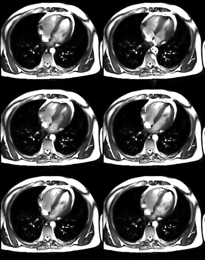
Nuclear magnetic resonance
Nuclear magnetic resonance (NMR) is a physical phenomenon based on the quantum mechanical properties of atomic nuclei. NMR also refers to the family of scientific methods that explore this phenomenon to study molecules (NMR spectroscopy), macromolecules (biomolecular NMR), as well as tissues and whole organisms (magnetic resonance imaging).
All nuclei with an odd number of protons + neutrons have intrinsic magnetic momentum and angular momentum, in other words, they have spin > 0. The nuclei most commonly used in NMR are protium (1H, the most sensitive isotope in NMR after the unstable tritium, 3H), 13C, and 15N, although isotopes of nuclei of many other elements (2H, 10B, 11B, 15N, 17O, 19F, 23Na, 29Yes, 31P, 35Cl, 113Cd, 195Pt) are also used.
NMR takes advantage of the fact that atomic nuclei (for example, within a molecule) resonate at a frequency directly proportional to the strength of an exerted magnetic field, according to the Larmor precession frequency equation, to subsequently perturb this alignment with the use of an alternating magnetic field, of orthogonal orientation. The scientific literature up to 2008 includes spectra over a wide range of magnetic fields, from 100 nT to 20 T. Higher magnetic fields are often preferred as they correlate with increased signal sensitivity, although for resonance imaging Magnetic fields in medicine are used to generate non-ionizing radiation. There are many other methods to increase the observed signal. The increase in the magnetic field also translates into a higher spectral resolution, the details of which are described by the chemical shift and the Zeeman effect.
The NMR phenomenon is also used in low-field NMR, ground-field NMR, and some types of magnetometers.
History
Discovery
Nuclear magnetic resonance was described and measured in molecular rays by Isidor Isaac Rabi in 1938. Eight years later, in 1946, Félix Bloch and Edward Mills Purcell refined the technique used in liquids and solids, so they shared the Nobel Prize in Physics in 1952.
Purcell had worked on the development of radar and its applications during World War II at the Radiation Laboratory at the Massachusetts Institute of Technology. His work during that project was to produce and detect radio frequency energy, and on absorptions of such RF energies by matter, preceding his co-discovery of NMR.
They realized that magnetically active nuclei, such as 1H (protium) and 31P, could absorb RF energy when placed in a magnetic field of a specific power and thus managed to identify the nuclei. Different atomic nuclei within a molecule resonate at different radio frequencies for the same magnetic field strength. Observing such magnetic resonant frequencies of the nuclei present in a molecule allows the trained user to discover chemical, structural, spatial, and dynamic information about the molecules.
The development of nuclear magnetic resonance as a technique for analytical chemistry and biochemistry paralleled the development of electromagnetic technology and its introduction to civilian use.
Physical principle
Nuclear spin
Hadrons (more specifically baryons) that make up the atomic nucleus (neutrons and protons), have the intrinsic quantum mechanical property of spin. The spin of a nucleus is determined by the quantum number of the spin I. If the combined number of protons and neutrons in a given isotope is even, then I = 0, i. and. there is no general spin; Just as electrons pair up in atomic orbitals, so do neutrons and protons pair up in even numbers (which are also spin ½ particles) to give an overall spin = 0.
A non-zero spin, I, is associated with a non-zero magnetic moment, μ:
- μ μ =γ γ I{displaystyle mu =gamma I}
where γ is the gyromagnetic constant. This constant indicates the signal intensity of each isotope used in NMR. Except in atomic disintegration, the nucleus separated from the electrons is not obtained, the electrons that revolve around the nucleus are distinguished by the four quantum numbers, quantum mechanics explains it
Values of spin angular momentum
The angular momentum associated with the nuclear spin is quantized. This means that both the magnitude and the orientation of angular momentum are quantized (ie, I can only take values in a restricted interval). The associated quantum number is known as the magnetic quantum number, m, and can take integer values from +I to -I. Therefore, for any nucleus, there are a total of 2I+1 states of angular momentum.
The z component of the angular momentum vector, Iz is therefore:
- Iz=m {displaystyle I_{z}=mhbar }
in which {displaystyle hbar } It's Planck's constant down.
The z component of the magnetic moment is simply:
- μ μ z=γ γ Iz=mγ γ {displaystyle mu _{z}=gamma I_{z}=mgamma hbar }
Behavior of spin in a magnetic field
Consider a nucleus with spin ½, such as 1H, 13C, or 19F. This nucleus has two possible states of spin: m = ½ or m = -½ (also called 'up' and 'down& #39;, or α and β, respectively). The energies of these two states are degenerate—meaning they are the same. Therefore the populations of these two states (i.e. the number of atoms in the two states) will be approximately equal under conditions of thermal equilibrium.
However, by putting this nucleus under a magnetic field, the interaction between the nuclear magnetic moment and the external magnetic field will cause the two spin states to stop having the same energy. The energy of the magnetic moment μ under the influence of the magnetic field B0 (main magnetic field) is given by the negative dot product of the vectors:
- E=− − B0⋅ ⋅ μ μ =− − μ μ zB0{displaystyle E=-{mathbf {B} _{0}}}{cdot {mathbf {mu } }=-mu _{z}B_{0}}}}}}
In which the magnetic field has been oriented along the +z axis (by convention).
Therefore:
- E=− − m γ γ B0{displaystyle E=-mhbar gamma B_{0}}
As a result, the different nuclear spin states have different energies in a magnetic field ≠ 0. In other words, we can say that the two spin states of a spin ½ have been aligned either for or against the field magnetic. If γ is positive (which is true for most isotopes) then m = ½ is in the low energy state.
The energy difference between the two states is given by the equation:
- Δ Δ E= γ γ B0{displaystyle Delta E=hbar gamma B_{0}}}
and this difference translates into a small majority of spins in the low energy state.
Resonance occurs when this energy difference is excited by electromagnetic radiation of the same frequency. The energy of a photon is E=h.. {displaystyle E=hnu }Where .. {displaystyle nu } It's his frequency. Therefore the absorption will occur when:
- .. =Δ Δ E2π π =γ γ B02π π {displaystyle nu ={frac {Delta E}{2pi hbar }={frac {gamma B_{0}}{2pi }}}}}}}}}
These frequencies typically correspond to the radio frequency range of the electromagnetic spectrum.
Nuclear Shielding
It might seem, from the above, that all the nuclei of the same nuclide (and therefore the same γ) resonate at the same frequency. This is not the case. The most important frequency disturbance for NMR applications is the 'screening' effect. exerted by the surrounding electrons. In general, this electronic shielding reduces the core's magnetic field (which determines the NMR frequency), because they are aligned away from Bo. As a result, the energy gap is reduced and the frequency required to reach resonance is also reduced. This NMR frequency shift, strongly influenced by chemical groups, is known as a chemical shift, and explains why NMR is a direct probe of chemical structure. If a core is more shielded, it will be offset towards 'high field' (smaller chemical shift) and if it is more unshielded, then it will be shifted towards 'low field' (higher chemical shift).
Unless the local symmetry is particularly high, the screening effect depends on the orientation of the molecule with respect to the external field. In solid-state NMR, the 'magic angle turn' (magic angle spinning) is necessary to dispel this orientational dependency. This is not required in conventional NMR since the rapid and disorderly movement of molecules in solution dissipates the anisotropic component of the chemical shift.
Fourier Transform Digitization
The natural recovery of the direction and sense of the spins once the radiofrequency is no longer applied, will generate emissions as a result of the energy release, which will be captured by the scanner's receiving antenna. These emissions have to go in accordance with the Dim-Phase, being the compilation of all these emissions the beginning of the magnetic resonance.
Once all the data extraction is finished, they will be processed in the frequency domain by using the Fourier transform, which will facilitate the reconstruction of the final image on the screen. The frequency of variation of a signal in space is called "K", that is, the data compiled in the domain of spatial frequencies is called K space.
The purpose of creating this space is to be able to apply Fourier's mathematical laws, which makes it possible to identify the place of origin of the emissions at a given moment and, therefore, their place of origin.
NMR Spectroscopy
NMR spectroscopy is one of the main techniques used to obtain physical, chemical, electronic, and structural information about molecules. It is a powerful series of methodologies that provide information on the topology, dynamics, and three-dimensional structure of molecules in solution and in the solid state. Likewise, in the years 1998-2001 nuclear magnetic resonance was one of the most used techniques to implement some principles of quantum computers.
MR Spectroscopy measures the activity of metabolites during cognitive processing. NAA (N-Acetyl Aspartate) peaks can be tracked in relation to the activation of an area of the brain during the requested task. Although it indirectly correlates with these processes, certain metabolic patterns have been found, such as a decrease in NAA peaks in the Hippocampus, related to a memory deficit, and a decrease in NAA peaks in the Temporal Lobe, related to epilepsy.
Most common applications
Magnetic resonance makes use of the resonance properties by applying radio frequencies to the atomic nuclei or dipoles between the aligned fields of the sample, and allows to study the structural or chemical information of a sample. MR is also used in the field of quantum computer research. Its most frequent applications are linked to the field of medicine, biochemistry and organic chemistry. It is common to refer to "magnetic resonance imaging" to the device that obtains magnetic resonance imaging (IRM, or MRI for Magnetic Resonance Imaging).[citation required]
Application in medicine
MRI is a technique used to diagnose diseases by obtaining images of the body. Despite the fact that there is no harmful effect on the patient, the practice is not recommended in pregnant women, unless its use is essential.
The machine used in magnetic resonance imaging, due to its size and technology, combines the advantages of high magnetic field equipment and open equipment. Thus, greater definition and better image quality are achieved, and the patient has a lesser feeling of claustrophobia and is also more comfortable. The duration of the test does not depend on the severity of the condition, but depends on the region to be studied.[citation needed]
- Development of exploration
Before starting the MRI, the healthcare staff will determine whether or not it can be done by filling out a questionnaire. Personal belongings will be deposited in a cabin. The health personnel will indicate the position to be placed on the table. Around the area of the body to be examined, antennas are placed, a device whose purpose is to improve the quality of the images. Finally the table will slide into the magnet and thus the exploration will begin.[citation needed]
- Development of the review
During the examination, the magnet will produce noises with different intensity, which can even be unpleasant, although before starting the examination, the health personnel can provide earplugs or headphones to reduce the noise. These noises are the product of obtaining the images. To obtain the highest quality images, it is essential to remain static, breathing calmly and following the instructions of the health personnel. Depending on the part of the body that has to be examined, you will have to hold your breath for a few seconds.[citation needed]
The magnet per se (read the magnetic field itself) cannot generate sounds since the human hearing system is insensitive to this physical phenomenon.
If there are noises, they will be the product of mechanical vibrations of ferromagnetic pieces or parts reached by the magnetic field while it is changing (see magnetostriction) to record the response of the atomic nuclei of the tissues under study. If there were no such variation (modulation) and the intervening fields were static, the dynamic behavior of atomic nuclei could not be recorded and consequently the sought data could not be obtained.
- Side effects
In connection with its use in medicine, sometimes the study requires the injection of drugs based on a chemical element known as gadolinium. The reason is that the gadolinium acts as a contrast medium that improves the quality of the MRI image. The chemical element is previously treated, binding it to chelators, to allow its elimination by the organism and to reduce its high toxicity. Gadolinium is responsible for a serious disease known as nephrogenic systemic fibrosis, a pathology that mainly affects people with kidney failure, the reason seems to be that the substance accumulates in large doses in the body of these people. Another worrying fact has recently been discovered, gadolinium also accumulates in significant amounts in the different tissues of people with normal kidney function.[citation needed]
Contenido relacionado
Scalar field
Phobia
Viscera


