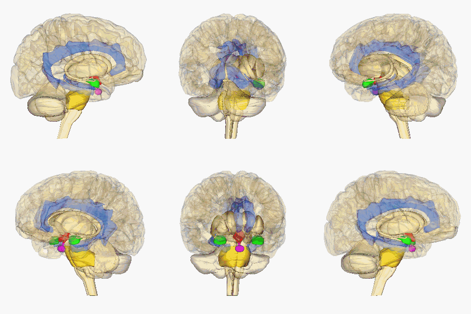
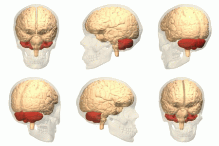
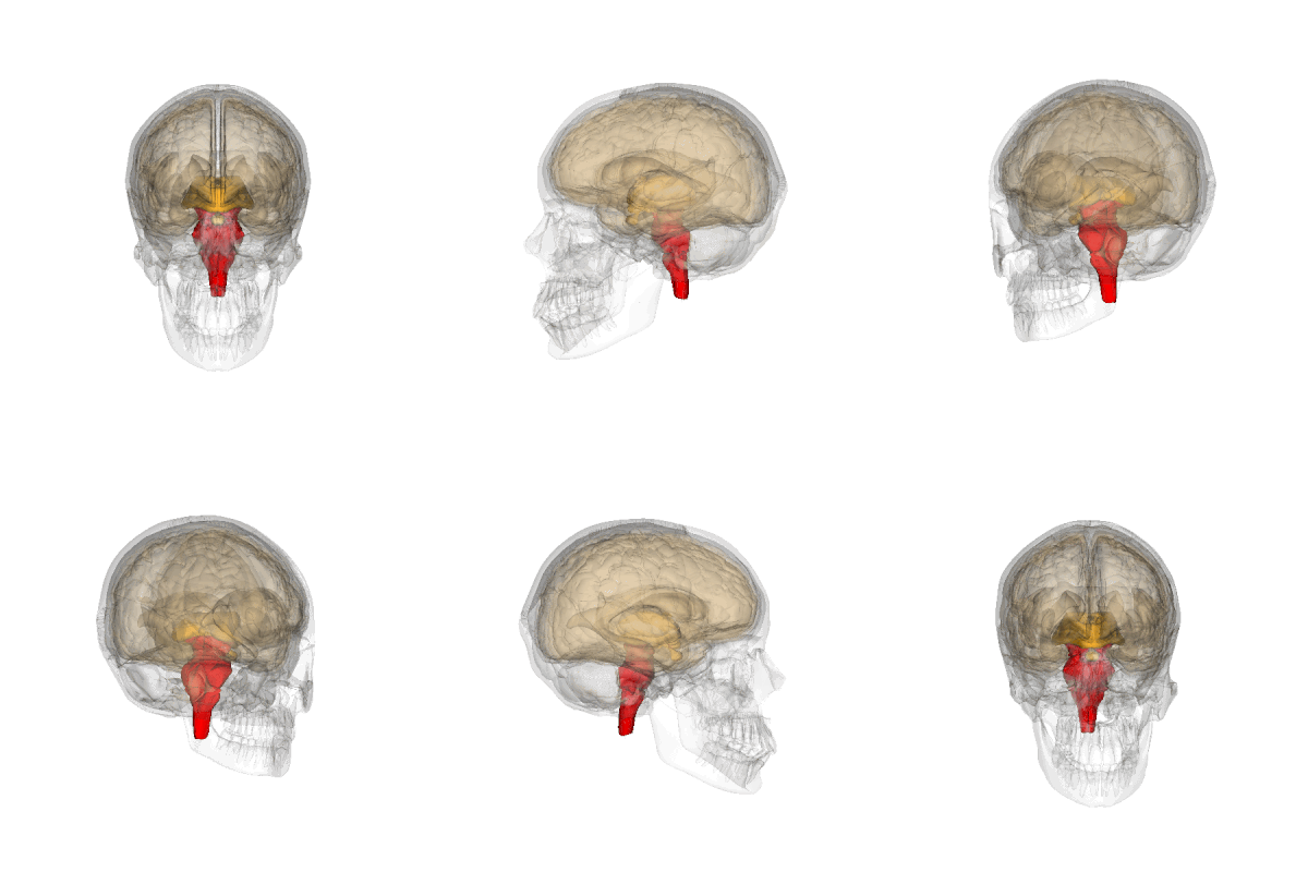
Neuroanatomy
Neuroanatomy is the study of the structure and organization of the nervous system. Comparative neuroanatomy is the science that analyzes and compares the nervous systems of different species. From the simplest systems to that of mammals and man.
The first known written record of a study of the anatomy of the human brain is Egyptian, the Edwin Smith Papyrus. The next major development in neuroanatomy came about a thousand years later, when the Greek Alcmaeon determined that the brain, not the heart, as was believed, governs the body and receives input from the senses. One of the founders of modern neuroanatomy was the discoverer of the neuron, the Spanish Santiago Ramón y Cajal, who was awarded the Nobel Prize in Medicine or Physiology in 1906.
Structural Neuroanatomical Division
The nervous system of vertebrates is made up of the brain and spinal cord (the central nervous system, or CNS) and the pathways of nerves that connect to the rest of the body (the peripheral nervous system, or PNS).
The central nervous system (CNS) consists of the brain, retina, and spinal cord, while the peripheral nervous system (PNS) consists of all the nerves outside the central nervous system that connect it to the rest of the body. Body.
The central nervous system is made up of brain regions such as the hippocampus that are critical for memory formation.
The nervous system also contains nerves, which are bundles of fibers that originate in the brain and spinal cord and branch several times to supply each part of the body. Nerves are made up primarily of the axons of neurons, along with a variety of membranes that line the nerve fascicles.
The brain and spinal cord are externally protected by the bony structures that are the skull and the vertebral column. Internally they are wrapped by three membranes: the dura mater, the arachnoid mater and the pia mater. They are also bathed by cerebrospinal fluid that fills the empty spaces and acts as a shock absorber, among other functions.
In order to specify anatomical locations, frequent references are made to conspicuous details of the brain such as fissures and orientation planes or section planes are used, which are generally «sagittal», «transverse» (or «coronal») and "horizontal".
The central nervous system is anatomically made up of:
- Cerebro
- Mesencéfalo
- Protuberance
- Cerebelo
- Bulbo raquíd
- Spinal cord (cervic, dorsal, lumbar, sacra and coccígea)
- Cranial nerves I and II
The peripheral nervous system is made up of:
- Cranial nerves III to XII.
- Spinal nerves (including 2 plexies: the brachial plexus and the lumbosacro plexus).
Functional Neuroanatomical Division
The peripheral nervous system is further subdivided into the somatic and autonomic nervous systems.
The autonomic nervous system also has two subdivisions: the sympathetic nervous system (SNS) and the parasympathetic nervous system (SNPS), which are important for the body's regulation of basic body functions, such as heart rate, respiration, digestion, temperature control, etc.
The sympathetic nervous system prepares the body to act in an emergency, and the parasympathetic nervous system prepares the body to conserve and restore energy.
Resources for Neurofunctional Research
Much of what we learn comes from looking at how "injuries" to specific areas of the brain affect behavior or other functions.
New resources have been improving the possibilities of observing the situation and aspects of brain function in living and healthy people. Computerized axial tomography, magnetic resonance imaging and positron emitters (PET) are image creators without "invading" the observed person.
The latter, with the help of appropriate injected products, makes it possible to observe the degree of activity of each brain area in different circumstances. In this way it is possible to determine with greater precision the areas involved in reasoning, memory, emotions such as love, fear, etc., and the routes carried out by the participating nervous stimuli are known.
Architecture of the spinal cord
It lies within the canal surrounded by the three meninges and cerebrospinal fluid. The architecture of the spinal cord is roughly cylindrical, beginning above at the foramen magnum in the skull, where it continues with the medulla oblongata, and ending below the spindle-shaped lumbar region at the conus medullaris, from which vertex forms a piamdrical prolongation descends, forming the terminal edge or filum terminalis.
Thirty-one pairs of spinal nerves are located along the course of the spinal cord, joined by anterior or motor roots, and posterior or sensory roots.
The structure of the spinal cord is made up of grey matter in its central portion, and white matter in its periphery.
In a cross section, the gray matter can be seen to form a silhouette similar to that of a butterfly, with its anterior and posterior gray cords joined by the gray commissure. The white matter is divided into anterior, lateral, and posterior white cords.
The architecture of the spinal cord changes according to its position.
Brain
It is situated in the cranial cavity and is continuous with the spinal cord through the foramen magnum. It is surrounded by three meninges. The brain is divided into three main parts, which are:
- Break it up.: later brain.
- Bulbo raquid.
- Protuberance.
- Brain.
- Mesencéfalo: medium brain.
- Tectum and tegmentum.
- Prosencéfalo: previous brain.
- Brain and brain.
Cellular neuroanatomy
The cellular basis of the nervous system is composed of neurons, glial cells, and extracellular matrix. There are neurons and glial cells of many types. Neurons are the information processing cells of the nervous system: they generate the sensation of our environment, produce our thoughts and cause our movements. They communicate with each other through electrical signals that run through their extensions: the axons and dendrites; interneuronal junctions are called synapses and are complex structures. Glial cells maintain homeostasis, myelin production, and provide support and protection to neurons in the brain. Some glial cells (astrocytes) can even propagate intercellular calcium waves over long distances in response to stimulation and release "gliotransmitters" in response to changes in calcium concentration. The extracellular matrix also provides support at the molecular level for brain cells.
Resources for neurocellular research
These resources are used in samples obtained in biopsies, necropsies and animals.
Staining is a technique used to enhance contrast by creating particular features in microscopic images.
In histochemistry uses knowledge about the biochemical properties of reactions of the chemical components of the brain, especially enzymes.
Immunocytochemistry is a special case of histochemistry that uses selective antibodies against a variety of chemical epitopes of the nervous system. It is able to selectively stain particular types of cells, axonal tracts, neuropils, glial processes or blood vessels, or certain specific intracytoplasmic or intranuclear proteins and other immunogenic molecules.
More complex techniques are also used, such as in situ hybridization that uses RNA probes, genetically encoded markers, and certain viruses that can replicate in brain cells and synapses.
Serial electron microscopy (electron microscope) is very useful.
See also
- Central nervous system
- Peripheral nervous system
- Sensory body
- Neuroembriology
Contenido relacionado
Cyclocarpales
Capparaceae
Araliaceae


