
Nervous system
The nervous system is a group of cells specialized in conducting electrical signals and is made up of neurons and glial cells. Neurons have the function of coordinating the actions of living beings within the animal kingdom by means of signals, from one place to another within the organism. Most multicellular animals have nervous systems with similar basic functions, although with different a highly variable degree of complexity. Only animals that do not have well-differentiated tissues and organs lack it, such as porifera (sponges), placozoans and mesozoans.
The nervous system captures stimuli from the environment (external stimuli) or signals from the organism itself (internal stimuli), processes the information and generates different responses depending on the situation. As an example, we can consider an animal that, through the light-sensitive cells of the retina, captures the proximity of another living being. This information is transmitted through the optic nerve to the brain, which processes it and emits a nerve signal that, through the motor nerves, causes the contraction of certain muscles in order to move in the opposite direction of the greatest danger.
Divisions of the Nervous System
.es.png)
- To study from the anatomical point of view the nervous system has been divided into central and peripheral. The central nervous system corresponds to the brain and spinal cord, while the peripheral nervous system includes the set of nerves that connect the central nervous system with the rest of the body. Within the peripheral nervous system, a sensitive or aferent nervous system is differentiated, in charge of incorporating the information from the receptors, and a motor or efferent system, which carries the output information to the effectors.
- From a functional point of view, it is usually distinguished between somatic and autonomous. The somatic nervous system is formed by the set of neurons that make voluntary actions possible, while the autonomous or vegetative nervous system is responsible for performing functions that are involuntaryly controlled, the latter includes the sympathetic nervous system, the parasympathetic and the entic nervous system that is found only on the wall of the digestive tract.
- Another way of studying it and from a more inclusive point of view, which encompasses most animals, is to follow the functional structure of the reflections that establishes the division between sensitivative or aferent nervous system, in charge of incorporating the information from the receptors, in the system of association, in charge of storing and integrating the information, and in the motor or efferent system, which carries the information out to the effectres.
Cells
Neurons are the cells that constitute the basic fundamental unit of the nervous system, they are interconnected in a complex manner and have the property of generating, propagating, encoding, and conducting signals by means of electrochemical gradients (electrolytes) at the level of axonal membrane and neurotransmitters at the level of synapses and receptors. The supporting or maintenance tissues are made up of glial cells (neuroglia) and a specialized vascular system.
Neurons

The neuron, like all cells, has a cytoplasm in which there is a nucleus and various organelles such as mitochondria and the Golgi apparatus. Its particularity lies in the fact that various branched extensions called dendrites and a single one called the axon start from the cell body. The dendrites receive the nerve signal in the direction of the cell body, while the axon emits it from the cell body to another neuron or a muscle cell, the axon can divide into thousands of branches, each of which carries information to a different cell. The basic structure of the nervous system is formed by networks of neurons interconnected by their dendrites and axons. The connection area between two neurons is called a synapse.
Morphological classification

2. Bipolar neuron
3. Multipolar neuron
4. Seudounipolar neuron
Based on the morphological division between the different anatomical parts of neurons and their various forms of organization, they are classified into four types:
- Unipolar, are cells with a single projection that part of the soma, are rare in vertebrates.
- Bipolarwith two projections coming out of the soma, humans are found in the olfactory epithelium and vestibular and cochlear nodes.
- Multipolar, are neurons with multiple dendritic projections and a single axonal projection, are characteristics of motor neurons.
- Seudounipolar, with a single projection but later subdivide in a peripheral and other central branch, are characteristics in most human sensitive nodes cells.
Physiological classification
Neurons are further classified into three general groups based on their function:
- Sensitive or aferential, usually located in the peripheral nervous system, are responsible for receiving very different types of stimuli both internal and external. This acquisition of signals is carried out by a wide variety of receptors:
- Nociception: Free endings charged with collecting information of tissue damage.
- Thermoreceptors: Sensible at temperature.
- Photoreceptors: They are sensitive to light, they are located in the eyes.
- Quimiorreceptors: They are those who catch chemicals such as taste (liquid-solid) and smell (gaseous).
- Mechanorreceptors: They are sensitive to rock, pressure, sound and gravity. They include cells responsible for the sense of touch, hearing and sideline of fish.
- Propioceptors: They are internal receptors located in the muscle bones and nerve endings that are responsible for collecting information for the body about the position of the muscles and tendons.
- Motors or drivers: Normally located in the central nervous system they are responsible for sending command signals by sending them to other neurons, muscles or glands.
- Interneuronas: Normally located within the central nervous system they are responsible for creating connections or networks between different types of neurons.
Nerve impulses
Neurons can communicate with each other thanks to electrical impulses that circulate through their extensions. The impulse is called an action potential and is unidirectional from the cell body to the axon. In the resting state there is a potential difference between the inside and the outside of the neuron since both spaces are separated by the cell membrane, this potential difference is called the resting membrane potential.
When an action potential or nerve impulse is generated, two consecutive things occur that affect the cell membrane, alter its permeability to Na+ and K+ ions and modify the resting membrane potential. First, the channels that facilitate the entry of Na+ into the cell (depolarization) open, then the membrane channels that allow K+ to leave the cell (repolarization) open. The action potential thus generated is transmitted unidirectionally through the axon until it reaches the next connection (synapse).

Synapse


1. Yellow: Sodium molecules
2. Red: Potassium molecules
3. Green: Neurotransmitter vesicles
The functional communication established between two neurons or between a neuron and a muscle cell is called a synapse. Through the synapse, the nerve impulse can circulate through several linked neurons. The neuron from which the impulse starts is called presynaptic and the one that receives it is called postsynaptic. Between the two there is a space called the synaptic space, which separates the membranes of the two neighboring cells. Two types of synapses can be distinguished:
- Chemical synapsis. This type of sinapsis is predominant in vertebrate animals, the presynaptic end is loaded with vesicles containing chemicals called neurotransmitters. For a nervous impulse to be transmitted, the first neuron must release the neurotransmitter into the synaptic space. The second neuron captures the neurotransmitter through specific receptors that once activated generate a new potential for action.
- Electrical synapses. In this type of sinapsis there are no neurotransmitters, the nervous impulse passes directly from the presynaptic neuron to the post-synaptic.
Neurotransmitters
A neurotransmitter is a chemical produced by neurons that is released into the synaptic gap of a chemical synapse by the action of a nerve impulse or action potential. It interacts with a specific receptor in the postsynaptic neuron where it produces a certain response that can be excitatory or inhibitory. Neurotransmitters are a fundamental aspect in the transmission of the nervous impulse and are of great interest in pharmacology, since many of the drugs that have some action on the nervous system act on them.
There are different substances that act as neurotransmitters, some of the most important are the following:
- GABA, acronym of gamma-aminobutyric acid.
- Serotonin, also called 5-hydroxytriptamine.
- Acetylcholine.
- Dopamine.
- Noradrenaline.
- Endorphine.
Glial cells

The glial cells (also known generically as glia or neuroglia) are cells of the nervous system that mainly carry out the function of support and protection of neurons. In humans they are classified according to their location or by their morphology and function. The various cells of neuroglia make up more than half the volume of the vertebrate nervous system. Neurons cannot function in the absence of glial cells.
Topographic classification
According to their location within the nervous system, whether central or peripheral, glial cells are classified into two large groups:
- The cells that make up the central blood are the astrocytes, oligodendrocytes, ependy cells, and the cells of the microgly, usually found in the brain, brain, and spinal cord.
- Cells that make up peripheral blood are Schwann cells, capillary cells and Müller cells. They are usually found throughout the peripheral nervous system.
Morpho-functional classification
Due to their morphology or function, glial cells include macroglial cells (astrocytes, oligodendrocytes), microglial cells (between 10 and 15% of glia) and ependymal cells.
Human Nervous System
It weighs around 2 kilograms and anatomically it can be divided into two distinct parts to facilitate its study: the central nervous system, which is made up of the brain and spinal cord, and the peripheral nervous system, which includes all the peripheral nerves, both motor nerves and sensory nerves.
Embryonic development
During embryonic development, the primitive neural tube gives rise to the formation of three brain vesicles called the forebrain, midbrain, and rhombencephalon. Subsequently, the forebrain divides and gives rise to the telencephalon and diencephalon, while the rhombencephalon gives rise to the metencephalon and myelencephalon. The midbrain remains undivided. In this way the five portions are constituted from which all the parts of the fully developed brain arise.
Taxonomy
The nervous system can be described according to its anatomy or according to its functionality:
| According to anatomy | According to function | ||||||||||||||||
|
| ||||||||||||||||
Central Nervous System

The central nervous system is made up of the brain and the spinal cord, it is protected by three membranes, the meninges. Inside it there is a system of cavities known as ventricles, through which cerebrospinal fluid circulates.
- The Right. is the part of the central nervous system that is protected by the skull bones. It is formed by the brain, brain and brain stem.
- The brain is the most voluminous part. It is divided into two hemispheres, one right and one left, separated by the interhemispheric scent and communicated by the silent body. The surface is called cerebral cortex and is formed by folds called constituted circumvolutions of gray substance. Subyacent to it is found the white substance. In central areas of the cranial volume there are areas of gray substance forming nuclei such as thalamus, rubber core and hypothalamus. Each brain hemisphere has several cysuras that divide the cerebral cortex into lobes:
- Front lobe. It is located in previous position.
- Temporary lobe. It is located in a lateral position behind the frontal lobe.
- Parietal lobe. It extends on the outer face of the hemisphere, under the temporal lobe.
- Occipital lobe. It is located at the back of the brain.
- The ceebelo is at the bottom and back of the brain, housed in the posterior brain pit next to the brain stem.
- The cerebral stem composed of mesencephalo, anular protuberance and the rachid bulb. Connect the brain with the spinal cord.
- The brain is the most voluminous part. It is divided into two hemispheres, one right and one left, separated by the interhemispheric scent and communicated by the silent body. The surface is called cerebral cortex and is formed by folds called constituted circumvolutions of gray substance. Subyacent to it is found the white substance. In central areas of the cranial volume there are areas of gray substance forming nuclei such as thalamus, rubber core and hypothalamus. Each brain hemisphere has several cysuras that divide the cerebral cortex into lobes:
- La spinal cord It is an extension of the brain, as if it were a cord that extends through the inside of the spine. In it the gray substance is found inside and white on the outside.


Peripheral Nervous System
The peripheral nervous system is made up of the cranial and spinal nerves, which emerge from the central nervous system and run throughout the body, containing axons of neural pathways with different functions, and the peripheral ganglia that are found in the path of the nerves and that contain neuronal bodies, the only ones outside the central nervous system.
- Them cranial nerves are 12 pairs that send sensory information from the neck and head to the central nervous system. They receive motor orders for the control of the skeletal musculature of the neck and head. These nervous tracts are:
- Par I. Olfactory nerve, with function only sensitiva quimiorreceptora.
- Part II. Optical nerve, with function only sensitive photoreceptor.
- Part III. Common eye motor nerve, with motor function for several eye muscles.
- Part IV. Pathetic nerve, with motor function for the greater oblique muscle of the eye.
- Par V. Trigem nerve, with facial sensitive function and motor for the muscles of chewing.
- Part VI. External abducens nerve, with motor function for the right muscle of the eye.
- Part VII. Facial nerve, with somatic motor function for facial and sensitive muscles for the older part of the tongue.
- Part VIII. Hearing nerve, collects auditory stimuli and equilibrium-orientation.
- Par. IX. Gloosopharyngeal nerve, with sensitive function quimiorreceptora (gusto) and motor for faringe.
- Par X. Neumogastric or vague nerve, with sensitive function and visceral type motor for almost the whole body.
- Par XI. Spinal nerve, with somatic motor function for the neck and back of the head.
- Part XII. Hypogloss nerve, with motor function for the tongue.
- Them spinal nerves 31 pairs and send information sensory (touch, pain and temperature) of the trunk and extremities, of the position, the state of the musculature and joints of the trunk and the extremities towards the central nervous system and, from it, they receive motor orders for the control of the skeletal musculature that are led by the spinal cord. These nervous tracts are:
- Eight pairs of cervical rachid nerves (C1-C8)
- Twelve pairs of chest rachid nerves (T1-T12)
- Five pairs of lumbar rachid nerves (L1-L5)
- Five pairs of sacral rachid nerves (S1-S5)
- A couple of coccygeous rachid nerves (Co)
Autonomic Nervous System
A less anatomical but more functional division is the one that divides the nervous system according to the role played by the different neural pathways, regardless of whether they run through part of the central or peripheral nervous system:
- The Somatic nervous systemAlso called nervous system of relationship life, is formed by the set of neurons that regulate voluntary or conscious functions in the body (e.g., muscle movement, touch, etc.).
- The autonomous nervous systemAlso called vegetative nervous system or visceral nervous system, is formed by the set of neurons that regulate involuntary or unconscious functions in the organism (e.g. intestinal movement, visceral sensitivity). In turn, the vegetative system is classified as sympathetic and parasympathetic, systems that have mostly antagonistic functions.
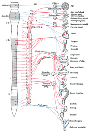 In blue is shown the parasympathetic inervation, in red the sympathetic inervation.
- The parasympathetic nervous system as a resting system gives priority to the activation of the peristal and secretive functions of the digestive and urinary system while promoting the relaxation of sphincters for the eviction of the excreta and urine; also causes bronchchoconstriction and respiratory secretion; promotes vasodilation to redistribute blood to the visceras and favors the counterpartous eye; and it produces miphiliasis to the next.
Unlike the sympathetic nervous system, this system inhibits the functions responsible for the behavior of escape by facilitating the decrease of frequency as of the force of the heart contraction.
The parasympathetic system tends to ignore the pattern of body metamerization by inervading most of the body through the vague nerve, which is emitted from the head (rachid lube). The nerves that inerve the same head are emitted from the mesencephale and bulb. The nerves that inerve the most distal digestive-urinary segments and sexual organs are emitted from the S2 to S4 medulla sections. - The sympathetic nervous system Being a system of escape or escape behavior gives priority to the acceleration and strength of heart contraction, stimulates the piloertion and sweating, favors and facilitates the activation mechanisms of the somatic nervous system for the timely voluntary muscle contraction, causes the bronchodilation of respiratory pathways to favor rapid oxygenation, propitiates vasoconstriction by redirecting the blood irrigation to muscles, heart and nervous system, best stimulates the adreduction
Instead, it inhibits the functions responsible for resting as intestinal peristalsis while increasing the tone of urinary and digestive sphincters, all of this to prevent the eviction of excreta. In males it ends sexual excitement through the ejaculation process.
The sympathetic system follows the body metamerization pattern by inervading most of the body, including the head, through the T1 to L2 medulla segments. - Enteric nervous system. The entheric nervous system consists of a set of neurons located on the wall of the digestive tract. It has an important function in the control of gastrointestinal motility. It consists of two nervous plexies: submucoso de Meissner and mientérico de Auerbach, which generate the patterns that cause gastrointestinal motility. In the case of an automatic mechanism other than will, the entic nervous system is included within the autonomous nervous system, but it is considered an independent entity of the sympathetic and parasympathetic.
- The parasympathetic nervous system as a resting system gives priority to the activation of the peristal and secretive functions of the digestive and urinary system while promoting the relaxation of sphincters for the eviction of the excreta and urine; also causes bronchchoconstriction and respiratory secretion; promotes vasodilation to redistribute blood to the visceras and favors the counterpartous eye; and it produces miphiliasis to the next.

Diseases
The nervous system can suffer numerous diseases of different origin: infectious, hereditary, degenerative, cerebrovascular (due to involvement of blood vessels), demyelinating or tumors.
- Infectious. They can be produced by bacteria, viruses, fungi or parasites. Some of the most frequent are meningitis and brain abscess.
- Hereditary. Among the hereditary diseases that affect the functioning of the nervous system is Huntington's disease.
- Degenerative. This group includes Parkinson's disease and Alzheimer's disease.
- Vascular. The involvement of blood vessels that bring blood to the nervous system can cause serious damage to nerve structures if these remain several minutes without receiving oxygen. This mechanism occurs in brain embolism and brain infarction.
- Dissylinizing. Multiple sclerosis occurs due to the involvement of the myelin pod that surrounds the axons that come from the neurons.
- Tumors. Different types of cancer can affect the nervous system in a primary way, including astrocytoma, oligodendroglioma and meningioma.
- Others. Epilepsy, cranial trauma.
Nervous system in animals
It is believed that the first neuron arose 600 million years ago, during the Ediacaran period, in diblastic animals such as cnidarians. The reflex act is the basic unit of integrated nervous activity and could be considered as the primordial circuit from which the rest of the nervous structures started. This circuit went from being made up of a single multifunctional neuron in the diblastic to two types of neurons in the rest of the animals called afferent and efferent. To the extent that intermediaries were added between these two groups of neurons with the passage of evolutionary time, such as interneurons and circuits of greater plasticity, the nervous system was showing a phenomenon of concentration in strategic regions, giving rise to the formation of the nervous system. central, with cephalization being the most complete feature of these phenomena.
Redundancy
In the transmission of signals, there are measures such as redundancy, which consists in the creation of alternate routes that carry part of the same information, guaranteeing its arrival despite any damage that may occur.
Myelination
Myelination of axons in most vertebrates and in some invertebrates such as annelids and crustaceans is another optimization measure. This type of coating increases the speed of the signals and decreases the caliber of the axons, saving space and energy.
Metamerization
Another important characteristic is the presence of metamerization of the nervous system, that is to say, that condition where a subdivision of the corporal structures is observed in units that are repeated with determined characteristics. The three groups that mainly display this quality are arthropods, annelids, and chordates.
| Two examples of metamerization | |||||||||
| |||||||||


Centralization
Centralization refers to the evolutionary tendency of neurons to group into localized centers of integration in which there are numerous cells that interact with each other to process stimuli and perform increasingly complex actions. This progressive centralization is what ended up giving rise to a central nervous system and a brain. Among the current animals that do not have centralization are jellyfish whose nervous system is a diffuse network of interconnected neurons without any central control point.
Cephalization
Cephalization refers to the evolutionary tendency of nervous tissue to clump together in the head area. This process has been favored by the existence of sense organs in the cephalic pole. Cephalization is already present in flatworms that contain cephalic ganglia that function as the brain and can be observed in arthropods, cephalopods and of course in all vertebrates.

Diblastic Animals
Diblastic or radiate animals, a paraphyletic group that includes both cnidarians and ctenophores, normally have a network of subectodermal plexuses without an apparent nerve center, but some species already present nerve condensates in a phenomenon that is understood to be the first evolutionary attempt to form a central nervous system. Some arrangements of these condensates, such as nerve rings in jellyfish, are reminiscent of later trends seen in cycloneurs.
Triblastic Animals
In triblastic or bilateral animals, a monophyletic group, there are two types of body plans called protostomes and deuterostomes that in turn have three types of nervous system arrangements: cycloneuros, hyponeuros, and epineuros. An essential difference is that in protostomes and deuterostomes the CNS is in inverted positions. For many years, these and other differences were considered to indicate essentially different body plans and CNS (by the relative position of the CNS, digestive system, and major circulatory vessel).
- Cycloneuro animals: the nervous system is arranged in a more or less circular way as in equinoderms.
- Hyponeuro animals: the nervous system is willing ventrally to the digestive system. It occurs in mollusks, arthropods and anelids.
- Epineuro animals: the nervous system is willing to dorsally with respect to the digestive system. It is given in the cords that include vertebrates and all mammals among them man.
Protostomous Animals
Protostomous animals, which are triblastic, such as flatworms, nematodes, mollusks, annelids, and arthropods, have a hyponeuronic nervous system, that is, it is a system made up of cerebral ganglia and ventral nerve cords. The ganglia that form the brain They are located around the esophagus, with periesophageal connectives that connect them to the nerve chains that run ventrally through the animal's body, in a lower position with respect to the digestive tract. Such a model of body plan is arranged in this way when an embryological process called gastrorrhaphy occurs in the gastrula.
Deuterostomous Animals
Deuterostome animals, which are triblastic, are divided into two groups according to their symmetry, radial or bilateral, or the arrangement of their nervous system, cycloneuros or epineuros. Within the cycloneuros are the echinoderms (radially symmetric) and the hemichordates. The nerve center is a ring located around the mouth (subectodermal or subepidermal). Within the group of epineuria are urochordates, cephalochordates and vertebrates in which they have a hollow and tubular nerve cord, dorsal to the digestive tract. From this cord, in more complex animals, the brain and brain develop. spinal cord. Such models of body plans are arranged in this way when embryological processes occur in the gastrula called isoquily in cycloneurs or nototenia in the case of epineuros.
| Fill | Superfilo | Changes in gastrula | Nervous system | Centralization | Metamerization | Cefalization | Mielinization |
|---|---|---|---|---|---|---|---|
| Copiers | Plastics | Specialization of gastrovascular cavity | Diffuse | No. | No. | 0 | No. |
| Cnidarios | Plastics | Specialization of gastrovascular cavity | Diffuse/Cicloneuro | No. | No. | 0 | No. |
| Platelmintos | Spiral prostomos | Specialization of gastrovascular cavity | Hiponeuro | Yeah. | No. | + | No. |
| Nematodes | Protomos ecdisozoos | Gastrorrafia | Hiponeuro | Yeah. | No. | + | No. |
| Artropods | Protomos ecdisozoos | Gastrorrafia | Hiponeuro | Yeah. | Yeah. | ++ | Crustáceos |
| Moluscos | Spiral prostomos | Gastrorrafia | Hiponeuro | Yeah. | No. | +++ | No. |
| Released | Spiral prostomos | Gastrorrafia | Hiponeuro | Yeah. | Yeah. | + | I'm sorry. Police |
| Let's go. | Deuteromos | Isoquilia | Cycloneuro | Yeah. | No. | 0 | No. |
| I have remembered | Deuteromos | Isoquilia | Cycloneuro | Yeah. | No. | + | No. |
| Corded | Deuteromos | Nototenia | Epineuro | Yeah. | Yeah. | +++++ | Vertebrates |
Nervous system by edge
Cnidarians
The phylum of cnidarians includes, among other organisms, hydras and jellyfish. They present the simplest and most primitive form of nervous system that receives the name of nervous network. In a neural network, neurons are dispersed without complex structural organization, and there is no brain.
Flatworms
The flatworm phylum includes some 20,000 species, including some parasitic species such as the tapeworm, or tapeworm, that lives in the human intestine. Its nervous system presents beginnings of cephalization and 2 longitudinal nerve cords that can be considered a primitive central nervous system. On the other hand, nervous tissue already contains numerous interneurons, that is, connection neurons between sensory and motor neurons that increase the complexity of the circuits.
Annelids
The group of annelids includes numerous species, one of the most characteristic being the earthworm. These animals have a nervous system made up of a double ventral nerve cord and two ganglia located in each metamer. They have a brain that is formed by the union of two dorsal ganglia that communicate through connectives to the ventral nerve cord.
Molluscs
Within the group of molluscs are the cephalopods (squid and octopus). They have a highly developed brain and sensory system. The brain is comparatively very large in relation to that of other invertebrates, which is why cephalopods achieve high memory and learning capacities.
The group of bivalves that includes clams and mussels has a less developed nervous system than that of cephalopods, probably due to their sedentary lifestyle. They lack a brain but have several ganglia that control various functions, including two cerebro-pleural ganglia on both sides of the esophagus that control the sensory organs and the mantle cavity (molluscs).

Arthropods
Arthropods are the most abundant and varied animals on earth, including insects, arachnids, and crustaceans. They have a well-developed nervous system that allows them to have a complex and coordinated behavior. Its central nervous system is of the ganglionic type and consists of a chain of segmental ganglia linked by a ventral nerve cord, some ganglia fused in the cephalic region and give rise to a brain.
Echinoderms
The echinoderm group includes the starfish and the sea urchin. These animals have a nervous system but do not have a brain that centralizes the activity. They have three nerve rings located in different planes around the digestive tract.
Vertebrates
The nervous system of vertebrates consists of a well-developed brain and spinal cord. The peripheral nervous system is made up of different nerves that connect to the central nervous system. These nerves are afferent (carry sensory information to the central nervous system) or efferent (carry motor orders from the brain to the organs). There are also peripheral ganglia that are groups of neurons linked to some of the nerves but should not be confused with the ganglion system of arthropods.
Nervous system in plants
The classical doctrine according to which the nervous system is exclusive to animals has been questioned in recent decades by the discoveries about the existence of electrical signals in plants and the use they make of them. Based on these discoveries, some scientists have proposed the need to create a scientific area called neurobiology of plants and the existence of a nervous system in plants. This proposal has caused a dispute in the scientific community between those who think it should be talk about the nervous system of plants and those who are against it. The immovable positions in the scientific debate on both sides have led to the proposal of a solution to the debate that consists of redefining the concept of nervous system using solely physiological and avoid phylogenetic criteria.
Contenido relacionado
Coix
Betulaceae
Sapindaceae