Nerve
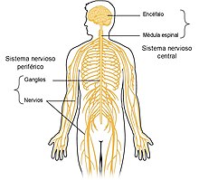
The nerves are structures that conduct nerve impulses located outside the central nervous system. They are formed by a set of grouped axons, each of which comes from a neuron. They can be motor or sensory, but most are mixed and contain both sensory and motor fibers. They originate in the spinal cord (spinal nerves) or leave directly from the brain (cranial nerves). In the human species there are 12 pairs of cranial nerves called cranial nerves and 31 pairs of spinal nerves, which tend to group together to form nerve plexuses.
Description
Nerves are bundles of nerve processes, or axons. They are cord-shaped and communicate the nerve centers of the brain and spinal cord with all the organs of the body. They are part of the peripheral nervous system. Afferent nerves carry sensory signals to the brain, for example from the skin or other organs, while efferent nerves carry motor signals from the brain to muscles and glands, causing muscles to contract and making movement possible. Nerve signals, also called nerve impulses, start from the cell body of a neuron and propagate rapidly down the axon to its end, where through the synapse, the stimulus is transmitted to another neuron, or to an effector organ, such as a fiber. muscle or a gland.
Structures
Each nerve is formed by the grouping of several hundred or thousands of axons that come together to form fascicles. Axons are extensions of neurons through which these cells come into contact with other neurons or with muscle fibers. In the human species, the individual diameter of the axons ranges between 0.1 and 20 micrometers, while the length varies between just a few centimeters and more than a meter in the axons that are part of the sciatic nerve and originating from the motoneurons of the anterior horn. from the spinal cord must reach the muscles of the leg and foot. Different components can be distinguished in the nerve trunks:
The nerve fibers that make up a nerve are surrounded by smooth tissue that receives different names depending on its location. The thin layer that surrounds each fiber is called the endoneurium, the individual fibers are grouped into fascicles covered by the perineurium, the complete nerve formed by the union of several fascicles is covered by the epineurium.
- Epineuro: It is the outermost layer of a nerve. It is a thick conjunctive layer, which holds the nerve fascicles. It is composed of connective cells and collagen fibers, mostly arranged longitudinally following the nerve. It contains some adipous cells and small blood vessels called vasa nervorum that provide the blood circulation of the nerve.
- Perineur: It is each of the concentric layers of connective tissue that wraps the fascicles of a nerve.
- Endomide: They are a fine phscicles of longitudinally arranged collagen fibers, along with some fibroblasts located between nerve fibers.
Each axon comes from a neuron. The cell membrane that covers the axon is called the axolemma. Most nerve fibers are covered by a myelin sheath made up of Schwann cells.
- Axolema. It is the cell membrane that covers the axon and separates it from the outside.
- Axoplasma. This is called the cytoplasm of the cell inside the axon.
- Schwann cells. Cells capable of manufacturing myelin that wraps some nerves of the peripheral nervous system.
As the nerve branches, the connective tissue sheaths become thinner. In the smaller branches the epineurium is absent, and the perineurium is indistinguishable from the endoneurium, being reduced to a thin fibrillar layer lined with flattened connective cells resembling endothelial cells.
Types of nerves
- According to their origin:
- Cranial nerves. Also called cranial pairs. There are 12 pairs of nerves that come directly from the brain.
- Rachemical nerves are 31 pairs of nerves that leave the spinal cord through the connecting holes. They can be divided into 5 cervicals, 12 dorsals, 5 lumbars and 6 sacrocoxygeos. Rachid nerves are grouped and give rise to several plexies: cervical plexo, brachial plexus, lumbar plexus and sacral plexus.
- According to his function:
- Sensitive or centripetal nerves: they are responsible for driving the external excitations to the nerve centers. They're pretty scarce. Generally nerve fibers are associated with motor fibers (centrifugals).
- Nerve motors or centrifugals: they bring to the muscles or to the glands the order of a movement or a secretion imparted by a nervous center.
- Mixed nerves: they work at the same time as sensors and engines. They are made up of fibers that carry external excitations to the nerve centers and orders of the muscles, from the centers to the periphery. As an example we can cite the glosopharynge that transmits to the brain the information of the sense of taste and produces at the same time the excitation of the tongue. All rachid nerves and various cranial nerves belong to this kind of nerve.
Types of nerve fibers
Nerve fibers that make up nerves transmit impulses by propagating action potentials. The speed of transmission depends on several factors: the diameter of the fibers and the existence or not of a myelin sheath that surrounds the axon. Fibers with a larger diameter and those that are surrounded by myelin have a higher conduction velocity. A small diameter unmyelinated fiber transmits only at 0.5 meters per second, while a large diameter and myelinated fiber can reach 120 meters per second. Myelin acts as an insulator, increasing conduction velocity and decreasing energy expenditure. It also has a protective function. For this reason, diseases called demyelinating diseases such as multiple sclerosis make nerve conduction too slow and ineffective.
According to the Erlanger and Gasser classification, the nerve fibers that make up the nerves can be classified into several types: A, B and C.
- Type A fibers, with myelin pod and subdivided into the types:
- Alpha: driving speed 70-120 m/s, diameter 12-20 microns, responsible for the contraction of the skeletal muscle.
- Beta: driving speed 30-70 m/s, diameter 5-12 microns, are sensitive fibers responsible for touch and pressure.
- Gamma: driving speed 15-30 m/s, diameter of 3-6 microns, responsible for the transmission to the muscular spindles.
- Delta: driving speed 12-30 m/s, diameter 2-5 microns, responsible for the transmission of acute pain located, temperature and part of the touch.
- Fibers B, with myelin pod, responsible for the preganglionar connection of the autonomous nervous system, driving speed 3-15 m/s, diameter below three microns.
- Fibers C, no myelin pod. They are responsible for the transmission of diffuse deep pain, smell, information of some mecanorreceptors, responses of the reflex arches and postganglionars of the autonomous nervous system, driving speed 0,5-2 m/s, diameter 0,4-1,2 microns.
Physiology
The nerve has two essential properties: excitability and conductivity. Excitability is the ability to react to chemical and physical stimuli, while conductivity is the ability to transmit excitation from one place to another.
Excitability
The neuron can be excited by a nerve center, by a natural stimulant such as light, or by an artificial stimulant such as an electrical discharge. The propagated stimulus is called a nerve impulse, and its passage from one point of the nerve fiber to another is nerve conduction.
Conductivity
Conductivity is the ability to transmit excitation from one place to another. This property allows a dendrite to transmit to a nerve center the excitation that comes from a peripheral pinprick. It also makes it possible for the motor centers of the brain to issue orders that are transmitted through the nerves to the skeletal muscles to cause their contraction and generate movements. For conductivity to take place, it is necessary that the nerve has not suffered any damage or degeneration and that its path has perfect continuity.
Neurons transmit electrical signals originating as a consequence of a transient change in permeability in the plasma membrane. Its propagation is due to the existence of a potential difference between the internal and external part of the cell called membrane potential. When the membrane potential of an excitable cell depolarizes beyond a certain threshold, the cell generates an action potential. An action potential is a very rapid change in membrane polarity from negative to positive and back again, in a cycle that lasts a few milliseconds. Nerve transmission is possible thanks to the existence of ionic channels in the membrane that covers the axon.
Main nerves of the human body
- Spinal nerves. They are a total of 31 pairs of nerves each with two parts or roots that join one another: one sensitiva and another motor. The sensitive part is the one that moves the information from the receptors to the spinal cord, while the motor part is the one that carries the impulses from the spinal cord to the corresponding effects. They are distributed as follows:
- 8 pairs of cervical nerves
- 12 pairs of dorsal or chest nerves
- 5 pairs of lumbar rachid nerves
- 5 pairs of sacred rachid nerves
- 1 pair of coccygeal rachid nerves
- Cranial nerves, also called cranial pairs, are 12 nerves that send sensory information from the neck and head to the central nervous system or transfer motor orders to control the skeletal musculature of the neck and head.
- I. Olfactory nerve. It is a sensory nerve only, leads the nervous impulses generated by the odoriferous substances from the nose to the brain.
- II Optical nerve. Exclusively sensory, convey visual information from the eye to the brain.
- III Oculomotor nerve. It has motor fibers that control the eye and parasympathetic movement that modify the diameter of the pupil.
- IV Troclear nerve. Its function is motor over one of the muscles that move the eyeball.
- V Trigem nerve. It is a mixed nerve that consists of a sensitive portion and another motor.
- VI Nervio abducens or External Eye Motor. It intervenes in eye mobility, it is only motor.
- VII Facial nerve. It is a mixed nerve with sensitive and motor fibers.
- VIII Nerve vestibulococlear. It conveys to the brain the auditory and sensory information from the inner ear.
- IX Gloosopharyngeal nerve. It's a mixed nerve. The sensitive portion carries signals from the tongue and the pharynx.
- X Nervio vago. It is sensitive and motor, it also provides parasympathetic fibers that act on different organs, including the stomach and the heart.
- XI Spinal nerve. It is an engine nerve that activates among others the sternocleidomastoid muscle, causing the spin of the head.
- XII Hypogloss nerve. It's a nerve engine for the musculature of the tongue.
Plexuses
The spinal nerves are grouped together to form plexuses. Different nerves arise from each plexus, some of the most important are listed below.
- Cervical plexus: major atrial nerve, phrenic nerve.
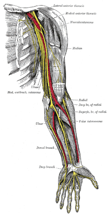
- Brachial plexus. It leaves the nerves destined to the shoulder and a superior member: axillary nerve, muscutaneous nerve, radial nerve, medium nerve, cubital nerve.
- Lumbar plexus: iliohipogastric nerve, ilioinguinal nerve, genitofemoral nerve, lateral femoral cutaneous nerve, obstructive nerve, femoral nerve.
- Plexo sacro: nerve of the inner shutter, nerve able, nerve of the femoral square, upper glyteus nerve, lower glyteus nerve, posterior cutaneous nerve of the thigh, sciatic nerve.
Development
Nerve growth normally ends in adolescence, but it can be stimulated again by a molecular mechanism known as the Notch signaling pathway.
Regeneration
If a neuron's axons are damaged, but not its soma, the axons can regenerate and remake synaptic connections with neurons with the help of guide cells. This is also known as neuroregeneration.
The nerve begins the process by destroying distal to the injury site, allowing the Schwann cells, basal lamina, and neurilemma near the injury to begin producing a regenerative tube. Nerve growth factors are produced which cause many nerve buds to appear. When one of the growth processes finds the regeneration tube, it permanently guides and accompanies it, helping it to grow rapidly towards its original destination. Nerve regeneration is very slow and can take several months to complete. While this process does repair some nerves, there will still be some functional deficit, as the repairs are not perfect.
Diseases
There are many diseases that affect or involve the nerves. A neuralgia is a symptom caused by a failure of the nervous system consisting of a sensory disorder or pain without affecting motor function. If it affects the peripheral nerves, it causes an alteration of the innervated area corresponding to the nerve. The cause of the lesion can be inflammation, an allergic reaction, poisoning, chronic alcoholism, certain metabolic diseases (such as diabetes mellitus), some types of kidney dysfunction, viral infections, and vitamin deficiencies. It can also be a mechanical injury, such as tears, fractures, compression injuries or gunshot wounds.
Cancer can spread by invading the spaces around nerves. This is particularly common in head and neck cancer, and in prostate and colorectal cancer.
Nerves can be damaged by physical injury, as well as conditions such as carpal tunnel syndrome and repetitive stress injuries. Autoimmune diseases such as Guillain-Barré syndrome, neurodegenerative diseases, polyneuropathy, Infection, neuritis, diabetes, or failure of the blood vessels surrounding the nerve cause nerve damage, which can vary in severity.
Multiple sclerosis is a disease associated with extensive nerve damage. It occurs when macrophages from an individual's own immune system damage the myelin sheaths that insulate the nerve axon.
A pinched nerve occurs when pressure is placed on a nerve, usually due to swelling from injury or pregnancy, and can lead to pain, weakness, numbness, or paralysis, such as carpal tunnel syndrome. Symptoms they can be felt in areas far from the actual site of damage, a phenomenon called referred pain. Referred pain can occur when damage causes impaired signaling to other areas.
The most common symptoms of nerve damage include:
- Numbness and tingling
- Pain puncture
- Ardor
- Muscle weakness
- Sensitivity to touch
- Paralysis
- Heat intolerance
- Digestive problems
- Dizziness or vertigo
Other animals
In vertebrates, the best known identified neurons are the giant Mauthner cells of fish and amphibians. Each fish has two Mauthner cells, located in the lower part of the brain stem, one on the left side and one on the left side. in the right. Each Mauthner cell has a crossing axon, innervating (stimulating) neurons at the same level of the brain and then traveling down through the spinal cord, making numerous connections as it goes. The synapses generated by a Mauthner cell are so powerful that a single action potential triggers a major behavioral response: Within milliseconds, the fish curves its body into a C shape, then straightens up, propelling rapidly forward. Functionally, this is a rapid escape response, most easily triggered by a loud sound wave or pressure wave impinging on the fish's lateral line organ. Mauthner cells are not the only neurons identified in fish; there are about 20 other types, including pairs of "Mauthner cell analogues" in each spinal segmental nucleus. Although a Mauthner cell is capable of eliciting an escape response on its own, in the context of ordinary behavior, other cell types often help shape the amplitude and direction of the response. In 2020, a group of researchers from the University of Bayreuth discovered that in zebrafish, these cells have the ability to quickly and completely regenerate if they suffer damage in the center of the axon, but die if damage to this structure occurs nearby. of the cell body. Such a type of repair is unknown in the nervous system of other species.
Mauthner cells have been described as command neurons. A command neuron is capable of driving a specific behavior on its own. Such neurons appear most frequently in the rapid escape systems of several species: the giant axon and the respective giant synapse of the squid, used for pioneering experiments in neurophysiology because Because of their enormous size, they participate in the rapid escape circuit of the squid. However, the concept of the command neuron has become controversial due to studies showing that some neurons that initially seemed to fit the description were only capable of evoking a response in a limited set of circumstances.
In radially symmetric organisms, there is no brain or centralized region of the head, but instead there are interconnected neurons distributed in neural networks that fulfill the function of the nervous system. These webs are found in Cnidaria, Ctenophora, and Echinodermata.
Contenido relacionado
Raven (disambiguation)
Dna viruses
Pathological types of breast cancer


