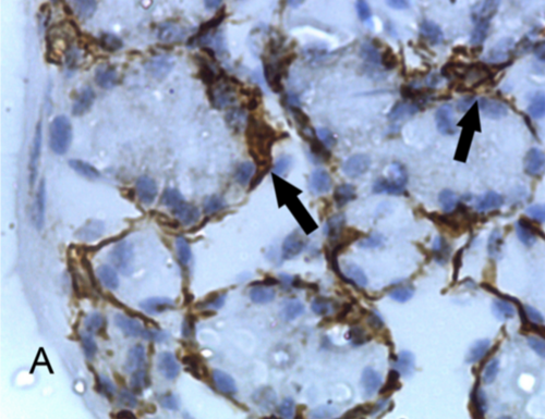
Myoepithelial cell
The myoepithelial cells are a normal component of the acini and ducts of the exocrine glands, where they are located between the secretory epithelial cells and the basement membrane. The function of these myoepithelial cells is multiple: they assist the progression of secretion through their contractions, they serve as a barrier between the connective tissue and the epithelium forming the basal membrane, they have supporting and paracrine functions of organization and polarity.
Anatomy
The term cell "epithelial#34; it was first used by Renault in 1897 in the parotid gland, but the cells were described by Zimmermann in 1898.
Myoepithelial cells are found in the epithelium of the glands, as a thin layer above the basement membrane and below the luminal cells.
They are present in sweat glands, salivary glands, mammary glands and lacrimal glands.
Microarchitecture
Myoepithelial cells are thin and spindle-shaped, similar to those of smooth muscle. They have an irregular cell nucleus and are found adjacent to the basal membrane.
They show a cytoplasm with a stellate appearance, with numerous processes that interdigitate with other similar processes of adjacent myoepithelial cells.
Ultrastructure
Under electron microscopy, myoepithelial cells show between four and six cytoplasmic processes that extend over the acinar cells, giving them a star-like appearance. The processes have thin, longitudinally oriented filaments 4–6 nm thick inside.
The cell body is small and dense, with a cytoskeletal network associated with filaments 10 nm in diameter.
The cytoplasmic organelles are perinuclear, and the plasma membrane parallels the basal membrane of the secretory parenchymal cells.
Micropyknotic vesicles are seen on the surface opposite the secretory cells in contact with the basal.
Myoepithelials show characteristics of both smooth muscle and epithelium. They are known for the presence of myofilament proteins that are related to those of smooth muscle, and intermediate filaments that contain cytokeratin related to desmosomes, which express affinity with the epithelium.
In vivo, myoepithelial cells are attached to the basement membrane by hemidesmosomes, and to luminal epithelial and other myoepithelial cells by desmosomes.
Myoepithelial cells are the barrier between secretory epithelial cells and the surrounding stroma in the duct system of the mammary gland. Several myoepithelial cell immunohistochemical markers in human tissue have been identified and validated. Pathologists use them as diagnostic tools to distinguish carcinoma in situ from invasive breast cancer.
Function
Myoepithelial cells, epithelial in origin, although with the particularity that they are capable of contracting in the manner of smooth muscle fiber. These myoepithelial cells cause the leakage of milk stored in the alveoli and expelled through the lactiferous ducts.
The function of these myoepithelial cells is to "milk" the secretory section, so that they contract and produce an accumulation of this secretory portion, which causes expulsion of the secretory product.
Replacement
Myoepithelial cells are epithelial in origin but are also capable of contracting in the smooth muscle cell manner.
Slow-cycling myoepithelial stem cells are normally inactive but can become active and function as an alternative source of cells in the glands.
Contenido relacionado
Monodon monoceros
Treponema
Sessile


