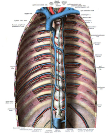
Mediastinum
The mediastinum is the anatomical compartment located in the center of the thorax, between the right and left lungs, behind the sternum and in front of the thoracic vertebrae. Below it is limited by the diaphragm that separates the thorax from the abdomen and above by the base of the neck. To understand its situation, the thoracic cavity can be divided into three spaces: two lateral ones, which are the pleuropulmonary cavities, each formed by a lung and its pleura, and a central space, the mediastinum.
Its importance is given because in this space are vital organs of the circulatory, respiratory and digestive systems, including the heart and the large blood vessels that enter and exit it. The mediastinum is a passageway for the esophagus, trachea, thoracic duct, and important nerves such as the vagus and phrenic nerves.
Content
Inside the mediastinum are the heart, the aorta artery, the superior vena cava, the inferior vena cava, the azygos and hemiazygos veins, the pulmonary arteries and pulmonary veins, the trachea and the main bronchi, esophagus, thoracic duct, thymus, lymphatics, lymph nodes, nerve nodes, phrenic nerve, vagus nerve, and recurrent laryngeal nerve.
The vertebral bodies, lungs, and pleural cavity are in the thorax but are not part of the mediastinum.
- Timo. It is a gland that plays an important role in the immunity and proliferation of T lymphocytes. It is located above the heart and behind the stern.
- Fonic nerve. Part of the spinal cord at the neck level and crosses the entire mediastinum to reach the diaphragm to which it inervates.
- Vague nerve. Part of the brain and follow a descending path, crosses the base of the skull and neck from where it enters the mediastinum and accompanies the esophagus to cross the diaphragm and enter the abdomen. One of its branches is the recurring laryngeal nerve that adopts an ascending path to reach the larynx.
- A chest conduct. It carries the lymph of most of the body. Part of the abdomen, crosses the diaphragm to enter the posterior mediastinum, ascends to the left of the descending aorta, crosses the whole median and flows to the cervical level in the internal jugular vein.
- Heart. It is located in the central portion of the lower mediastinum, above the diaphragm, in front of the vertebral bodies and behind the stern. It is wrapped in the pericardial sac and flanked by the lungs and pleura.
- Arteria aorta. It is the main artery and gives numerous branches that distribute oxygenated blood to all organs. Its path in the chest passes through different regions of the mediastinum, part of the left ventricle of the heart, first ascends and reaches the upper mediastinum where it emits branches for the head, neck and higher limbs, then descends and is placed in the posterior mediastinum until it crosses the diaphragm and leaves the chest.
- Vena cava inferior. It originates from the union of the two primitive illogical veins at the level of the 5th lumbar vertebra. From there, go through the abdomen in the ascending sense, cross the diaphragm and enter the abdomen median until reaching the right atrium of the heart where it flows.
- Vena cava superior. Bring the blood from the head, neck and upper limbs to the right atrium. It measures between 6 and 8 cm long and around 21 mm in diameter.
- Come here. It is an important venous throne of about 0.9 cm in diameter. It originates in the lumbar region, ascends by the retroperitoneum and enters the thorax placing itself on the right side of the posterior mediastinum, continues its ascent until it reaches the level of the fourth thoracic vertebra, where it flows into the upper vena cava. On your journey you receive the blood from the hemiceous vein that circulates parallel during a part of your journey.
- Esophagus. It is a hollow tubular organ that measures about 30 cm long and puts in contact the mouth cavity with the stomach. On its way from the neck to the abdomen, it crosses the chest, placing itself in the chest median back, ahead of the spine and behind the trachea.
Limits
The mediastinum is located in the center of the thorax, between the two lungs from which it is separated by the pleura. Its anatomical limits are:
- Upper limit: neck base.
- Lower limit: diaphragm.
- Previous limit: stern and coastal cartilage.
- Rear Limit: anterior and lateral surface of the chest vertebrae.
Divisions of the mediastinum
Numerous classifications of the mediastinum spaces have been carried out by anatomists, surgeons and radiologists that do not coincide and vary considerably between different authors, some are highly complex and distinguish up to nine compartments. Below are some of the most used that divide the mediastinum into three or four spaces.
Upper and lower mediastinum
Many authors divide the mediastinum into superior and inferior. The superior is a single space, but the inferior is subdivided into three: anterior, middle, and posterior.
Superior mediastinum: located above the heart. Contains the thymus, the upper half of the superior vena cava, the aortic arch, and its branches.
Inferior mediastinum: contains the heart and is divided into three parts: anterior, middle, and posterior.
- Previous average: is the smallest part of the mediastinum and is located in front of the heart, between this and the breastbone, contains the lower end of the thymus.
- Medium average: is the most important subdivision, since it places the heart and the pericardium that envelops it. Also pulmonary arteries and veins, ascending aorta and major bronchus.
- Later mediastin: it is behind the heart and in front of the vertebral bodies of the eight lower dorsal vertebrae. It contains the esophagus and the chest descending aorta.
It should be understood that some structures that pass through the mediastinum, including the esophagus, are found in more than one subdivision.
Anterior, middle and posterior mediastinum
A second classification divides the mediastinum from the radiological point of view into three zones: anterior, middle, and posterior. This division proposed by Felson uses the lateral chest radiograph as a reference. The anterior and middle mediastinum are separated by an imaginary line that extends from the posterior border of the silhouette of the heart to the anterior border of the trachea. The posterior and middle mediastinum are separated by a line located one centimeter behind the anterior margin of the vertebral bodies.
Galli classification
The Argentine anatomist Eugenio Antonio Galli proposed a different classification taking the esophagus as a reference, considering the anterior mediastinum to the space located in front of the esophagus and posterior to the rest.
- Previous mediastin. It can be divided into antero-superior or tracheo-thymic-vascular and mediastinal antero-inferior or cardio-pericardium, occupied by the heart.
- Later halfstine. The main elements of the posterior mediastinum are the esophagus, chest aorta and chest duct.
Diseases
The mediastinum can be affected by different diseases:
- Mediastinitis. It is characterized by inflammation in the mediastinum, usually caused by germs that colonize this anatomical region. Germs can be reached through several pathways, for example by perforation of the esophagus, or by extension of lung infectious processes.
- Medium-stynic fibrosis, also called fibrous mediastinitis.
- Medium-stynic emphysema, also called neumomediastino.
- Midstine tumours. In turn they are divided into.
- Benign tumors.
- Mediastinal cancer. It can be primitive of mediastinum such as thymoma and lymphoma or secondary by dissemination or extension from other organs.
- Medium-stynic syndrome. It is a set of symptoms that occur by the compression of structures that form the median, may be due to inflammatory or tumor processes. Different possibilities can be distinguished within the mediastinal syndrome, depending on the affected structure.
- Upper vena cava syndrome.
- Lower vena cava syndrome.
- Tracheal syndrome.
- Nervous compression syndrome:
- Fonic nerve. By compression of this nerve, it causes pain that is radiated on the shoulder.
- Recurrent laryngeal nerve. It causes alterations of the voice (phone).
- Nice nerves. It causes Claude Bernard-Horner's syndrome.
- Esophageal syndrome. It causes compression disphagia of the esophagus.
Contenido relacionado
Fertilization
Lung cancer
Goodpasture syndrome


