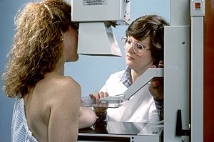
Mammography
The mammography or mastography consists of a diagnostic X-ray image exploration of the mammary gland, using devices called mammographs (in doses of around 0.7 mSv). These devices have specially adapted X-ray emission tubes to achieve the highest possible resolution in the visualization of the internal fibroepithelial structures of the mammary gland.
Origin
The beginnings of mammography or mammography as a radiological method go back to 1913 when Alberto Salomón, a German surgeon, was the first to use radiography to study breast cancer and is considered the inventor of breast radiology; he X-rayed mastectomy specimens (which had been removed from 3,000 patients) to determine the extent of the tumor, distinguish the difference between non-cancerous and cancerous, and its multiple types.
In 1930, American physician and radiologist Stafford L. Warren published "A Roentgenologic Study of the Breast," a study in which he produced stereoscopic X-ray images to track changes in breast tissue such as outcome of pregnancy and mastitis. In 119 women who subsequently underwent surgery, it correctly found breast cancer in 54 of 58 cases.
In 1933 Alberto Baraldi introduced the roentgenneumo-mastia (aerogram), which consists of injecting air into the periglandular and retromuscular tissues, allowing not only to detect the presence of a tumor, but also its characteristics and the relationship with the adjacent planes.
In 1937 Frederick Hicken presented a new diagnostic method which he called a mammogram and which is based on the introduction of contrast media into the milk ducts
In 1945 Raúl Leborgne in Uruguay gave impetus to the method, developed the breast compression technique to produce better quality images and characterized micro calcifications.
Ten years later, Robert Egan of the M.D. Anderson of the University of Texas, further improved mammography technology by using an industrial film that required less X-ray dose and produced better quality images, tumors appeared white and normal tissue dark gray. Egan examined the breasts of 1,000 women who were healthy and did not suspect breast cancer and found breast cancer in 238.
In the 1960s, the first randomized screening trials began with the study of the New York Insurance Plan, followed by the study of two Swedish counties, carried out by Lazlo Tabar, and others carried out in different countries. These trials demonstrated that it was possible to reduce mortality from breast cancer thanks to these programs.
In 1976, the American Cancer Society began recommending mammography as a method of early detection of breast cancer. At the time, ACS stated that women under the age of fifty could benefit from mammograms and recommended annual mammograms for women age fifty and older.
In 1993, the American College of Radiology developed a Breast Reporting and Data System, or BI-RADS, which standardized the way doctors reported mammography results.
In 2000, digital mammography was approved, which is faster and has lower doses of radiation.
In 2011, digital tomosynthesis (3D images) was implemented, which improved the accuracy of mammography. Digital tomosynthesis imaging uses a lower radiation dose and allows the user to view the breasts in three dimensions, as well as look at each layer of breast tissue separately instead of the entire breast at once, and reduces the number of false-positive results.
Applications
The ability to identify small lesions has led to the use of mammography in systematic reviews to detect tumors before they can be palpable and clinically manifest (mammographic screening). This diagnosis, made at a very early stage of the disease, is usually associated with a better cure prognosis, as well as the need for less aggressive treatment to control the cancer.
In many countries the routine mammography of women is recommended as a screening method for early diagnosis of breast cancer. The United States Preventive Services Task Force recommends mammograms, with or without clinical breast examination, every 1–2 years in women age 40 and older. In conjunction with clinical studies, a relative reduction in breast cancer mortality has been found of 20%. Starting in 2000, mammograms became controversial, when results of two high-quality studies were published.
Radiologists use a standard method to interpret and communicate mammography results, which is now considered the universal language in the diagnosis of breast pathology. When they detect a suspicious lesion of cancer, they classify it within a BI-RADS (Breast Imaging-Reporting and Data System) category, the first stages I and II are benign, stage III is probably benign, while stages IV and V increase the risk of cancer. probability that they are malignant. This system makes it possible to standardize the terminology of the mammographic report and to categorize the lesions, establishing the degree of suspicion and assigning the attitude to be taken in each case. On many occasions, mammography can reveal malignant lesions without being clinically palpable.
| Category | Definition | Action |
|---|---|---|
| BIRADS 0 | Insufficient | Other procedures and/or comparison with previous studies are required |
| BIRADS 1 | Negative | Regular annual follow-up |
| BIRADS 2 | Blessings | Regular annual follow-up |
| BIRADS 3 | Probably Benigno | Strict monitoring 6-12-24-36 months |
| BIRADS 4 | Sugestive of malignity | It should be considered to take histologic material from the injury by some method of biopsy |
| BIRADS 5 | Highly suspected of Malignity | Biopsy and treatment |
| BIRADS 6 | Carcinoma confirmed | Final treatment |
False negatives
Mammography is false negative (no cancer) in less than 10%. This is partly due to obscuration by dense or very dense tissue that hides the cancer, and because the appearance of cancer on mammograms has a large overlap with the appearance of normal tissue.
Mammography Techniques
Before the test, it is important to comply with the instructions of the health personnel. The proper way to appear for this exam is freshly bathed, with shaved armpits, without deodorant or cream, with two-piece clothing.
4 basic X-rays are needed for tissue evaluation (two for each breast).
First: Cephalocaudal, or CC (where the ray strikes from top to bottom). The patient stands in front of the mastograph, discovers her breast and the radiologist will be the one to position it. The breast will be left on a plate, taking care that the skin does not form folds and the nipple is completely in profile, to the extent that the patient's anatomy allows it. If this is not possible, it will be very helpful to place markers to avoid any confusion during the study. A compressor is lowered little by little until the tissue expands. Next, the X-ray will be captured, checking that the shoulder and chin do not cast any shadows.
Second: Medial Lateral Oblique, or MLO (in which the mastograph is obliqued at 45 degrees). The patient stands on one side of the apparatus. You are asked to raise your arm and rest it on the opposite side. In this position, the pectoral muscle will be evaluated, so a bit of the axillary area is included, leaving the compressor below the clavicle. As in the previous phase, it must be ensured that there are no folds in the skin, that the nipple is in profile and that the compression is gradual.
The automatic system of the devices allows the pressure of the breast to be released as soon as the X-ray is fired.
Mammographer or mastographer
Mammograms are performed with specific imaging equipment because breast cancer, in its early stages, looks very similar to healthy breast tissue, and lower-energy X-rays must be used to highlight the differences.
They are X-rays with photon energy in the range of 12 and 30 kEV, so a special X-ray tube and a high-frequency generator are used to improve image quality. The tube has a molybdenum (Mo) anode, although it can also be tungsten (W) or rhodium (Rh). Focal spot size is important (smaller is better), as microcalcification imaging requires small focal spots (0.3 or 0.1mm can be chosen). As for the shape of the point, it is usually rectangular but it is preferable that it be round.
The window of the X-ray tube, which is made of beryllium, has a very important function due to the low voltage used, so that it does not attenuate the radiation beam. After the window, adequate filtration (in type and thickness) for the radiation beam should be placed, according to the characteristics of the patient's breast.
The breast compression system is used to flatten the soft tissue as much as possible and decrease its optical density. Thus, the tissue is brought closer to the image receptor, scattered radiation is reduced and image quality is increased, which favors the detection of small, low-contrast lesions and high-contrast microcalcifications. The use of grids is accepted, although the dose received by the patient increases, because it implies a great improvement in the contrast in the image.
The mammography machine includes an automatic exposure control (AEC) that measures the intensity and quality of the rays at the image receptor. This measurement allows you to assess the composition of the breast and select the correct blank/filter combination. Currently they are used as screen/film image receptors and digital detectors.
Screen/film combinations are unique to mammography and use a special cassette with a single fluorescent screen with a single emulsion film of a very fine cubic grain (0.5 to 0.9 µm to produce higher contrast) and an emission screen characteristic light. The chassis has a front cover made of low atomic number material to achieve low attenuation. The image receptors are CCD (charged coupled device) devices made of silicon or photostimulable selenium and the amount that the equipment has affects the quality of the image.
Tomosynthesis mammography
Lately, a new type of mammograph has entered the market, which acquire images while the ray tube rotates. This causes an approximate three-dimensional reconstruction of the breast, being able to distinguish superimposed lesions than in a conventional mammogram in addition to clearer images. The consequences derived from this new system are a lower number of repeat mammograms as well as a reduction in biopsies, which leads to a more effective diagnosis of the patient. There are clinical studies where fewer false positive cases were detected with 3D than with 2D. Its operation is: the acquisition of a series of low dose images along a small acquisition angle. A reconstruction algorithm is applied to the set of projections and different "sections" of the breast are obtained. These cuts are always parallel to the platter and play back immediately on a high-definition screen.
The acquisition angle is what has taken the longest to optimize, since it can be thought that for large angles the reconstruction is most faithful to reality (as occurs when the study is by computed tomography). Well, in tomosynthesis it is not like that since the acquisition panel has a restriction that is that it cannot rotate according to the position of the ray tube for a normal incidence. In this way, parts of the breast can be lost if the angulation is excessive or too much "shadow" by capturing the image so that two parts of the acquisition panel acquire the same patient tissue.
The acquisition time must be as short as possible because if the patient moves, a true-to-life reconstruction is not obtained.
There are two modes of image acquisition on a tomosynthesis mammograph. One is "step and shoot", that is, the ray tube stops when each image is acquired. This technique has some advantages, which are that the ray emitting focus does not move when acquiring a new image. However, it significantly increases the acquisition time of the study. The other acquisition mode is continuous mode, in which the ray tube is emitting radiation while moving. In turn, the acquiring panel is also continuously obtaining images.
Mammography-contrast tomosynthesis
Contrast-enhanced mammography helps in staging breast cancer.
Recently, mammography with tomosynthesis has incorporated this new technique, which consists of injecting the patient with an iodinated contrast (the same one used in scanner tests) to later perform the mammography. That contrast will be picked up by the breast tumors and will be represented in the image as a light bulb on in a dark room.
It allows an increase in the sensitivity and specificity of the test, helping to detect hidden lesions by providing functional information.
In certain cases, it could be an alternative/complement to Breast Magnetic Resonance.
Contenido relacionado
Principal memory
Highway
Pharmaceutical specialty


