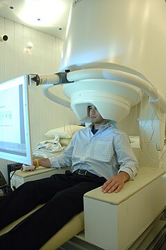
Magnetoencephalography
Magnetoencephalography (MEG) is a non-invasive technique that records functional brain activity by capturing magnetic fields, making it possible to investigate the relationships between brain structures and their functions. The possibility of such recordings is determined by neuronal postsynaptic activity and by the synchronous activation of millions of neurons, which generates a uniform, differentiated and localized brain activity, capable of being recorded by magnetometers located along the cranial convexity.
Operation
One of the recording techniques of magnetic fields of biological origin with the highest incidence and scientific relevance is Magnetoencephalography (MEG). The capacity of the MEG, both in analysis and organization of the information received, is so great that it allows assessing brain activity in milliseconds and organizing functional brain maps with delimitation of the brain structure in a space of small centimeters, and even cubic millimeters.. This allows generating functional maps of brain activity capable of being organized and represented temporally and spatially. In particular, the MEG records the postsynaptic activity generated by the apical dendrites of the pyramidal cells whose justification from the neurophysiological point of view can be found in the postsynaptic potentials (PPS) that are potentials with a slower kinetics, lasting between 10 and more than 100 ms. PPS cause low frequency neuromagnetic activity (between 10 and 100 Hz).
If we analyze the process carefully, we will verify that the initial excitation of a region of the cytoplasmic membrane produces the input of current (transmembrane current or Imemb). This region is called a sink. This current must form a closed circuit, which will propagate through the interior (intracellular current or Intra) of the axoplasm both anterogradely and retrogradely, so that it will find membrane areas through which it will exit, while depolarizing them. These regions of current output are called sources. From these sources the current will propagate through the extracellular space (extracellular or volume current, Ivol). This mechanism with two types of current (intracellular and volume) is responsible for the generation of magnetic fields. Since it is the same circuit, it is evident that the magnitude of both currents will be the same (Iintra = Ivol). However, the volumes through which they propagate are not equal, and this difference will be very important when determining magnetic fields.
Advantages
There are different techniques for studying brain activity that we can compare with the MEG:
- In the face of techniques that measure or value the brain structure, Magnetic Resonance (MRI) or Computed Axial Tomography (CT), MEG gives us information on the functional processes of cerebral anatomy with less spatial resolution but with greater temporary resolution.
- Regarding the techniques that measure or study brain metabolism, such as single photon emission tomography (SPECT) and postitron emission tomography (PET), they provide information on different vascular and metabolic changes underlying neuronal activity with a very limited temporary resolution and far from the actual time of functional processes.
- And comparing it with techniques that study or measure bioelectric processes such as Electroencephalography (EEG), the EEG has a temporary resolution close to the MEG, but the spatial resolution is very limited. On the other hand, the signals recorded by the EEG are affected by the different degrees of resistance of tissues that pass to the external electrode, which entails difficulties and imprecisions in interpreting the location of the different brain sources that generate the electroencephalographic signal. On the contrary, MEG records primary electrical activity, whose associated magnetic fields do not suffer from attenuation, distortion or behavioral modification problems.
Contenido relacionado
Computed axial tomography
Visual evoked potentials
Philadelphia chromosome
