Lymph node
The lymph nodes, lymph nodes, lymph nodes or lymph nodes are oval or reniform structures (with kidney-shaped), encapsulated, which are structurally part of the lymphatic system and functionally part of the immune system. They are located along the course of the lymphatic vessels forming chains or clusters. Its size is variable from millimeters to a couple of centimeters. They are distributed throughout the body, being found more abundantly in the armpits, in the groin, in the neck, in the abdomen (mainly in the mesentery) and around the large blood vessels.
The lymph reaches the lymph node through the afferent lymphatic vessels, it circulates slowly through the network or labyrinth that makes up its internal structure of connective tissue, which acts as a filter, allowing close contact between antigens from microorganisms (bacteria and viruses) or other foreign substances with B and T lymphocytes, thus developing a humoral or cellular immune response. Once the lymph is filtered through the nodes, it leaves through the efferent lymphatic vessels towards the lymphatic circulation, propagating the immune response (antibodies and activated cells) to the whole organism.
Structure
Lymph nodes are generally less than 1 cm in diameter, but can grow with increased activity, are kidney-shaped, and are made up of a connective tissue capsule from which emerge trabeculae that divide the node internally together with reticular tissue forming the structural integrity of the ganglion. Afferent lymphatics enter the node and efferent lymphatics exit the node.
The interior of the ganglion has three functional compartments:
- Network of lymphatic breasts that are the continuation of the aferent lymphatic vessels and continue with the efferent vessels.
- Network of blood vessels by which blood cells (especially lymphocytes) access the node.
- The lymph node parenchyma that is divided into a cortex and a marrow.
Capsule
Each lymph node is surrounded by dense irregular collagenous connective tissue, which is thickest at the site of the slit or hilum through which blood vessels enter and exit, and where efferent lymphatics exit in scant numbers.. Afferent lymphatics enter abundantly at various points in the convex capsular zone. From the capsule, trabeculae extend into the interior of the ganglion in varying numbers depending on the size of the ganglion (the larger the more trabeculae), which maintain its structure.
Rind
The cortex is the area of the ganglion parenchyma attached to the capsule and is divided into compartments by connective tissue trabeculae. The cortex is subdivided into two areas: the external and the internal. The outer cortex is made up of primary and secondary lymphoid follicles. Primary lymphoid follicles are round and are composed of naïve or resting memory B lymphocytes. Secondary lymphoid follicles have two zones: the germinal center that contains B lymphocytes activated by the presence of an antigen, and the mantle or growing zone made up of smaller (with little cytoplasm) and inactive lymphocytes, which extend like a mantle around to the germinal center oriented towards the side of the capsule. In addition to B lymphocytes, there are macrophages, veil cells (type of monocyte), and follicular dendritic cells, which present antigens to B lymphocytes.
The inner cortex, or paracortex, is composed of diffuse lymphoid tissue and is rich in T lymphocytes. Interdigital dendritic cells and macrophages are also located. Dendritic cells can retain many antigens by having a large surface area with numerous dendrites that are cytoplasmic processes.
Marrow
The medulla is located in the central part of the lymph node, it is made up of medullary cords of lymphatic tissue that surround the medullary sinuses through which the lymphatic fluid flows, which in this area converge to form the efferent lymphatic vessels. The spinal cords contain mainly macrophages, plasma cells and their precursors, and mature lymphocytes; these cells are passed to the lymph to later enter the blood circulation.
Lymphatic and blood circulation
The afferent lymphatic vessels, which carry the lymph, enter the node through the convex side of the capsule. The lymph then reaches the subcapsular or marginal sinus (between the capsule and the parenchyma), continues through the labyrinth of cortical sinuses, crossing the cortex parallel to the trabeculae and then through the very tortuous medullary sinuses, reaching the hilum. and leaves through the efferent lymphatic vessel.
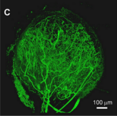
The arteries enter through the hilum of the lymph node and divide into arterioles that go through the trabeculae. Some leave them in the form of capillaries that circulate through the spinal cords. Other arterioles reach the cortex where they form a capillary network and then return via postcapillary venules through the paracortex and then the medulla, where in the medullary cords they join to form larger venules that accompany the arteriolar course towards the hilum.
Function
The main function of the lymph node is to allow interaction between antigens and lymphocytes. It is the main place where lymphocytes proliferate to achieve the immune response. Antigen-presenting cells travel from infected tissue to nodes via the lymphatic circulation, entering the node via afferent lymphatics. In the ganglion, lymphocytes are activated by contact with antigens. Once activated, they exit through the efferent lymphatic vessels, pouring into the bloodstream through the thoracic duct, where they reach the infected tissue to perform their function. On the other hand, the lymphocytes reach the ganglion mainly through the blood vessels, specifically through the postcapillary venules; only between 10 and 30% of the lymphocytes arrive through the afferent lymphatic vessels. After 12 hours of their arrival, if the circulating lymphocytes do not find antigens, they return to the circulation through the efferent lymphatic vessels.
Other cells present in the ganglion also perform their functions: macrophages engulf about 90% of the antigens that enter the ganglion. If it is the first contact with the antigen, the response begins with the activation of naïve helper T lymphocytes located in the paracortex; 48 hours later they are transformed into lymphoblasts from which clones of effector and memory helper T lymphocytes are generated in five days. Some B lymphocytes present in the paracortex are also activated and, together with some T lymphocytes, migrate to the primary follicles of the cortex where they generate lymphoblasts, from which those with greater affinity for the antigen will later be selected to amplify the response to a new contact.
When the body is fighting an infection, the immune response described involves cell multiplication inside the ganglion, increasing the size of the germinal centers of the cortex. The reaction is more notable when the response is mainly cellular (by T lymphocytes). It also increases cell recirculation through the lymphatic system. All this produces a characteristic swelling of the nodes.
Pathologies
Lymph nodes can also enlarge when they contain metastases of cancer cells, then called metastatic lymph nodes.
Contenido relacionado
Anger
Stress test
Urination


