Liver
The liver is an organ that is present in both humans and other vertebrate animals. The human liver has an average weight of 1500 g, it is located in the upper right part of the abdomen, below the diaphragm, it secretes bile essential for the digestion of fats, it also has many other functions, among them the synthesis of plasmatic proteins, vitamin and glycogen storage and detoxifying function. Its main cells are hepatocytes. It is responsible for removing different substances from the blood that may be harmful to the body, including alcohol, making them harmless. The absence of liver or its lack of function is incompatible with life. It also has the ability to regenerate.
Etymology
The word «liver» does not derive from its homonym in Latin jecur, nor from the Greek hepatos. It comes from the Latin expression ficatum jecur which literally means "liver fattened with figs". In ancient times, the inhabitants of Rome had the custom of feeding certain birds with figs in order to obtain a gastronomic delight, since the liver of these animals acquired a delicious flavor in this way. Over time ficatum jecur came to mean simply liver and the expression was shortened, first becoming ficatum, then fégado and finally liver. Therefore liver and fig have the same etymology in Spanish.
Liver Anatomy
General aspects
The liver is triangular in shape, red-brown in color, with a smooth surface and a soft, depressible consistency. In the human adult it measures on average 26 cm wide, 15 cm high and 8 cm thick at the level of the right lobe, its approximate weight is 1.5 kg.
Location
The liver is located in the upper right region of the abdomen, below the diaphragm, occupying the right hypochondrium and a part of the epigastrium. Under normal conditions it does not exceed the limit of the costal margin. It fills the space of the diaphragmatic dome, where it can reach up to the fifth rib, and is close to the heart from which it is separated by the diaphragm. It is covered by a fibrous capsule, Glisson's capsule, on which the peritoneum is applied.
Faces
The liver is surrounded by the visceral peritoneum and has two faces:
- Anterosuperior face. It has a convex shape and is in contact with the diaphragm that separates it from the lung bases and the phrenic face of the heart.
- Post-ferior face. Also called visceral face because in it the liver is related to structures located on the right side of the abdomen, many of which leave an impression on the lower face of the right lobe of the liver. Thus, we have from the back to the front the conical impression determined by the hepatic angle of the colon, the duodenal impression marked by the duodenum, glued to the cystic pit where the gallbladder is housed, the less marked renal impression formed by the upper pole of the right kidney and later a very deep groove marked by the lower vena cava. On the lower side of the left lobe are the gastric impression and the slag of the esophagus on the back edge.
At the base of the liver is the gallbladder and the hepatic hilum, which is the entry area for the portal vein, the hepatic artery, and the exit of the hepatic duct. The structure of the liver will follow the divisions of the hepatic portal vein. After division of segmental branches, the branches of the portal vein, accompanied by those of the hepatic artery and divisions of the hepatic ducts, lie together in the portal space.
Liver lobules
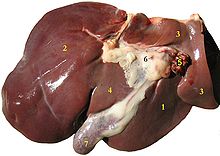
The liver is divided by the falciform ligament into two main lobes, right and left. There are two other smaller lobes, the quadrate lobe and the caudate lobe, which for many anatomists belong to the left lobe, although other texts consider the liver to have four lobes.
- Right lobelocated to the right of the falciform ligament;
- Left lobe, spread over the stomach and located to the left of the falciform ligament:
- Square lobe, visible only on the lower face of the liver; it is limited by the umbilical groove to the left, the vesicular bed to the right and the hilio of the liver behind;
- Spiegel lobe (closed lobe), located between the back edge of the hilio hepatico ahead, the vena cava behind.
There are frequent anatomical variants such as Riedel's Hepatic Lobe where there is a right infracostal prolongation that can be confused with hepatomegaly (enlarged liver).
Hepatic segments
Couinaud's classification divides the liver into eight segments that are functionally independent. Each of these segments has a branch of the hepatic portal vein, a branch of the hepatic artery, and a venous outlet branch that supplies the veins. segments 5, 6, 7, and 8 correspond to the right lobe, 2, 3, and 4 to the left lobe, and 1 to the caudate lobe.
Ligaments
The liver is covered by the visceral peritoneum, it has several connections with the parietal peritoneum that are called liver ligaments, which are not really true ligaments, but fibrous tracts that support the liver and support it on the adjacent structures. These hepatic ligaments are the following:
- Round liver ligament. It comes from the obliteration of the umbilical vein, binds the liver to the umbilical area of the previous abdominal wall.
- Coronary League. Join the back portion of the diaphragmatic face of the liver with the diaphragm, extend to both sides with the left and right triangular ligament that have the same function.
- Falciform League. Join the diaphragmatic face of the liver to the diaphragm and the previous abdominal wall. It marks the division between the right lobe and the left.
- Gastrohepatic ligament. Join the lower curvature of the stomach to the liver
- Venous duct ligament. It is the fibrous remnant of the venous duct that during the fetal period connects the umbilical vein directly with the lower vena cava.
- Hepatoduodenal ligament. It unites the duodenum to the hepatic hill and acts as support of the porta vein, the hepatic artery and the main bile path.
Blood circulation of the liver
Blood reaches the liver through the portal vein and the hepatic artery.
The portal vein system makes up 70-75 percent of the blood flow and contains nutrient-rich, low-oxygenated blood from the gastrointestinal tract and spleen.
Arterial blood arrives through the hepatic artery, a branch of the celiac trunk that contains oxygenated blood.
Blood from both sources mixes in the hepatic sinusoids and leaves the organ through the hepatic veins, also called suprahepatic, which finally drain into the inferior vena cava.
Lymphatic drainage of the liver
Lymphatic drainage from the liver is carried out by vessels that empty into the inferior vena cava or into lymph nodes that follow the reverse course of the hepatic artery.
Innervation of the liver
The liver receives nerves from the celiac plexus, the left and right vagus nerves, and also the right phrenic nerve, via the diaphragmatic plexus. The nervous contribution also comes from the celiac plexus that innervates the liver, a mixture of sympathetic and parasympathetic fibers. These nerves reach the liver along with the hepatic artery.
Liver histology
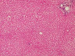
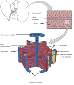
Classically, the hepatic lobule is considered the functional unit of the organ; a human liver contains between 50,000 and 100,000 lobules. Each lobule is three-dimensional (3D) in the shape of a hexagonal prism and in the central sector there is a long longitudinal central vein. In a two-dimensional (2D) histological section, the centrolobular vein is found in the center of the hexagon and the portal spaces in the corners. Between the corners of the hexagon and the center are the hepatic sinusoids and hepatocytes that are arranged in a radiating pattern around each centrilobular vein. In the hepatic lobule, arterial and venous blood from the portal spaces mix to flow into the central vein of each lobule. Within the liver lobule the following structures can be distinguished:
- Porta or triad spaces: they are triangular areas located in the angles of the hepatic lobes, formed by a laxo conjunctive estroma; they contain inside a branch of the hepatic artery, a branch of the porta vein and a bile conductillo; the bile produced by the hepatocytes is poured into a network of tubes into the hepata cells.
- Hepatic sinusoids: they are capillaries that are arranged between the foils of hepatocytes and where they converge, from the periphery of the lobulillos, the branches of the hepatic artery and the porta vein; the blood flows from the triads to the central vein, circulating in a centripetal form; the wall of the sinusoids is formed by a basal layer of In the sinusoids, the liver and porta circulation flow. These drain their content into the central hepatic vein, from it to the right and left hepatic veins, and finally to the lower vena cava.
- Disse Space: it is a narrow perisinusoidal space, which is between the wall of the sinusoids and the foils of hepatocytes, occupied by a network of reticular fibers and blood plasma that freely bathes the surface of the hepatocytes. In the space of Disse there is the metabolic exchange between hepatocytes and plasma where the abundant liver lymph is formed. In this space, hepatic star cells or Ito cells are also found in a starry form and their function is to store vitamin A.
Liver cells
The main cells that form part of the liver lobule are the following:
- Hepatocytes: they constitute about 80% of the weight and 65% of the cellular population of the liver tissue. They are polyemdrical cells with 1 or 2 polyploid spherical nuclei and a prominent nucle. They present the acidophilic cytoplasm with basophile bodies, and are very rich in oreganols. In addition, the cytoplasm contains inclusions of glucogen and fat. The plasma membrane of the hepatocytes has a sinusoidal domain with microvellosities that looks towards the space of Disse and a lateral domain that looks at the neighboring hepatocyte. The plasma membranes of two contiguous hepatocytes delimit a channel where the bile will be secreted. The presence of multiple orgánulos in the hepatocyte is related to its many functions: protein synthesis, metabolism of carbohydrates, formation of bile, catabolism of drugs and toxics and metabolism of lipids, purins and gluconeogenesis.
- Kupffer cells: they are fixed macrophages belonging to the mononuclear phagocytic system that are attached to the endothelium and that emit their extensions to the space of Disse. These cells eliminate from blood circulation, through the process of fagocytosis, all kinds of strange, unnecessary or altered particles, including aged erythrocytes and bacteria. They also act as antigen-representative cells and activate the immune response of T lymphocytes.
- Endothelial cells: These cells tap the light of the sinusoids, have a fenestrated cytoplasm (with pores) through which they penetrate the components of the blood in the direction of the sinusoidal membrane of the hepatocytes.
- Starry or Ito liver cells: They are star-shaped and possess the ability to store lipids and vitamin A, which constitute the main reserve of this vitamin of the organism. After liver damage, starry liver cells, which are the main responsible for the fibrogenic process, are activated by acquiring contactial, proliferative and profibrogenic properties. During the healing process, these cells produce a lot of extracellular matrix proteins, mainly type I collagen.
- Pit Cells: They are lymphoid cells resident in the liver similar to Natural killer cells. They have cytotoxic capacity.
- Colangiocitos or Chain Cells: Form the wall of the small ducts through which the bile circulates.
Physiology of the liver
The liver is present in vertebrates in the form of an organ and in some invertebrates in the form of a gland. It is the most voluminous viscera in the anatomy and one of the most important in terms of the metabolic activity of the organism. It performs unique and vital functions, including plasma protein synthesis, detoxifying function, and storage of vitamins and glycogen. It also removes many substances from the blood that can be harmful to the body, transforming them into harmless ones. The main functions of the liver are summarized below.
Bile production
Bile is necessary for the digestion of food, it contains bile salts formed by the liver from glycocholic acid and taurocholic acid, which in turn are derived from the cholesterol molecule. Bile is excreted into the bile duct and stored in the gallbladder from where it is expelled into the duodenum when food is ingested. Thanks to bile it is possible to absorb the fats contained in food.
Metabolism
The metabolic functions of the liver are very numerous.
- Metabolism of carbohydrates.
- Gluconeogenesis is the formation of glucose from some amino acids, lactate and glycerol;
- Glucogenolisis is the fragmentation of glucogen to release glucose in the blood;
- Glucogenogenesis or glucogenesis is the synthesis of glucogen from glucose.
- Metabolism of lipids.
- Cholesterol synthesis. Cholesterol manufactured by the liver is intended for different purposes, it forms part of the cell membranes and participates in the synthesis of bile acids.
- Triglyceride production.
- Conversion of glucids and proteins in fatty acids.
- Protein metabolism.
- Albumin synthesis
- Lypoprotein synthesis to transport fatty acids through the blood.(VLDL, HDL, LDL)
- Synthesis of transport proteins such as transferrin and ceruloplasmin.
- Synthesis of coagulation factors, such as fibrinogen (I), protrombin (II), coagulation factor V, proconvertin (VII), coagulation factor IX and coagulation factor X.
- Synthesis of non-essential amino acids. Amino acids are the constituents of all proteins, the liver can only synthesize non-essentials, the essentials must be obtained from the proteins of the diet.
- Synthesis of enzymes such as aminotransferase and alanine aminotransferase, essential for transamination.
- Synthesis of peptideal hormones, including angiotensingen.
- 1-antitriptyline alpha synthesis.
Immune Function
- In hepatic sinusoids there are a large number of Kupffer cells, which are macrophages resident in the liver that make bacteria, viruses and macromolecules strange to the organism.
- The liver is the organ that produces most of the proteins that form the complement system, which is made up of about 18 glucoproteins found in the serum and are activated sequentially in cascade. This system plays an important role in the immune response.
- The liver produces the C-reactive protein, an acute phase reactant whose synthesis significantly increases in inflammatory processes.
Blood detoxification
- Ethanol metabolization thanks to the alcohol-dehydrogenase enzyme. This enzyme is mainly located in the liver although it is also present in other tissues.
- Neutralization of numerous toxins.
- Metabolization of most drugs. For example, paracetamol is metabolized by the liver by uniting with glucuronic acid by removing it this way through the urine.
- Ammonium transformation into urea. This is an important detoxifying process, as urea is less toxic than ammonia and is easily eliminated through the urine.
- Metabolization of bilirubin. bilirubin is a toxic substance that comes from the degradation of hemoglobin. The liver eliminates it through bile after conjugating it with glucuronic acid.
Storage of substances
- Glucogen (an important reservoir of approximately 150 g);
- Vitamins, including vitamin A, vitamin D and vitamin B12
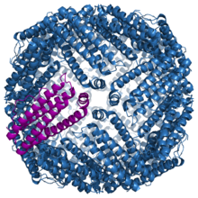
- Minerals, ferritin-shaped iron, copper, etc.
Hematopoiesis
In the first 12 weeks of intrauterine life, the liver is the main organ of red blood cell production in the fetus. From the 12th week of gestation, the bone marrow assumes this function.[citation needed]
Liver diseases
Some of the liver diseases are:
- Virus hepatitis: Hepatitis A; Hepatitis B; Hepatitis C; Hepatitis D; Hepatitis E.
- Hepatic cirrhosis;
- Autoimmune diseases: primary sclerosing colangitis, primary biliary cirrhosis and autoimmune hepatitis.
- Diseases per deposit: Hemochromatosis and Wilson disease.
- Congenital diseases that cause increased bilirubin: Gilbert syndrome, Crigler-Najjar syndrome, Rotor syndrome and Dubin-Johnson syndrome;
- Hepatic steatosis, including non-alcoholic liver steatosis and non-alcoholic steatohepatitis.
- Vascular diseases: Budd-Chiari syndrome, porta vein thrombosis.
- Hepatocarcinoma (liver cancer).
- Others: Alcoholic hepatopathy, liver abscess, liver fascioliasis.
Liver in non-human animals
Dogs and cats
The liver in mammals has a structure and function very similar to that of humans, however it cannot metabolize the same substances. In dogs and cats, certain drugs such as paracetamol cannot be easily metabolized by the liver, so they are toxic with very small doses. On the other hand, cats can present a specific liver disease that does not exist in other animals, lipidosis. feline liverwort.
Birds
In small vertebrates and especially in birds, it is common to refer to this viscus as "liver" or "liver".
Contenido relacionado
Isachne
Cryptophyta
Aegilops


