Lichen
A lichen is traditionally defined as a holobiont made up of a fungus (mycobiont) and one or several photosynthetic populations of algae or cyanobacteria (photobionts) spread extracellularly in the mycelium of the mycobiont considered as the host or ex-inhabitant. Although lichens are considered to be the best known mutualistic interactions, it is difficult to define what a lichen is because of its symbiotic nature.
The mycobiont-photobiont interaction must present emergent properties and the formed thallus must be morphologically different from that of the separate species. When the photobiont is the host, it is said to be a mycophycobiosis but sometimes there is holobionts that cannot be easily categorized as lichen or mycophycobiosis, known as border lichens where it is not possible to define a host. Recently, more members of the symbiosis have been found and there is a vision holistic view of the lichen as a microhabitat where various species of fungi from the Dikarya clade, microalgae, and bacteria coexist in an intricate symbiotic system.
According to the nature of this association, numerous structural types of lichens can be distinguished: from the simplest, where fungus and algae come together by chance, to the most complex, where the mycobiont and the phycobiont give rise to a thallus morphologically very different from the one they form separately, and where the alga is found forming a layer under the protection of the fungus. Lichens are multicellular organisms, exceptionally resistant to adverse environmental conditions and capable, therefore, of colonizing very diverse ecosystems. The protection provided by the fungus against desiccation and solar radiation, and the photosynthesis capacity of the alga give the symbiont unique characteristics within living beings. The synthesis of compounds only present in these organisms, the so-called lichen substances, allow a better use of water and light and the elimination of harmful substances.
Taxonomy and classification
The group of organisms that we call lichen-forming fungi is a polyphyletic group, that is, coming from a multitude of different ancestors, which has evolved towards the same pattern based on different relationships; Even so, there is no classification for this group fully accepted by all experts. The classification of Ozenda and Clauzade (published in 1970) attends in the first place to the type of fungus that forms the symbiosis. In this way, two classes are differentiated: ascolichens and basidiolichens. depending on whether the fungus is an ascomycete or a basidiomycete, respectively. Within the first group there are, in turn, two subclasses pyrenolichens and discolichens depending on whether they have perithecia or apothecia. Ascolichens make up 96% of lichens, with very few basidiolichens.
Lichens are classified by the fungal component. The lichen species receive the same scientific name (binomial name) as the fungal species on the lichen. Lichens are being integrated into fungal classification schemes. The alga has its own scientific name, which is unrelated to that of the lichen or fungus. There are between 13,500 and 17,000 identified lichen species. Nearly 20% of known fungal species are associated with lichens.
The "lichenized fungus" it can refer to the whole lichen or just the fungus. This can cause confusion without context. A particular species of fungus can form lichens with different species of algae, giving rise to what appear to be different lichen species, but are still classified (as of 2014) as the same lichen species.
Previously, some lichen taxonomists placed lichens in their own division, Mycophycophyta, but this practice is no longer accepted because the lichen groups belong to separate lineages. Neither ascolichens nor basidiolichens form monophyletic lineages in their respective fungal divisions, but they do form several main groups that form solely or mainly lichens within each division. Lichens arose independently from fungi that associated with algae and cyanobacteria at various times throughout evolutionary history.
Ascolichens (are classified in the classes Arthoniomycetes, Coniocybomycetes, Collemopsidiomycetes, Lichinomycetes, Lecanoromycetes, Eurotiomycetes, Leotiomycetes) and basidiolichens (are classified in the classes (Atractiellomycetes, Tremellomycetes, Agaricomycetes).
Even more unusual than basidiolichens is the fungus Geosiphon, a member of Glomeromycetes that is unique in that it encloses a symbiotic cyanobacterium within its cells, Geosiphon is not generally considered that it is a lichen, and its peculiar symbiosis went unrecognized for many years. The genus is most closely related to endomycorrhizal genera. Ascolichens of the order Verrucariales also form marine lichens with the brown algae Petroderma maculiforme and have a symbiotic relationship with the marine algae Blidingia minima, where the algae are the dominant components. The fungi are believed to help seaweeds resist drying out when exposed to air. In addition, lichens can also use yellow-green algae (Heterococcus) as their symbiotic partner.
Evolutionary history
Fossil remains of lichens are extraordinarily rare. It is known in the world of paleobotany that the incomplete fossil record cannot show at all the reality of the flora of the time to which it belongs; For this reason, it is necessary to deduce from the few traces preserved and from the phylogeny at what moment many of the plant groups appear and in this case the symbiosis between an alga and a fungus.
The oldest specimen identified as a lichen, Thuchomyces lichenoides, dates from the Precambrian. It would be a marine species according to the sediments in which its fossils were found; and although the mycobiont has been identified, the evidence for the existence of a photobiont associated with it is inconclusive. Even with this, the Rhynie Chert site has given an example of a fossil lichen called Winfrenatia reticulata, of extraordinary scientific value, since it places this group in the Devonian era; therefore, this fossil can be considered as the oldest known for the group. Another representative of the group that appeared in the Middle Devonian is Spongiophyton, although its affiliation is doubtful. Fossil remains from the Carboniferous, Permian and Triassic are missing and even the Paleogene are very scarce and inconclusive. From the Eocene the epiphic species Strigula is known, from the Oligocene the species, Anzia sp, Calicidum sp and Chaenotheca and from the Miocene Chaenothecopsis bitterfeldensis.
Coevolutionary considerations and phylogenetic studies linking current distributions and continental movements have suggested that the lichen lifestyle is very old. It seems likely that many of the present families, genera, and in some cases species evolved in Permian/Triassic times, about 190-280 million years ago, from a few earlier species. There are theories that maintain that lichens could be the first species to colonize the terrestrial environment, a theory that is too controversial and with still very inconsistent evidence.
In the attempt to present the orders of the Ascomycota division together in relation to their biology, it was found that the order Peltigerales occupies a more or less central position. It was also observed that this order has a particular significance in the evolution of Ascomycetes, since it includes some genera that are essentially terrestrial, although capable of spreading over rough bark and trees; that is, they generally occur in primary habitats, which have existed before the rise of phanerogams. Cyanobacteria happen to be one of the first photosynthetic organisms; present on earth since the Precambrian, which is why this type of photobiont could have been present for the fungi capable of associating with it from the evolutionary beginning of the group.
Symbiosis
The basis of symbiosis is the uptake of nutrients by the fungus from the algae; For this, in almost all the lichens studied, some form of penetration of the fungus into the algae cells has been found, which is achieved by means of haustoria. Two types of haustoria or penetrating organs of the fungus are differentiated: intracellular and intramembranous. In crustacean or crustose lichens (which form crusts) and in some more highly structured forms the penetrations are generally intracellular where the haustoria penetrate the protoplast of the fungus. gonidial layer (where the alga is found); the wall of these haustoria is thinner than that of the rest of the hyphae so that it is easier for them to penetrate the plant cell. In the morphologically more evolved lichens, the haustoria are intramembranous; in these cases they penetrate the wall of the gonidial layer but not the cytoplasm, leaving an invagination in the wall of the alga. The lichen obtains its food from the substances synthesized by the alga through photosynthesis; In this process, the carbohydrate called ribitol is synthesized, which is transferred to the fungus by diffusion. Inside the hyphae, this ribitol is modified to mannitol, which is not transferable from the fungus to the alga. In this way, the mycobiont ensures its food and the alga continues to synthesize. When the photobiont is a cyanobacterium, the synthesized carbohydrate is glucose, which the fungus also modifies to mannitol. The alga for its part obtains from the fungus the necessary protection against desiccation, an increase in its water absorption capacity thanks to the characteristics of the fungus hyphae. In short, symbiosis allows algae or cyanobacteria to colonize ecosystems where, due to extreme weather, they could not develop on their own.
Lichen metabolites are usually a mixture of those produced by the alga for its own functioning and those produced by the fungus. There are few cases in which substances proper to the lichen are produced that none of the bionts could produce on their own; among these substances are lichen substances, a very heterogeneous set of lichen-specific products produced by the fungus, many of them called lichen acids. Around 200 types of lichen substances are known, although research is constantly adding new types, which is why it is thought that many are exclusive to a single species, meaning that it is the symbiosis itself that produces them. It is worth clarifying that these substances do not appear in the lower cortex or in the gonidial layer and in almost all cases they appear in the form of tiny crystals and granulations arranged on the surface of the hyphae. The function of lichen substances is not entirely clear; it is thought that they can act as a deterrent to herbivores or as protection against bacteria and other pathogens. It is also possible that they play an important role in the absorption of water or increasing the permeability of the algal membrane, thereby allowing the entry of metabolites into the cell interior. It is known that they act as protection against various contaminants, UV radiation and even radioactivity. Some of them are simply debris from the day-to-day cellular activity of the symbiont organism. These lichen metabolites are the basis of many taxonomic studies on these organisms.
Types of bionts
It can be said that the formation of symbioses, especially mutualists, by fungi with all kinds of photobionts is a characteristic that has given an enormous evolutionary advantage to the species that form them, at least this can be deduced from the data that about species that form some kind of symbiosis we have; thus, of the total of 64,200 species of fungi, around 30% (19,000 species) have opted for this type of association, more than 8% form symbiosis such as mycorrhizae and 21% form lichen associations.
Among the major groups of fungi, the number of species involved in lichen associations is enormous; For example, 98% of these fungi belong to the Ascomycota subdivision, but of the forty-six known orders of fungi, sixteen form lichens and only six of them form only lichens, that is, they are not known as free-living fungi. There are relatively few exemplary basidiolichens Dictyonema, Multiclavula, and Omphalina and about fifty genera of conidial fungi.
A priori, it can be affirmed that from the evolutionary point of view the formation of lichen symbiosis is a very favorable evolutionary strategy and that it is in constant evolution in some fungi, for example in Hymenomycetes or Helotiales, although it seems that it is also being lost in others, such as Caliciales and Lecanorales.
The phycobiont is usually a chlorophyte, more rarely a cyanophyte; sometimes there are triple associations, as in the genus Lobaria, an ascolichen, with a chlorophyte as a phycobionta, which, however, presents special structures, called cephalods, in which an association with a cyanophycea (Nostoc). These cephalods develop in those lichen symbionts in which an extra symbiosis with a cyanophycea is necessary to be the main symbiont alga unable to fix the atmospheric nitrogen necessary for the fungus Due to their dual nature, lichens present characteristics of both the fungus and the algae, despite presenting their own particular characteristics.
The widespread presence of a third component of the symbiosis, a yeast of the division Basidiomycota belonging to the genus Cystobasidium, has been suggested in the outer cortex of stratified lichens. Its function and importance within the lichen symbiosis has yet to be investigated.
Photobionts
About forty genera of algae and cyanobacteria that act as photobionts in lichen symbiosis are currently known. Of these, three genera are the most frequent, Trebouxia, Trentepohlia and Nostoc, the first two being green algae and the third being cyanobacteria.
Eukaryotic photobionts are known as phycobionts while cyanobacterial photobionts are known as cyanobionts; the vast majority of phycobionts are green algae (Chlorophyta division) that have chlorophyll a and b and only two genera (of the Heterokonta clade) have chlorophyll a and c. The metabolic transfer between photobionts and mycobionts is highly dependent on the type of photobiont present. Thus, when it is a green algae, the shared carbohydrate has alcohol groups (ribitol) while in lichens with cyanobacteria it is glucose.
The identification of cyanobacterial photobionts in lichenic thallus is sometimes impossible because the morphology of the cyanobacterial thallus changes due to the presence of the mycobiont, therefore the filamentous forms, as occurs with the genus Dichothrix, are usually grossly misshapen; only the branching filaments of genera such as Stigonema can be identified in the lichen thallus.
Cyanobacteria also do not show all the phases of their life cycle when they are part of a lichen, which makes their identification even more difficult, as occurs in genera such as Chloroccidiopsis and Myxosarcina; the observation of all the vital forms of this type of organism is usually essential for its correct taxonomization at the species level, it is necessary in the work with phycobionts the isolation of the alga and its subsequent culture in free life. The type of vegetative reproduction is also a tremendously important character in taxonomy, thus species with heterocysts (such as Nostoc) increase the production frequency of these when they are in symbiosis compared to when they live freely.; Also, the size of the cells is usually greater in species in symbiosis, as occurs with Gloeocapsa in lichens of the genus Lichinella or Peccaria, this increase is usually be interpreted as a result of the presence of a large number of haustoria of the fungus inside the algal cell and the close union of these with the plant.
In green algae the organization of the thallus is always very simple when they act as photobionts, only structural forms are known in cocci, sarcinas and filaments where the filamentous structures are usually very small in size.
Identifying the green algae that form the lichen is often easier than when working with cyanobacteria. On many occasions it is not necessary to isolate the alga and cultivate it to be able to identify it at least up to the genus level. To refine the species, cultures are essential because the morphology of the chloroplasts and various stages of the algal life cycle are modified or absent in the symbiosis with the fungus.
Mycobionts
Lichen-forming fungi are in most cases obligate symbionts and are not capable of living isolated in the environment; they only prosper when they find a suitable photobiont, in isolated culture they show their imperfect form, being able to produce asexual spores but practically never produce organized reproductive structures like non-lichenized fungi or thallus structure that resembles that of lichen.
Currently, there is no evidence to confirm that the fungi that are part of the lichen symbiosis are morphologically different from those that live freely. It is true that some spherical organelles have been found in some groups of Ascomycotas, of a protein nature and function It is unknown that they are not found in free fungi and that some authors have attempted to assign functions related to symbiosis, but the latest research suggests that they may rather be organelles of the lichen-forming Ascomycota species in which it was found and not due to the symbiosis with the alga.
These concentric bodies have subsequently been located in the cytoplasm of mycelia, ascogen cells, paraphyses and all kinds of cells in plant pathogenic fungi and saprobes, especially in dry environments, so it is likely that they have a storage function for substances or resistance to desiccation, necessary, yes, in lichenized fungi.
The main difference between lichenized and non-lichenized fungi is of course the type of nutrition they provide. Many fungi appear in nature only as part of lichens, although in culture they have been isolated from the algae and have been able to survive.
Cultures of these lichen-forming fungi in isolation have almost in all cases given a very different phenotype from that expressed when they form the symbiosis, a large proportion of these fungi have presented a characteristic thallus formed by agglutinated cell masses with growth filamentous only on the periphery; However, some of the most interesting groups, such as the Perigerales, are only capable of growing when they are part of a lichen. In this way, spore cultures of various species of this group have been cultivated isolated from the algae, and although they have germinated their Mycelium has been unable to develop, only after the addition of a compatible photobiont has the now lichen grown and with it the fungus.
The third component
A study published in 2016 revealed the presence of a third component of the symbiosis, a yeast from the Basidiomycota division, in numerous species of lichens. Originally, this study sought to locate the genetic cause of the greater presence of the lichen substance called vulpinic acid in Bryoria tortuosa than in Bryoria fremontii, two species so close phylogenetically that they are currently considered phenotypic varieties. The analysis of the metagenomic rRNA of the lichen cortex showed, together with the genome of the ascomycete Bryoria and the cyanobacterium Trebouxia (the bionts that form this symbiosis), the presence of a genome of a basidiomycote of the genus Cystobasidium. This yeast appeared in samples of both species, although it did so more profusely in the variety Bryoria tortuosa. Despite the fact that microscopic observation could not reveal the presence of this yeast, the fluorescent in situ hybridization technique made it possible to locate the spherical cells of between 3 and 4 μm in diameter of the new biont embedded in a polysaccharide matrix from the external zone of the yeast. cortex. The masking of yeast cells in the polysaccharide matrix could explain why this had not been observed before.
The protocol was repeated in up to 52 species of lichens belonging to very diverse clades and localities, and the presence of Cystobasidium was found in all of them. The function of this basidiomycotic biont is still unknown, although it is believed that it may be related to the production of secondary metabolites, such as vulpinic acid from Bryoria and its abundance in different individuals would be the cause of the different known phenotypes. in the same species. It is possible that this biont is even more important for symbiosis than the simple production of lichen substances, which could explain why many lichens grown in vitro from only their two known bionts exhibit aberrant morphology. or poorly developed in the cortex.
Organization of the lichen thallus
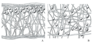
Homomer thallus is the one in which the photobiont and mycobiont are uniformly distributed. Gelatinous lichens are the ones that mainly have a homomeric thallus structure. These lichens are capable of absorbing more water than non-gelatinous lichens relative to their dry weight. This means that the exchange of gases is very limited in these organisms since it is possible that the thallus is saturated with water and therefore the diffusion of carbon dioxide (CO2) is seen. enormously difficult. For this reason, in these lichens the presence of CO2 is a limiting factor for photosynthesis. Several CO2 concentration methods have recently been described that allow its accumulation in the thallus for later use by the photobiont.
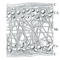
Heteromerous thallus are those in which the photobiont and mycobiont occupy different strata within the lichen. The thallus is divided into several layers, on the one hand a superficial cortex appears, with very tight fungal hyphae where, in general, traces of the algae are never found. Next, the so-called gonidial layer appears, with lax hyphae mixed with algae cells. It is the region where photosynthesis occurs by the alga and its interaction with the fungus is made more evident by the presence of the haustoria. Finally the marrow with loosely packed hyphae of the fungus. Gaseous exchange in this type of lichen is carried out through structures called pores, ciphelas and pseudociphelas, more or less depressed areas of the cortex with lax hyphae of the fungus that leave a narrow pore. On some occasions, the scars left by the isidia also act by allowing the passage of CO2 into the medulla.
Morphology of the thallus in the lichen symbiont
There are few examples of lichens whose morphology is determined by the photobiont (it occurs, for example, in the genera Coenogonium, Ephebe, Cystocoleus or Racodium), in most cases it is the mycobiont that sets the growth guidelines for the symbiont. The development of a certain type of thallus is important to know the relationships that will be established in the symbiont, for this reason, the group has traditionally been divided in the morphology of its thallus into scabby, foliose, fruticose and compound lichens. Despite this classification, there are other possible biotypes in lichens, such as the so-called gelatinous ones that some authors, however, include in the above types.
Scabby stem
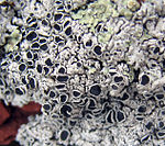
The so-called scabby thallus are those that grow strongly attached to the substrate, to the point that it is impossible to separate them from it without destroying it. The characteristics of the thallus of this type of lichen allow them to survive in very extreme environments and on exposed rock surfaces. They have both homomeric and heteromeric organization, especially on the margins of those with large areoles or in intermediate species with foliose lichens. They do not have lower crust; Its growth is marginal and many times several individuals can overlap and form characteristic patchy structures; the simplest form of organization is present in genera such as Lepraria in which the hyphae of the mycobiont surround small groups of algae with no possibility of differentiating them, the thallus has a powdery appearance as if affected by leprosy, which is why the genus takes the name; In the epiphytic lichen genus Vezdaea, the photobiont is organized in small granules or soredia barely one millimeter in diameter.
The structure of endolithic lichens (which live inside rock microfissures) or endophloeodic (under the cuticle of plant leaves) is much more complex; in many cases there is differentiation between the superior cortex and the rest of the thallus; various genera such as Buellia and Lecidea even develop stratification of the thallus in the absence of the substrate; This last genus locates the photobiont in the medulla and can extend up to two millimeters inside the sandy grains of the rocks in which they live, even introducing many more as in the species Lecidea sarcogynoides from South Africa. where various individuals have penetrated up to 9.6 millimeters deep every hundred years.
Scab lichens, which live strongly attached to the surface of rocks, can present very diverse morphologies. In this way we find species with unrestricted margins, outlined that hardly differ from the substratum. Lichens with well defined borders, lighter or darker in color than the rest of the individual and well differentiated from the environment. Figured thallus, radially lobed and with the edges loosely attached to the substratum and may even separate from it. Finally, the areolate thallus have a division on their upper face with numerous grooves that delimit portions or areoles, the grooves reveal the innermost area of the individual, dark in color.
Epilitic lichens are the most abundant among the scabby lichens, their thallus is usually perfectly limited on its margin or with imprecise limits. An areolate thallus is one in which the symbiont is distributed in multiple portions (areoles) of polygonal shapes. In the dry season each of the areoles of this organism are perfectly distinguishable while in the wet season they are not; the areoles develop from a primary thallus by extension towards its entire circumference. The most complex thallus within the scabs is called scaly where the areoles grow until they partially separate from the substrate, forming the characteristic scales that give the phenotype its name.
Leafy stem
Foliose lichens are those in which the thallus is partially detached from the substrate and not in as close a relationship with it as in the previous ones. The thallus can be homomeric or heteromeric. The most usual is that they have a dorsiventral organization, distinguishing between ventral and dorsal areas. Within this type of lichens there is an enormous diversity in terms of shapes, organization and sizes.
Laciniate lichens are those that have the typical structure of foliose lichens; they adhere to the substratum in almost all their extension and in most of the species have lobes whose distribution in the thallus is the most varied, radial, alternate, etc. In some species the lobes may be inflated by further growth of the medulla. They are the lichens that reach the largest sizes within the group and present a wide range of colours, consistency and shapes, examples of which are the genera Xanthoparmelia, Physcia and Solorina. A lichen is called umbilicate when it has a circular thallus with a single anchor, called umbilical, to the substrate in the center as occurs in Umbilicaria.
A very particular type of foliose lichens grow in deserts and have an interesting hygroscopic movement: in times of drought they are capable of rolling up on themselves to show as little surface area as possible and thus avoid drying out exposing their lower surface formed by hyphae of the fungus, also in a rolled state they are capable of being transported by the wind, normally to shady places such as stone bases or bushes awaiting the arrival of moisture; this occurs in species such as Xanthomaculina convoluta or Chondropsis semivirdis.
Frutic stem
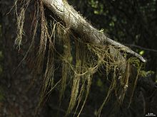
The thallus of fruticose lichens is elongated, cylindrical or very narrow, in all cases it resembles a hair. They generally present a single point of attachment to the substrate, leaving the rest of the organism far from it; they can branch, sometimes very profusely; They have apical or intercalary growth and can be solid or hollow, in the case of homomers, and flattened in heteromers. However, there are exceptions to this general morphology. Thus Sphaerophorus melanocarpus has dorsiventral symmetry although the width of the lobes is very reduced. The size of these lichens is highly variable depending on the species. For example, the genus Usnea grows several meters, while other species barely grow a few millimeters. The attachment to the substrate is carried out by means of special fixation structures that in some species degenerate at maturity, leaving the individual free of the substrate. Examples of this type of lichens are those belonging to the genera Stereocaulon and Roccella.
Heteromorphic talli
Some genera develop two types of morphology in their thallus, for example in the genus Cladonia the thallus on which a vertical reproductive part called podecio and a part vegetative horizontal in the form of scales or crust. They are called compound thallus; in them a difference is made between a horizontal thallus or primary thallus attached to the substrate and another vertical or secondary carrier of the fruiting bodies. It is possible that in the adult state the primary thallus is lost as occurs in Cladina. In some species the secondary thallus is made up of carpogenic tissue, which means that it is part of the fruiting body, while in other species the development of the fruiting body takes place at the end of the secondary thallus, not itself being part of them.
Playback
Lichens can reproduce asexually from portions of the thallus with representatives of the two bionts in the so-called taline fragmentation or from specialized structures called soredia (which in turn are grouped into soralia) and isidia.
A soredia is a group of hyphae of the fungus surrounding a few algal elements that detaches from the surface of the lichen thallus to be disseminated either by the wind or by rain splashes; this structure completely lacks internal organization. Isidia, on the other hand, are structured in the same way as the lichen thallus; They are portions of the thallus that develop on the surface, preserving the layered structure and cortex, and that can be detached easily. These types of asexual reproduction are the only processes by which the entire structure of the lichen is disseminated and not just one of its components. It should be noted that these structures are created exclusively by the fungus, as this is the only component that needs symbiosis for its survival, or at least the one that has the greatest advantage in union. The evolutionary advantage implied by the formation of these structures reveals the goodness of symbiosis for the mycobiont and its need to ensure a photosynthetic organism for its survival.
The rest of the reproductive structures formed in the symbiont do not involve the two bionts. Probably due to as yet unknown limitations imposed by the mycobiont, the photobiotic element is unable to reproduce on its own while it is part of the symbiosis. The fungus, for its part, is capable of reproducing asexually and sexually according to the characteristics of the group to which it belongs, ascomycete, basidiomycete or another. The fungal reproductive components are capable of disseminating in search of an alga to associate with, which will be the same species with which it previously formed the symbiosis or a different one, depending on the specialization of the fungus.
Sexual spores formed in ascomycotes are characteristically found in perithecia and apothecia; in lichens these structures can be formed exclusively by the fungus or have part of the algar layer participating in them, in both cases the structures produced in the hymenium spread in search of a new photobiont or develop a free-living fungus, except in fungi they are incapable of living outside of symbiosis.
The fungus can also develop asexual reproductive structures on its own; a single species of lichen (Micarea adnata) presents the structure called sporodochia formed by a series of branched or simple conidiophores similar to acervuli and synemas. The most common asexual reproductive structure is the pycnidium, an open or closed cup-shaped or sphere-shaped receptacle that contains a large number of conidiophores that produce conidia inside. This particular type of spores that are produced in large numbers are able to remain in the environment for a long time waiting to find the right alga or cyanthophyte with which to associate, as occurs with sexual spores.
Ecology
These organisms are primary colonizers in almost all known ecosystems, their ability to adapt to environments with scarce nutrients makes them capable of developing early and beginning the formation of soil for the subsequent arrival of other plant organisms. Lichens are very specific organisms with respect to the substrate and the conditions of the environment in which they develop. It is possible to find lichen symbionts in environments extremely hostile to life such as polar or desert areas where the characteristics provided by symbiosis allow their development. According to an experiment carried out by the European Space Agency in 2005, two species of Antarctic lichens were able to survive in space without any type of protection. This great capacity for survival has allowed the various species of lichen symbionts to colonize and thrive in virtually all terrestrial ecosystems.
There are several species of marine lichens corresponding to several rows of Ascomycote fungi and which, in a large part of the cases, have the alga Trebouxia as a photobiont. These lichens have as their main habitat the intertidal zone where the beating of the waves prevents the growth of many algae, in this way the symbiosis would benefit the alga not so much in terms of water storage but in terms of mechanical protection. It is possible to recognize in these communities a vertical zonation of the environment where the organisms develop, in this way there are species that are always submerged and others that are only at the highest moments of high tide.
Lichens are used as a biological indicator of air quality due to their longevity and because they obtain most of their nutrients from the air, which makes them very sensitive to impurities present in the environment, such as their susceptibility to the presence of sulfur dioxide in it. Young stems are much more sensitive to this environmental contamination than fully developed ones. The production of soredia is notably reduced in contaminated media, therefore the propagation of these organisms is significantly reduced. Sulfur dioxide is primarily responsible for the acidification of rainwater (acid rain) necessary for the growth of these organisms; The response of the thallus to this type of contamination is the creation of hydrophobic lichen substances and the reduction of the surface exposed to rain, so that photosynthesis is reduced and with it growth, apart from the amount of usable water. The recovery, however, of these organisms has turned out to be spectacular once environmental conditions return to normal. Thus, according to studies carried out, after the elimination of a large part of the environmental pollutants from the city of London at the end of the 20th century, lichens have progressively returned to colonize those habitats that were appropriate for them; similarly, in a Latin American country by reducing lead in fuel and improved traffic patterns lichen cover improved.
In the tropics, an experiment was carried out with the lichen Hypogymnia physodes, which is used in Europe as a standard species to assess air pollution. Its experimental transplant from Reutlingen (Federal Germany) to San José (Costa Rica), showed that it survives for at least three and a half to ten months and that it reacts to the tropical environment, acquiring the color characteristics of the native species.
Contenido relacionado
Ailuropoda melanoleuca
Ependymal cell
Aizoaceae


