
Kidney

The kidneys are the main organs of the human urinary system. They are responsible for the excretion of waste substances through urine and have other very important functions, including regulating the balance of the internal environment of the organism (homeostasis), controlling the volume of extracellular fluids, the osmolarity of blood plasma, the electrolyte balance and the pH of the internal environment. In addition, the kidney makes hormones such as erythropoietin, which regulates the production of red blood cells, and renin, which regulates blood pressure. The kidneys are paired organs shaped like a bean, bean, or bean. In humans they are located in the posterior part of the abdomen on either side of the vertebral column; each kidney is about 12 cm long and 6 cm wide and weighs between 125 and 150 grams in an average adult. The absence of kidneys or their failure to function is incompatible with life, which is why patients with severe renal failure require the use of dialysis procedures (artificial kidney) or a kidney transplant to continue to live.
Anatomy
The kidneys in humans are located in the posterior part of the abdomen. There are two, one on each side of the spine. The right kidney lies behind the liver and the left kidney lies below the diaphragm and adjacent to the spleen, separated from these organs by the posterior parietal peritoneum. Above each kidney is an adrenal gland. Asymmetry within the abdominal cavity caused by the liver results in the right kidney being slightly lower than the left. The kidneys are located behind the peritoneum, in the retroperitoneum, between the last thoracic vertebra and the first three lumbar vertebrae (T12 to L3). The upper poles of the kidneys are partially protected by ribs 11 and 12. Each kidney is surrounded by two layers of fat (perirenal and pararenal) that help protect them.
Organization
The weight of the kidneys is approximately equal to 1% of a person's total body weight. The kidneys have a concave side and a convex side. In the inward-facing concave portion is a central region called the hilum through which the renal artery enters the kidney and exits the renal vein and ureter.
In the human kidney, two distinct areas can be distinguished, an external area of a lighter color called the cortex and an internal area called the renal medulla. The renal medulla contains between 8 and 18 cone-shaped structures called renal pyramids. At the apex of each pyramid is the renal papilla very close to the hilum. The ureter starts from the renal hilum through which urine passes to the urinary bladder from where it is discharged to the outside through the urethra.
Blood Supply


Each kidney receives its blood supply from one of the two renal arteries that branch from the abdominal aorta. The blood supply to the two kidneys under normal conditions corresponds to approximately 22% of cardiac output, the blood supply to the kidneys being closely linked to blood pressure.
On entering the hilum of the kidney, the renal artery divides into segmental arteries that branch into smaller interlobular arteries between the renal papillae that give rise to the arcuate arteries, which run along the boundary between the medulla and the renal cortex. The arcuate arteries give off smaller branches called radial cortical arteries or interlobular arteries. The branches of these cortical arteries are the afferent arterioles that form the glomerular capillaries that drain into the efferent arterioles. The efferent arterioles are divided into the peritubular capillaries that supply blood to the cortex and the vasa recta, which are capillaries that supply blood to the renal medulla. Venous return follows a reverse path to arterial through the interlobular veins, arcuate veins, and interlobular veins that ultimately drain into the renal vein.
Histology
Renal cortex
It is the external part of the kidney and is approximately 1 cm thick, brownish-red in color and easily distinguishable when cut from the internal or medullary part. It forms an arch of tissue located immediately under the renal capsule. The renal cortex contains 75% of the glomeruli and the proximal and distal tubules. It receives 90% of renal blood flow and its main function is filtration, reabsorption and secretion.
Kidney medulla
The renal medulla is made up of between 8 and 18 conical formations that are called renal pyramids or Malpighian pyramids. The broad base of each pyramid faces the renal cortex, and its apex, or papilla, points inward, discharging into the minor calyx, which funnels into the renal pelvis. The pyramids appear hatched because they are made up of straight parallel segments of renal tubules. Between one renal pyramid and another are Bertin's columns, structures that are composed of the same tissue as the renal cortex, however due to their location they are considered part of the medulla.
Nephron


At the microscopic level, the kidney is made up of between 800,000 and 1,000,000 functional units, called nephrons. It is in the nephron that the filtration of blood plasma and the formation of urine actually take place; The nephron is the basic constituent unit of the renal organ. In each kidney there are 250 collecting ducts, each of which collects urine from 4000 nephrons.
The structure of the nephron is complex, it is composed of a renal corpuscle in communication with a renal tubule. The renal corpuscle is a spheroidal structure, made up of Bowman's capsule and the hair ball contained inside or glomerulus. The tubule where the glomerular filtrate is discharged is divided into three parts: proximal convoluted tubule, loop of Henle, and distal convoluted tubule.
The function of the nephron is divided into three fundamental steps:
- Filtration: It is the first step in the production of urine, it is that the water accompanied by many of the substances present in the blood penetrate the light of the capillaries of the renal glomérulo, and the surrounding podocytes, to enter the capsule of Bowman and the renal tubule. The daily filtration volume is about 180 litres, however 99% of this liquid is reabsorbed later and passes back to the blood. The filtration process is selective, so that the medium and high molecular weight proteins are held in the blood, while the water and electrolytes are easily passed, so that they are found in the proximal contoured lobe to a concentration similar to that of the blood before reabsorption is started.
- Tubular reabsorption: It is the process by which most of the water and many of the dissolved substances of importance to the body are reinstated into the blood. It takes place mainly in the proximal contoured tubules, but also in the ase of Henle and in the distorted tubules.
- Secretion: It is the opposite to reabsorption; at this stage some blood components are eliminated by active secretion of renal tubule cells. Secretion is not synonymous with excretion; in secretion substances are actively eliminated in the light of the tubulum. A secretion mechanism eliminates, for example, H hydrogen ions+, which contributes to maintaining the pH of the blood at appropriate levels. It is also eliminated by ammonia secretion (NH4)+) and some drugs.
Juxtaglomerular apparatus
The juxtaglomerular apparatus is a small structure that is located in each of the nephrons between the afferent and efferent arteriole, next to the distal convoluted tubule. It is made up of renin-secreting juxtaglomerular cells, macula densa cells, and Goormaghtigh cells or extraglomerular mesangial cells.
Collector duct system
Glomerular filtrate flows from the distal convoluted tubule of the nephron to the collecting duct system of the kidney. Each collecting duct receives the distal convoluted tubules of nearby nephrons as tributaries. The collecting ducts are grouped together, giving rise to increasingly thick ducts that finally open into the renal papilla, where they are called Bellini ducts or papillary ducts. In the collecting ducts, the antidiuretic hormone acts by increasing the permeability to water and facilitating its reabsorption, concentrating the urine when necessary.
Kidney Physiology

The kidneys filter the blood from the circulatory system and urinate metabolic waste products from the body, including urea, uric acid, creatinine, potassium, and phosphorus. Urine production takes place through a complex system that includes filtration, reabsorption, and secretion mechanisms. Every day the kidneys produce about 180 liters of glomerular filtrate that are concentrated in only 1 or 2 liters of urine. Urine continually descends from the kidney through the ureters to the urinary bladder, where it is stored until it is expelled through the urethra.
Kidney Functions
- Excrete waste substances through the urine. The main function of the kidney is the production of urine, through which the body removes waste substances from metabolic reactions of the organism. Some of the substances eliminated by urine are the urea and ammonia product of the desalination of amino acids, uric acid from metabolization of nucleic acids and creatinine that comes from phosphocreatin present in the muscle fibers.
- Regular homeostasis of the body.
- Regular plasma volume. The kidneys have the ability to keep the plasma volume within a desirable range, controlling the concentration of the urine, saving water when necessary and avoiding dehydration.
- Regular the ionic composition of the blood. The kidneys are able to increase or decrease the elimination of various ions through the urine, including potassium (KK)+), sodium (Na+), chlorine (Cl-), calcium (Cat+), and phosphate (HPO4--). This regulation is of great importance, small elevations or decreases in blood potassium levels, for example, can cause severe heart function disorders.
- Keep the osmolarity of the blood. The kidney regulates the loss of water and the concentration of ions in blood, thus maintaining a constant osmolarity of the blood in values of about 300 milliosmoles per litre. In the presence of the antidyuretic hormone (ADH; also called vasopressin), the collector tubes of the kidney become permeable to the water and facilitate its reabsorption, thus concentrating the urine and reducing its volume. Conversely, when the body should remove excess water, for example after drinking excess liquid, the production of anti-diauretic hormone decreases and the colector duct becomes less permeable to the water, making the urine diluted and abundant. The inability of the body to reduce the production of anti-diauretic hormone appropriately, a condition known as inadequate secretion of the anti-diauretic hormone (SIADH), causes water retention and dangerous dilution of body fluids. The shortfall in the production of anti-diarrhetic hormone, or the inability of the collector ducts to respond to it, causes insipid diabetes that has an excessive amount of urine and a tendency to dehydrate.
- Regulates blood pressure. The kidney plays a very important role in keeping blood pressure stable by secreting the hormone renin that raises blood pressure when necessary.
- Regulation of acid-basic balance. The kidney maintains stable the pH of the blood by means of a mechanism by which it eliminates variable amounts of hydrogen ions (H +) through the urine, conserving on the contrary the bicarbonate ions (HCO3-).

- Secreting hormones.
- Eritropoyetin, which stimulates the production of red blood cells by the bone marrow.
- Renin, which regulates blood pressure. When the yuxtaglomerular device detects that there is low renal plasma or hypoxia, the kidneys release renin to activate the renin-angiotensin-aldosterone system that generates peripheral vasoconstriction that increase blood pressure, guaranteeing, in theory, greater renal flow.
- In the kidney, vitamin D is transformed into active or calcitriol form, essential to maintain calcium in the bones.
- Calicreine.
- Metabolic function.
- Gluconeogenesis. It consists of glucose synthesis from amino acids and other precursors during prolonged fasting. The 90 % of gluconeogenesis takes place in the liver and only 10% in the kidney.
Diseases that affect the kidneys
Almost all kidney diseases act on the nephrons and cause them to lose their filtering capacity, a process called kidney failure. Kidney failure can happen quickly, often as a result of kidney trauma or poisoning, but nearly all kidney pathologies slowly and silently destroy the nephrons, causing chronic kidney failure that progressively worsens over years or decades. The two most common causes of chronic kidney failure are diabetes and high blood pressure. The medical specialties that study the kidneys and the diseases that affect them are nephrology and urology.
List of kidney diseases

- Kidney tubular acidosis. It is due to an alteration in renal tubules that causes metabolic acidosis.
- Kidney agenesis consists of the absence of kidney from birth. It can be unilateral or bilateral.
- Kidney calculus. Also called renal litiasis or nefrolitiasis and consists of the existence of a calculus or stone within the kidneys or urinary tracts (ureters or bladder). Acute pain caused by kidney stones is called nephrical colic.
- Kidney cancer. There are different varieties, the most common form is renal carcinoma. Wilms tumor is a type of kidney cancer that occurs mainly in childhood.
- Glomerulonefritis. Disease that initially affects the function of renal glymerulo, there are numerous variants, including IgA nephropathy, post-strep throat glomerulonephritis and fast progressive glomerulonephritis.
- Hydronephrosis. It is a dilation of the renal cholector system due to difficulty in eliminating urine, caused by the existence of a mechanical obstacle at some point of the urinary apparatus, or a functional alteration.
- Kidney failure. It is the final consequence of many kidney diseases.
- Diabetic nephropathy. It is the kidney disease caused by diabetes mellitus.
- Hypertensive nephropathy. High blood pressure can damage the kidney function.
- Pielonefritis. Bacterial infection that affects the kidney and urinary tract.
- Polycystic kidney. Genetic and progressive disease that can seriously damage kidney function.
- Alport syndrome. Genetic disease that affects the kidney and other organs.
- Kidney trauma. It usually associates with an impact on the abdomen.
The kidney in animals

Invertebrates
Invertebrates may possess excretory organs that are sometimes referred to as 'kidneys', but, even in Amphioxus, these are never homologous with vertebrate kidneys, and are more accurately known by other names, such as nephridia.
Vertebrates
In most vertebrates, the primitive kidney (mesonephros) persists into the adult, though usually fused with the metanephros; in amniotes however, the mesonephros only exists in the embryo.
Primitive Vertebrates
In the most primitive vertebrates, hagfish and lampreys, the kidney is remarkably simple: it consists of a row of nephrons, each of which empties directly into Wolff's duct.
Fish and amphibians
The kidneys of fish and amphibians are typically narrow, elongated organs that occupy a significant part of the trunk. The collecting ducts of each group of nephrons usually open into the Wolffian duct. However, the situation is not always so simple; in cartilaginous fish and some amphibians, there is also a shorter duct, similar to the amniote ureter, which drains the posterior (metanephric) parts of the kidney, and joins the Wolffian duct in the bladder or cloaca. In fact, in many cartilaginous fishes, the anterior portion of the kidney may degenerate or cease to function entirely in the adult.
Reptiles
Reptile kidneys consist of a number of lobes arranged in a broadly linear pattern. Each lobe contains a single branch of the ureter at its center. Reptiles have relatively few nephrons compared to other amniotes of a similar size, possibly due to their lower metabolic rate.
Birds
Birds have relatively large, elongated kidneys, each of which is divided into three or more distinct lobes. The lobes are composed of several irregularly arranged smaller lobules, each centered on a branch of the ureter. Birds have small glomeruli, but about twice as many nephrons as mammals of similar size.
Mammals
The human kidney is quite similar to that of other mammals. Distinctive features of the mammalian kidney, compared with that of other vertebrates, include the presence of renal pelvis, renal pyramids, and a renal cortex clearly distinguishable from the medulla. This last feature is due to the presence of elongated loops of Henle; these are much shorter in birds, and not really present in other vertebrates. Only in mammals does the kidney acquire its classic bean shape, although there are some exceptions, such as the multilobed kidneys of pinnipeds and cetaceans.

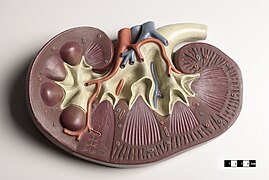
Teaching model of a mammal kidney.

Learning model of a mammal kidney and adrenal gland.
Teaching model of a mammal kidney.


Contenido relacionado
Harvard Medical School
Pinidae
Hildegard of Bingen