Karyotype
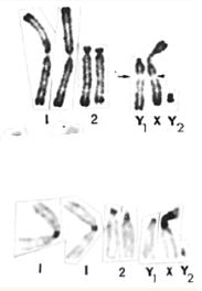
The karyotype (other than an idiogram) is the chromosomal pattern of a species expressed through a code, established by convention, that describes the characteristics of its chromosomes. Because they are often linked in the clinical setting, the concept of karyotype is frequently used to refer to a cariogram, which is a schematic, photo, or drawing of the chromosomes of a metaphase cell arranged according to their morphology (metacentric, submetacentric, telocentric, subtelocentric and acrocentric) and size, which are characterized and represent all individuals of a species. The karyotype is characteristic of each species, as is the number of chromosomes; Human beings have 46 chromosomes (23 pairs because we are diploid or 2n) in the nucleus of each cell, organized into 22 autosomal pairs and 1 sexual pair (XY male and XX female). Each arm has been divided into zones and each zone, in turn, into bands and even the bands into sub-bands, thanks to marking techniques. However, it may be the case, in humans, that there are other patterns in the karyotypes, which is known as chromosomal aberration.
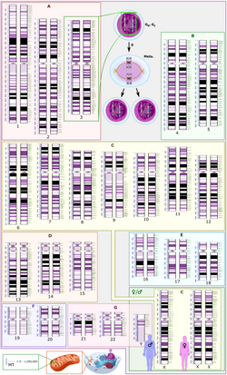
Chromosomes are classified into 7 groups, from A to G, based on their relative length and the position of the centromere, which defines their morphology. In this way, the human karyotype is formed as follows:
- Group A: There are chromosomal pairs 1, 2 and 3. They are characterized by being very large, almost metacentric chromosomes. Specifically, 1 and 3 metacentric; 2 submetacentric.
- Group B: There are chromosomal pairs 4 and 5. These are large and submetacentric chromosomes (with two very different arms in size).
- Group C: There are chromosomal pairs 6, 7, 8, 9, 10, 11, 12, X. They are submetacentric medium chromosomes.
- Group D: There are chromosomal pairs 13, 14 and 15. They are characterized by medium acrocentric chromosomes with satellites.
- Group E: There are chromosomal pairs 16, 17 and 18. They are small chromosomes, metacentric 16 and submetacentric 17 and 18.
- Group F: There are chromosomal pairs 19 and 20. This is small and metacentric chromosomes.
- G: There are chromosomal pairs 21, 22. They are characterized by small and acrocentric chromosomes with satellites.
Karyotyping can analyze numerical and structural abnormalities, something that would be very difficult to observe using Mendelian genetics.
Requirements for the karyotype study
First of all, the maximum conditions of sterility must be observed. In addition, the following must be met:
- Cells must be found in division. To do this, it is better to incubate the sample in the presence of inducing products of mitosis (mitogens), as is the case of phytohemoaglutinin.
- Cells should stand in prometafase, using colchicin, which interferes with the polymerization of mitotic spindle microtubes.
- In order to achieve good chromosomal separation, cells must undergo osmotic shock. For this purpose, a hypothetical medium (0.075M KCl) is used, which causes increased volume of cells.
- Cells have to be fixed.
- The staining of chromosomes must be done to make them identifiable.
Staining
The study of karyotypes is possible due to staining. Usually a suitable stain is applied after the cells have been arrested during cell division by a colchicine solution. For humans white blood cells are most frequently used because they are easily induced to grow and divide in tissue culture.
Sometimes observations can be made when cells are not dividing (interface). The sex of a neonate fetus can be determined by observing cells at the interface (see amniotic puncture and Barr body).
Most (but not all) species have a standard karyotype. Humans normally have 22 pairs of autosomal chromosomes and one pair of sex chromosomes. The normal karyotype for the female contains two X chromosomes called 46 XX, and the male one X and one Y chromosome, called 46 XY. Any variation from this standard karyotype can lead to developmental abnormalities.
Cytogenetics laboratories use various chromosome banding techniques. In this regard, the staining method for quinacrine bands (Q bands) stands out. It was the first to be used, requires a fluorescence microscope, although its use is no longer as widespread as that of Giemsa bands (G bands). To produce these G bands, Giemsa staining is applied after partially digesting the chromosomal proteins with trypsin. Reverse bands (R bands) require heat treatment and reverse the normal black and white pattern seen in Q bands and G. This method stands out for its great utility in staining the distal ends of chromosomes. There are other staining techniques such as C bands and NOR (nucleolar organizer region), the latter staining specifically certain regions of the chromosome. Thus, C bands stain constitutive heterochromatin, which is normally located near the centromeres, and NOR staining marks the satellites and stalks of acrocentric chromosomes.
The high-resolution bands suggest staining of chromosomes in prophase or early metaphase (prometaphase) before reaching maximum condensation. Prophase and prometaphase chromosomes are more elongated than metaphase chromosomes; For this reason, the number of bands observed, for the set of chromosomes, increases from 300-450 to almost 800. This allows less clear anomalies to be detected, which are not usually seen with conventional bands. To obtain this type of bands, another requirement must be added to perform the karyotype. It is a component used in chemotherapy, methotrexate that together with colchicine is added before staining.
Karyotype study method
- Taking peripheral blood and separation of white blood cells (T lymphocytes)
- Incubation in the presence of products that induce mitosis (mitogens), such as phytohemaglutinine. Mitogens are added to cells so that they grow properly until they form a monolayer. They are then collected by separating them from the flask through a scraper.
- Stop mitosis in metaphase (using colchicin, which interferes in the polymerization of mitotic mitubules).
Both the mitogen addition step and the colchicine addition step are critical steps for karyotyping.
- Step by a hypothetical medium that makes the cells swell
- Deposit a drop of preparation between cover and cover (on which pressure is made to disperse chromosomes)
- Fig, dye and photograph the nuclei exploded (10-15; 30 in mosaics). They are looked at from 10 to 15 nuclei because there may be many false positives because we have added mitogens (so that the cells divide quickly and quickly) and colchicin, which can cause mutations and irregularities in chromosomes. If the 10-15 cores are not the same can be because of these substances or that we are in front of a mosaic organism (so we should look at more nuclei)
- Currently there are catching devices and analysis programs that produce cariotype automatically with the data obtained.
A count of at least 12-25 cells in metaphase is necessary. This is because if, for example, when counting a nucleus, chromosome 21 is missing, it may be that it is a mosaic and that the rest of the cells do present that chromosome. Another option, and more likely, is that it is an effect of the addition of mitogen, since this compound alters the normal process of cell division, favoring aneuploidies. A last option could be that the chromosomes overlap and when proceeding to analyze the karyotype, only one chromosome is counted when there are actually two. For all these reasons, a minimum of 12-15 metaphase cells should be counted that are widely separated on the slide.
Classic karyotype
In the classic karyotype, a Giemsa solution is usually used as a stain (specific for the phosphate groups of DNA) to color the chromosome bands (G-bands), less frequently the use of the Quinacridine dye (binds to regions rich in adenosine-thymine). Each chromosome has a characteristic band pattern that helps to identify it.
Chromosomes are arranged so that the short arm of the chromosome faces the top and the long arm faces the bottom.
Some karyotypes name the short arms p and the long ones q. In addition, the different stained regions and subregions are given numerical designations according to their position relative to these chromosome arms.
For example, Cri du Chat syndrome involves a deletion on the short arm of chromosome 5. It is written as 46,XX,5p-. The critical region for this syndrome is the deletion of 15.2, which is written as 46,XX, del(5)(p15.2)
Spectral karyotype
Spectral analysis of karyotypes (or SKY) is a molecular cytogenetics technology that allows the study and visualization of the 23 pairs of chromosomes simultaneously.
Fluorescently labeled probes are made for each chromosome by labeling specific DNA from each chromosome with different fluorophores. Because there are a limited number of spectrally distinct fluorophores, a combinatorial labeling method is used to generate many different colors.
The spectral differences generated by combinatorial labeling are captured and analyzed using an interferometer attached to a fluorescence microscope.
The image processing program then assigns a pseudocolor to each spectrally different combination, allowing the visualization of colored chromosomes.
This technique is used to identify chromosomal structural aberrations in cancer cells and other pathologies when Giemsa banding or other techniques are not precise enough.
This type of technique will improve the identification and diagnosis of chromosomal aberrations in prenatal cytogenetics as well as in cancer cells.
Digital karyotype
Digital karyotyping is a technique used to quantify DNA copy number on a genomic scale. These are locus-specific DNA sequences from across the genome that are isolated and enumerated.
This method is also known as virtual karyotyping.
Observations on karyotype
- The chromosomes suffer large variations in their size throughout the cell cycle, from being very little compacted (interface) to being very compacted (metafase).
- Centeromer position difference.
- Differences in the basic number of chromosomes may occur due to successive displacements that remove all genetic material from a chromosome, causing it to be lost.
- Grade and distribution differences in heterochromatin regions. Heterochromatin, is an inactive form of condensed DNA located above all on the periphery of the nucleus that is strongly stained with the colorations, taking darker coloration than chromatin.
Variation of these chromosomes is frequently found:
- Between sexes.
- Between gametes and the rest of the body.
- Among the members of a population.
- Geographical change.
History
Levitsky was the first to define a karyotype as the phenotypic appearance of somatic chromosomes, in contrast to their gene content. This concept continued to be studied with the work of Darlington and White. The research and interest in the study of the karyotype led to the question: how many chromosomes does a human diploid cell contain?
In 1912, Hans von Winiwarter demonstrated that man had 47 chromosomes in spermatogonia and 48 in oogonia, concluding a mechanism of XX/XO sex determination. Years later, in 1922, von Winiwarter was not sure if man's chromosome number was 46 or 48. A deeper study was needed to answer this question.
- Cells were used in cultivation.
- Cells pretreated in a hypothonic solution, which makes chromosomes expand and increase in size.
- With a solution of the colchicin stop the mitosis process in the metaphase.
This took until the mid-1950s, when it was generally accepted that the karyotype of man includes only 46 chromosomes. In great monkeys, the karyotype is 48 chromosomes, so it was explained that chromosome 2 in humans was formed by a fusion of hereditary chromosomes, thus reducing their number.
Diversity and evolution of the karyotype
Although DNA replication and DNA transcription are highly standardized in eukaryotes, the same cannot be said for their karyotypes, as they are highly variable between species in chromosome number and detailed organization despite having been built with the same macromolecules.
This variation provides the basis for a range of studies that might be called developmental cytology.
In some cases there are even significant variations within species. In a 2000 review Godfrey and Masters conclude: "In our view, it is unlikely that one process or the other could independently account for the wide range of karyotype structures that are observed. But used in conjunction with other phylogenetic data, fissioning Karyotyping may help explain dramatic differences in diploid numbers between closely related species, which were previously unexplained.
- Changes during development
Over time, some of the organisms were eliminating the presence of some components of their nucleus, as well as heterochromatin.
- The elimination of chromosome; In some species (moscas) chromosomes are eliminated during development.
- The decrease in chromatin; In this process (in some copepods) part of chromosomes are emitted out into some cells. This is a process where the genome is carefully organized, where new telomers are organized and built and where certain regions of heterochromatin are lost
In Ascaris suum, all somatic cell precursors experience decreased chromatin.
- X-inactivation; Inactivation of an X chromosome is carried out during the early development of mammals. In the placental mammals, inactivation is random between the two Xs, but in marsupials it is the patherno X chromosome that is inactive.
Sometimes there are cases where some chromosomes are abnormal, resulting in a disorder for the new descendant.
- Number of chromosomes in each series
An example of variability between closely related species is that of the muntjac (a mammal of the cervid family that lives in India and Southeast Asia), which was investigated by Kurt Benirschke and his partner Doris Wurster where they showed that the diploid number of the Chinese muntjac (Muntiacus reevesi) was found to be 46 and all were telocentric. When the karyotype of the Indian muntjac (Muntiacus muntjak) was studied, they found that the female had 6 chromosomes and the male 7.
- "They just couldn't believe what they saw... They kept quiet for two or three years because they thought something had gone wrong with their tissue cultivation... But when they got a couple more specimens, they confirmed their findings."
The number of chromosomes in the karyotype between unrelated species is highly variable.
The lowest record belongs to the nematode Parascaris univalens, where the haploid number is n = 1; the highest record might be somewhere among ferns, with the Adder's Tongue fern Ophioglossum ahead with an average of 1262 chromosomes.
The highest record for animals could be among the shortnose sturgeon Acipenser brevirostrum with 372 chromosomes.
The existence of supernumerary or B chromosomes means that the number of chromosomes can vary even within the same population. (The supernumerary chromosome is located in the place of the normal chromosome 21. The formula of this triple chromosome can be XXY or XYY).
Polyploidy: the number of receptors in a karyotype
Polyploidy (more than two sets of homologous chromosomes in cells) occurs mainly in plants. It has been of great importance in the evolution of these according to Stebbins. The proportion of plants with polyploid flowers is 30-35% and in the case of grasses a much higher value, around 70%.
Polyploidy in lower plants (ferns and psilotales) is also common. Some fern species have reached levels of polyploidy well above the highest known levels in flowering plants. Polyploidy in animals is much less common, reaching importance in some groups. In humans there have been cases of triploid (69, XXX) and even tetraploid (92, XXXX) embryos and fetuses, which with a large percentage ended in miscarriage.; in the rare case of neonates with this chromosome load, their life expectancy did not exceed a few days postpartum due to various alterations in all their organs.
Endopolyploidy occurs when the adult tissues of cells have stopped dividing by mitosis, but the nuclei contain more of the original somatic chromosomes.
In many cases, endodiploid nuclei contain tens of thousands of chromosomes (they cannot be counted exactly). Cells do not always contain exactly multiples (powers of two), which is why the increase in the number of chromosome sets caused by reproduction is not entirely accurate.
This process (mainly studied in insects and some higher plants) may be a developmental strategy to increase the productivity of tissues that are very active in biosynthesis.
This phenomenon occurs sporadically throughout the eukaryotic kingdom from protozoa to man; This is diverse and complex, and it serves differentiation and morphogenesis in many ways.
See paleopolyploidy for investigation of duplication of ancient karyotypes.
Aneuploidy
The term is mainly used when the number of chromosomes varies within the cross-breeding population of species. This can also be used within a closely related group of species.
Classic examples in plants are the genus Crepis, where the gametic number (= haploid) forms the series x = 3, 4, 5, 6, and 7; and Crocus, where each number from x = 3 to x = 15 is represented by at least one species. Evidence of various kinds shows that evolutionary trends have gone in different directions, in different groups.
Closer to home, great apes have 24x2 chromosomes, where humans have 23x2.
Human chromosome 2 was formed by mixing ancestral chromosomes, reducing the number. Aneuploidy is not normally considered -ploidy but -somy, such as trisomy or monosomy.
Aneuploidies are named as follows: number of times it is repeated followed by the word “somia” followed by the chromosome number involved. The origin of this mutation may come from nondisjunction in meiosis I or II.
Chromosomal abnormalities
These abnormalities can be numerical (presence of extra chromosomes) or structural (translocations, large-scale inversions, deletions, or duplications).
Numerical abnormalities, also known as aneuploidy, refer to changes in the number of chromosomes, which can lead to genetic diseases. Aneuploidy can be seen frequently in cancer cells. In animals, only monosomies and trisomies are viable, since nullisomies are lethal in diploid individuals.
Structural abnormalities often result from errors in homologous recombination. Both types of abnormalities can occur in gametes and thus will be present in all cells of an affected person's body, or they can occur during mitosis and give rise to individual genetic mosaics that have normal and some abnormal cells.
Chromosomal abnormalities in humans:
- Turner syndrome, where there is only one X chromosome (45, X or 45 X0)
- Klinefelter syndrome, is given in male sex, also known as 47 XXY. It is caused by the addition of an X chromosome.
- Edwards syndrome, caused by a trisomy (three copies) of chromosome 18.
- Down syndrome, caused by chromosome trisomy 21.
- Patau syndrome, caused by chromosome trisomy 13.
Trisomy 8, 9, and 16 have also been found to exist, although they usually do not survive after birth. There have been no human cases of trisomies on chromosome 1, since they all end in miscarriage and are never born.
There are some disorders that result from the loss of a single piece of chromosome, including:
- Cri du Chat (catch) where there is a short arm in chromosome 5. The name comes from the cry caused by newborns resembling the maullide of a cat due to a larynx malformation.
- Deletion syndrome given by the loss of a part of the short arm of chromosome 1.
- Angelman Syndrome; 50% of the cases are missing a long arm of chromosome 15.
These chromosome abnormalities can also occur in cancer cells from a genetically normal individual. A well-documented example is the Philadelphia chromosome or the so-called Philadelphia translocation, which is a genetic abnormality associated with chronic myeloid leukemia (CML).
This abnormality affects chromosomes 9 and 22. 95 percent of chronic myeloid leukemia patients present this abnormality, while the rest of the patients suffer cryptic translocations invisible to preparations using the G-band method or other translocations that affect another or other chromosomes in the same way that happens with chromosomes 9 and 22.
Parts of two chromosomes, 9 and 22, swap places. The result is that part of the Breakpoint Cluster Region (BCR) gene on chromosome 22 (region q11) is fused with part of the ABL gene on chromosome 9 (region q34). The ABL gene takes its name from "Abelson", the name of a leukemia-causing virus that precursors of a protein similar to the one produced by this gene.
Nomenclature
Since 1995, different symbols have been used to describe the anomaly suffered by a specific chromosome or a karyotype, following the rules imposed by the ISCN (English acronym from the International System of Human Cytogenetic Nomenclature). That is, the chromosomal formula reflects the simplified description of a karyotype. In the chromosome formula, the total number of chromosomes (including the sex chromosomes) is recorded, followed by a comma, after which the sex chromosomes are written. If there are numerical or structural aberrations of the autosomes, they are written below, after another comma. When there is a mosaic, that is, two or more different cell populations coexist, the karyotypes corresponding to each one are written separated by a bar; The one with the least number of chromosomes is written first, followed by those with the highest number. Some of the symbols and abbreviations used to describe karyotypes are:
- p: short arm of chromosome.
- q: long arm of chromosome.
- +: gain of a complete chromosome.
- -: loss of a complete chromosome.
- tel: telomer.
- r(): chromosome in ring. Between parentheses we put the chromosome involved.
- deletion of. In the first parenthesis we put the chromosomes in which deletion occurs, and in the second where deletion occurs.
- ins: insertion.
- dup: duplication.
- inv()(): investment. In the first parenthesis we put the chromosomes in which the investment occurs, and in the second the ends of the inverted segment.
- t: translocation of.
- sea: DNA fragment that is not known from where it comes from, and is named the name "marking chromosome".
- dic: dicentric chromosome.
- dn: chromosomal aberration not inherited from the parents but assorted de novo.
- h: region of heretochromatin.
- i: isochromosome, i.e. chromosome that has the two arms equal either two arms or two arms 1.
- .ish: cariotype studied by FISH.
- mat: rearrangement of a chromosome of maternal origin.
- path: rearrangement of a chromosome of paternal origin.
- psu dic: pseudodicentric chromosome, that is, in which only one of the centers is active.
- tri: trisomy.
- trp: tripling a portion of a chromosome.
Examples of chromosome formulas
- Euploid numerical saws: the total number of chromosomes is multiple of the haploid number.
- 69,XXY: abnormal cariotype, with 69 chromosomes (triploid), 2 chromosomes X and a chromosome Y.
- Numerical aneuploids: the total number of chromosomes is not multiple of the haploid number, that is, there is one or more chromosomes of less or more.
- 45,X: monosomy in which there are 45 chromosomes when having a single chromosome X. It's typical in person with Turner Syndrome.
- 47,XXY or 47,XY,+X: man with an additional X chromosome (Klinefelter syndrome) which causes the individual not to develop the secondary sexual characters.

- 47,XX,+21: woman with Down syndrome.

- Mosaicism: existence of several different cell populations in the same individual.
- 45,X/46,XY mosaic with two cell lines, one with 45 chromosomes and one single X and one with 46 chromosomes, one X and one Y.
- Structural sawing: are those in which one or more chromosomes change their own structure by the addition or loss of genetic material, by altering their form or the pattern of bands. These changes are called reorganizations and always relate to chromosomal breakage. In the chromosomal formula it is necessary to specify, after the total number of chromosomes followed by a comma, the type of reorganization with the corresponding abbreviation.
- 46,X,i(X): 46 chromosomes, with a normal X chromosome and an isochromosome X.
- 46,XX,del(7)(q1q3): woman with a deletion of band 1 to band 3 of the q arm of chromosome 7.

- 47,XX,+mar: woman with a fragment of DNA that is not known where she comes from.

- 46,XY,inv(11)(p11p15): male with an investment within chromosome 11, from the p11 band to the p15 band.

Contenido relacionado
Hybrid
Frankeniaceae
Atomic physics


