Joint (anatomy)
The anatomical structure that allows the union between two bones or between a bone and a cartilage is called articulation. The joints are stabilized by ligaments that join the bone ends and have mobility thanks to the muscles that are inserted in their vicinity. The part of anatomy that deals with the study of joints is arthrology.
The most important functions of the joints are to constitute points of union between the components of the skeletal system (bone, cartilage), and to facilitate mechanical movements, providing elasticity and plasticity to the body. Some joints are not mobile, such as those established between the bones of the skull, however they are of great importance because they allow the protection of the brain and at the same time make possible its growth during childhood.
Introduction
The adult human body is made up of approximately 206 bones, which are rigid and contain a large amount of calcium salts that give them their hardness. They have five main functions: support, protection, movement, calcium reservoir and hematopoiesis (formation of blood cells).
Bones form the skeleton, which is divided into two:
- Axial skeleton: formed by head, neck and trunk bones (cranium, ribs, stern, vertebrae and sacral).
- Appendicular skeleton: formed by bones of the members included those who form the breast and pelvic waist.
Bones are linked by joints that allow movement, there are different types of joints, but one of the most important are the synovial joints, represented among others by the hip, knee, shoulder, and interphalangeal joints of the hands and feet. They are highly mobile and are formed by a cavity filled with synovial fluid and lined by the synovial membrane. The ends of the bones that form it are covered by articular cartilage. The entire set is externally reinforced by a fibrous joint capsule that gives it greater stability.
Synovial fluid
Synovial fluid is found in small amounts within the joint capsule and bathes the surfaces that make up the joint. It has the function of nourishing and lubricating the cartilage, reducing the friction of the joint surfaces, thus facilitating movement. It is produced by the synovial membrane and has a high content of hyaluronic acid.
Synovial membrane
Lines the internal surface of the joint. It forms numerous folds and villi, so its extended surface is very large. It contains several types of cells, type A synoocytes, cells similar to macrophages that clean metabolic debris present in the joint, and type B synoocytes that synthesize hyaluronic acid, a mucin that provides synovial fluid with viscosity and lubricating properties.
Joint cavity
Corresponds to the space between the articular surfaces of the bones. It is filled with synovial fluid and surrounded by the synovial membrane.
Articular cartilage
It plays a very important role in synovial joints such as the knee and shoulder, it is made up of hyaline cartilage and covers the surface of the bones, its thickness being between 2 and 4 mm. It has the function of transmitting and absorbing loads and providing a suitable surface for the sliding of the articular surfaces. It does not have its own blood vessels, so nutrients reach it through the synovial fluid. The ability to regenerate if you suffer injury or wear due to overload is low. It is made up of special cells called chondrocytes surrounded by extracellular matrix. The extracellular matrix is made up of water (65-80%), collagen (10-20%) and proteoglycans (10-15%), which gives it resistance to compression forces. Chondrocytes are responsible for producing the structural components that make up cartilage.
Subchondral bone
It is the part of the bone that is adjacent to the articular cartilage.
Joint capsule
The joint capsule is a structure formed by dense connective tissue that surrounds the joint and gives it stability, firmness and flexibility, closely joining the ends of the bone. Inside the joint capsule is the synovial fluid.
Classification
For their study, joints can be classified according to their structure or function:
- For its structure (morphologically):
Morphologically, the different types of joints are classified according to the tissue that joins them into three categories: fibrous, cartilaginous, and synovial.
- For its function (physiologically):
Physiologically, the human body has various types of joints, such as synarthrosis (not mobile), amphiarthrosis (with very limited movement -for example, the spine-) and diarthrosis (greater range or complexity of movement).
| Articles | Diarrhothosis | Great mobility | Shoulder, knee, hip |
| Anphirtrosis | Low mobility | vertebral bodies | |
| Overview | Null mobility | Skull bones |
Classification by structure
Joints can be classified according to the tissues from which they are made. There are three types: synovial, fibrous and cartilaginous:
Synovials
Synovial joints allow a wide range of motion and account for the majority of joints in the extremities. They are divided into 6 groups:
- Alticulation of hinge, gynglym or troclear: The hinge joints are synovial joints where the joint surfaces are moulded in such a way that only the movements on a shaft (monoaxial), can only perform two types of bending and extension movements. For example, the humerus-cubital joint on the elbow, the phoenic-rotulian on the knee and the joints between the proximal and medium phalanges and between the middle and distal phalanges on the fingers of hands and feet.
- Articles in pivot or trocoids or trochus: They are synovial joints where the joint surfaces are shaped similar to a pivot and only allow movements in the longitudinal axis and the only permitted movements are lateral rotation and medial rotation movements. For example, the atlantoaxial (atlas-axis) of the neck that allows the head to turn.
- Flat, slide or arthrodia: They are synovial joints that are characterized because their joint surfaces are flat and only allow slide movements. For example acromioclavicular joint and intercarpal joints.
- Products in saddle, selar or reciprocal lace: they receive their name because their shape is similar to that of a saddle. For example, the one between the first metacarpal and the trapeze bone (rapezometarcarpiana) and the sternoclavicular joint.
- Condiloid or ellipsoidal substances: it forms where two bones are irregularly attached and one bone is concave and another convex. Examples are temperoomaxilar, occipitoatloidea, metacarpo falángicas and metatarsofalángicas.
- Spherical or enatrosis: they have ball shape and receptacle and are characterized by free movement in any direction, for example coxofemoral (cadera) and escapulohumeral (hombro). They allow movements in more than 3 axes or planes (multiaxial) and make possible the movements of abduction, aduction, bending, extension, internal rotation and external rotation.
| Name | Synonym | Examples | |
| Synovial articles | Alticulation in hinge | Trocleartrosis | Humerocubital culture |
| Articulation in pivot | Trocoids | Articulation between the atlas and the axis in the neck | |
| Flat coating | Artrodia | Articulation acromioclavicular | |
| Articulation in saddle | Reciprocal lace | Articulation trapeciometacarpiana | |
| Condiloid Articulation | Condilartrosis | Temporomaxillary | |
| Spherical Articulation | Enatrosis | Coxofemoral Articulation |
Fibrous
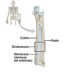
They are those in which the ends of the bones are united by fibrous tissue. These types of joints have very little mobility. An example of a fibrous joint is the sutures that join the bones of the skull. A particular type of fibrous joint is the syndesmosis in which two bones are joined by a sheet of fibrous tissue, as occurs in the interosseous membrane of the forearm that joins the ulna to the radius. A particular case is dentoalveolar syndesmosis, also called gomphosis, which is a fibrous joint, without movement under normal conditions, that is established between the root of a tooth and the alveolar process located in the jaw.
Cartilaginous
In this type of joint, the cartilaginous tissue serves as a union between the bone ends, they do not have an articular cavity as in synovial joints, and the movement they can allow is small. An example is the intervertebral discs formed by fibrocartilaginous tissue that join the vertebral bodies of the spine together. The resulting structure is very resistant and has a great capacity to absorb forces, but it is not lacking in flexibility, which is why the spine in its set has an appreciable degree of mobility.
Classification by function
Diarthrosis
The term diarthrosis comes from the Greek dia, separation, and arthron, joint. They are the most numerous in the skeleton. They are characterized by the diversity and amplitude of the movements that allow the bones. They have articular or lining cartilage on both parts of the joint. A typical example of diarthrosis is the glenohumeral joint, the joint that joins the humerus to the scapula. Contouring the glenoid fossa is the marginal labrum or glenoid labrum. The two articular surfaces are joined by the capsule that attaches around the glenoid fossa of the scapula and the anatomical neck of the humerus. The capsule is externally reinforced by extracapsular ligaments and internally it is upholstered by the synovium. They are the most mobile and fragile since they are less resistant and more covered.
Amphiarthrosis
This type of joint is held together by elastic cartilage and has very little mobility. An example of amphiarthrosis is the joints between the vertebral bodies in the spinal column. The symphysis is a subtype of joint whose characteristics are intermediate between diarthrosis and synarthrosis because they can present an articular cavity within the interosseous ligament, such as the pubic symphysis and sacroiliac joint.
Synarthrosis
Synarthrosis are joints that have very little mobility. The joints between the bones that form the skull are called sutures and are a good example of synarthrosis. Depending on the type of tissue that serves as a joint, they are divided into:
- Syndrosis, when the binding tissue is cartilizing. The cost-sternal articulation between the first rib and the stern is an example of synchondrosis. The cartilage plates found in the metaphysis of the long bones during childhood are also considered synchondrosis and allow the longitudinal growth of the bone, this type of sincondrosis has no movement function, when the cartilage is totally osified and the longitudinal growth of the bone is completed, the syncondrosis is transformed into synstosis.
- Synfibrosis, when the binding tissue is fibrous as in the sutures between the bones of the skull.
Joint diseases
There are different diseases that can affect the joints. Here are some of the most common:
- Arthritis. It is defined as inflammation in the joint. It can obey many causes. There are various pathologies that have arthritis.
- Rheumatoid arthritis.
- Gouty arthritis. It is produced by the uric acid deposit in the joints.
- Septic arthritis. It is caused by the invasion of the joint by an infectious agent, usually a bacterium.
- Psoriasic arthritis.
- Systemic erythematous lupus arthritis.
- Juvenile idiopathic arthritis. It is the most common form of persistent arthritis in childhood.
- Ankylopytic spondylitis.
- Reactive arthritis.
- Arthrosis. Also called osteoartrosis or osteoarthritis, although in fact it is not an arthritis, it is characterized by a progressive deterioration of the joint cartilage and is accompanied by alterations of the synovial membrane and the subcondral bone. Preferably affects knees, hips, hand joints and spine.
Contenido relacionado
Ipomoea sweet potatoes
Esox lucius
Apnea


