Human tooth

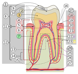
17. Periodont 18. En dice: 19. libre o interdental 20. marginal 21. alveolar 22. Periodontal League 23. Elveolar eggs 24. Irrigation and inervation: 25. dental 26. periodontal 27. through the alveolar channel
The tooth (from lat. dens, dentis) is a hard anatomical organ, embedded in the alveolar processes of the maxillary bones and mandible through a special type of articulation called gomphosis, in which different structures that make it up are involved: dental cementum and alveolar bone, both joined by the periodontal ligament. The tooth is composed of mineralized tissues (calcium, phosphorus, magnesium), which give it hardness. Together they form the temporary dentition (or "milk" teeth) and the permanent dentition.
The temporary dentition (deciduous) consists of 20 teeth, whose appearance begins at approximately six months of life and its replacement by permanent dental organs lasts until approximately 12 years of age.
The permanent dentition begins to erupt from approximately six years of age, and will replace the teeth of the first dentition until reaching adolescent age. The permanent dentition consists of 32 teeth. At the age of 16 to 25 years, the third molars, also called "wisdom teeth or wisdom teeth", whose retention within the jaws is very frequent, can erupt.
Teeth are structures of mineralized tissue that begin to develop from embryonic life, and begin to erupt in the first six months of life, which help the process of chewing food for good digestion. The tooth performs the first stage of digestion and also participates in oral communication.
Basically, two parts can be recognized in the tooth, the dental crown, the part covered by dental enamel and the dental root that is not visible in a healthy mouth.
The teeth, ordered from the center towards the jaws are: incisors that cut, canines that tear, premolars that crush and the molars that grind.
Tissues of the tooth
Dental enamel: it is a tissue formed by hydroxyapatite and proteins (in a very low proportion). It is the hardest tissue in the human body and in the world. In areas where the enamel is thinner or has worn away, it can be extremely sensitive. The enamel is translucent, insensitive to pain because there are no nerve endings in it. With fluoride, fluorhydroxyapatite crystals are formed, which is much more resistant than hydroxyapatite to the attack of dental caries.
Dentin: mineralized tissue, but to a lesser extent than enamel. It is responsible for the color of the teeth. It contains tubules where extensions of the odontoblasts are projected, called Thomes' fribs, which are the cause of sensitivity. The physical properties of dentin are: Color, radiopacity, translucency, elasticity, hardness and permeability.
Root cementum: highly specialized connective tissue. It is a hard, opaque and yellowish layer that covers the dentin at the level of the root of the tooth. It is responsible for joining the dental organ with the alveolar bone through the periodontal ligament.
Dental pulp: mesodermal tissue is made up of a soft tissue that contains blood vessels (arteries and veins) that carry blood to the tooth and nerve fibers that give the tooth sensitivity. These nerves cross the root (of the tooth) through fine channels. Its main cell is the odontoblasts (they are cells of both the pulp and the dentin), these make dentin and are the ones that maintain the vitality of the dentin. The odontoblasts have processes known as Odontoblastic Processes or Thomes' fribs, which are housed in the dentinal tubules. Together with the dentin it forms the dentin-pulp organ.
The vascular-nerve package is housed in the dental pulp, which is made up of a nerve fillet, a vein and an artery, giving it the necessary irrigation and innervation. The functional activities of the pulp are: temperature inducing, formative, nutritive, sensitive, defensive and repairing.
Periodontum: Set of ligaments that fix the tooth inside the bone socket of the maxilla. Basically they are the structures that give support and sustainability to the tooth.
Supporting structures of the teeth
The periodontal tissues that make up the periodontium are all those tissues that surround the tooth.
The periodontium contributes exponentially to the vitality of the dental organ. The periodontium is made up of the following structures:
Gingiva: is the part of the buccal mucosa that surrounds the neck of the teeth and covers the alveolar bone.
Periodontal ligament: is a connective tissue structure that surrounds the root and attaches it to the alveolar bone. Among its functions are the insertion of the tooth to the alveolar bone and the resistance to the impact of blows. It also has mechanoreceptive properties, being able to transmit the forces exerted on the tooth to the adjacent nerves.
Dental cementum: it is the mineralized structure that covers the root dentin, compensates for the physiological wear and tear in passive eruption and, above all, the attachment to the gum fibers and the periodontal ligament.
Alveolar bone: is the part of the maxilla and mandible bone where the teeth are housed. Alveolar bone is called the bone of the maxillae and mandible that contains or lines the sockets or alveolar processes, in which the roots of the teeth are maintained.
Morphological structure
- Crown: is the part of the tooth that is covered by enamel. We can observe in the mouth the functional part of the dentary organ. This portion of the tooth is permanently exposed to the mouth.
- Neck.: called the cervical zone, is the union of the crown with the root and is placed in the marginal gum.
- Raíz: This part of the tooth is not visible in the oral cavity as it is embedded in the alveolar process, inside the bone, and is covered by the dental cement. Serve as anchor. The teeth usually have between one and three roots, depending on whether they are incisive (one root), canines (1), premolars (1 or 2) or molars (two or three, in exceptional cases more than three)
Tooth development
Dental development is a set of very complex processes that allow the eruption of teeth by histological and functional modification of totipotent embryonic cells. The possession of teeth is common to many very different species, their dental development is quite similar to that of humans. In humans, the presence of enamel, dentin, cementum, and periodontium is required to allow the environment of the oral cavity to be conducive to development, which occurs during fetal development.
Temporary (deciduous) dentition
Until 6 or 7 years of age, the human species only has 20 teeth, the so-called temporary dentition or deciduous dentition, commonly called milk, which will be replaced by a total of 32 teeth that constitute the definitive dentition or permanent dentition, with four groups of teeth with specific functions.
The function of these first teeth is to prepare the food for its digestion and assimilation in stages in which the child is in maximum growth; they serve as an eruption guide: they maintain the space for the permanent dentition; they stimulate the growth of the jaws with chewing; phonation: the anterior teeth are involved in the creation of certain sounds.
Permanent dentition
After the deciduous dentition the milk teeth are pushed out by a second dentition. These first teeth fall out (exfoliate) naturally, leaving the permanent teeth to emerge.
Depending on the shape of the crown and therefore its function, there are four types of teeth:
- Incisive (8 teeth): previous teeth with sharp edge. Its main function is to cut food. They possess a conical crown and root only. The upper incisors are bigger than the lower ones.
- Canines (4 teeth): with the shape of pointed cuspid. They are called fangs in the other animals. They are located next to the incisors and their function is to tear food.
- Premolar (8 teeth): they have two pointed cuspides. They facilitate the crushing of food.
- Moor (12 teeth): they have wide cuspides. They have the same function as premolars. The crown of this type of teeth can have four or five prominences, like two, three or four roots. It's the biggest teeth.
Functions of teeth
The functions of the teeth are:
- Masticatoria
- Cosmetic
- Aesthetic
- of preservation of the arch
The dental shape determines the function of each tooth within the chewing movements. For a good function, the teeth must be well positioned, the contacts between teeth of different arches, upper and lower, are just as important as the contacts between adjacent teeth, the latter are called interproximal contacts and protect the dental papilla since they prevent food from being stored in it when chewing, avoiding packing, gingival trauma from hard foods and, therefore, the increase in bacterial plaque.
Functions of the interproximal contact point:
- Stabilizes the tooth in its alveoli and the dental arches.
- Prevents the packaging of food and therefore protects from possible gingivitis, periodontitis, caries, etc.
- Protects the Toothpaste by diverting food that in chewing go to Toothpaste.
Dental malpositions have altered contact points, which is a risk factor for various oral pathologies.
Percentages of the function according to the tooth:
- Masticatoria: Incisivos: 10%, Caninos 20%, premolars 60%, molars +90 %
- Cosmetic and Cosmetic: Incisive: 90%, Canines 80%, Premolars 40 %, Molars 10 %
Dental groups
There are two large dental groups: the anterior group, made up of incisors (central and lateral) and canines, and the posterior group, made up of premolars and molars.
- Previous Panel: They have four surfaces and an incisal edge. The superior incisives largely determine the facial aesthetic of the individual. Canines determine expression and facial appearance.
- The chewing function is to cut, the incisives, and to tear, the canines for their strong anchor in the bone and their position in the arches, in addition, the canines, contribute to the stability of the entire arch.
- Incisives possess what is called Incisal guide, this is that in the protrusion jaw movements, the jaw moves forward, the lower incisives contact the superiors sliding the incisal edge of the lower incisives by the palate face of the upper incisives and thus the later, premolar and molar sectors, are separated so that undesirable and harmful contacts are avoided. This is rooted to prevent lesions in the later teeth.
- The canines possess the Canine guide, in the laterality movements, the jaw moves to the sides, the canines on the side towards which the jaw moves contact and slides the cusp of the lower canine on the palate face of the upper canine so that the later, premolar and molar sectors are separated by preventing harmful shocks between their cuspides in these movements.
- The previous group helps to produce dental and lipdental sounds.
- Subsequent group: have four faces and an occlusal surface. This group is not as important in the aesthetic function as the previous group has, even so the subsequent dendentary losses lead to bone loss, resulting in the collapse of the skin and facial muscles.
- Premolars have a chewing function of tearing and crushing, molars, thanks to their later position in which the masticatory muscles, which are four: masetero, temporal, external Pterigoideo and internal Pterigoide, can apply great forces to produce effective crushing. Them molars are teeth with a higher number of cuspides and a higher chess surface although their cuspides are less sharp than those of premolars or canines.
- Premolars sometimes collaborate with the canines in the canine guide, when this happens is called group function and consists of avoiding subsequent contacts in laterality movements either with a good canine guide or, failing that, with the help of the premolars with a good group function.
Plaque bacteria
It is a population of cells (mainly bacteria) that grow attached to a surface wrapped in an exopolysaccharide matrix that protects them both physically and chemically, forming a thin, sticky, translucent and soft layer called microbial biofilm. The danger is that the microbial biofilm can cause caries and periodontal diseases. The biofilm of bacterial plaque is the determining factor for the onset of gingivitis and/or periodontitis. Other risk factors may contribute to the progression and severity of the lesions.
Dental diseases
Cavities
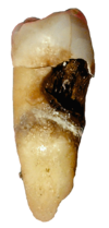
It is a multifactorial infectious disease (diet rich in carbohydrates, poor oral hygiene, incorrect brushing technique, etc.) that causes loss of hardness of the teeth, initiated by demineralization of the dental surface. It is generated by the action of the acids produced by the bacteria existing in the dental plaque that attack and destroy the enamel, the dentin, and to a greater degree, it reaches the pulp, forming a pit or fissure inside the tooth.
Teeth can also be "chipped" by the consumption of carbonated drinks (soft drinks or sodas), due to the acids and sugars they contain (sucrose). If these types of drinks are consumed, it is important to rinse your teeth with water and use toothpaste and fluoride rinses (moderately) since in excess it also causes stains on the teeth (mostly in children).
Periodontal diseases
They are those infectious diseases that inflame and destroy the supporting structures of the teeth, causing dental pain.
1- Gingivitis: It consists of inflammation and bleeding of the gums as a result of a bacterial infection. It bleeds because where there is an infection, the body sends more blood with leukocytes to fight the infection; as there is more blood "under pressure" so to speak, it is more likely that brushing breaks the capillaries of the gums and therefore they hurt and bleed.
2- Periodontitis: It occurs when the tissue that joins the bone with the teeth is destroyed. The teeth begin to loosen due to inflammation of the gum. It can be broadly considered as: mild, moderate and advanced periodontitis. The latter is the most serious of periodontal diseases and is sometimes known as chronic periodontitis. Ulcers appear that allow the exit of the infected material towards the periodontal membrane and the alveolar bone, which results in a slow and progressive destruction of this bone.
3-Toothache
Dental pain caused by one of the pathologies mentioned, a lesion in the mouth and this in turn to the teeth.
Oral hygiene
Oral hygiene consists mainly in the use of a toothbrush, since this partly removes the accumulation of a biofilm or biofilm (previously considered only bacterial plaque). Toothbrushing is advised by a specialist in periodontics, who is the expert in guiding the brushing technique of each patient, due to his individual condition, the same technique could not serve all people. The use of mouthwashes or mouthwashes is of great hygienic value, due to their chemical protection, especially rinses with fluoride content, they manage to help the work of remineralizing the enamel that daily food tends to erode, the presence of alcohol in mouthwashes is It is associated with an intensification of the disease since alcohol is a bacterial fixative, therefore it adheres to dental plaque more strongly and promotes caries and periodontal disease in the long term. Chlorhexidine-based mouthwash has antimicrobial properties, quite important in the control of periodontal disease as well as powerful anti-caries.
Dental floss should also be used, it is considered to provide 40% of hygiene, that is, almost half, together with brushing, the flossing technique is quite simple and fast once the training has been acquired that the same dentist can guide, there are different types of dental floss. Waxless floss is considered the cleanest dental floss for hygiene, used in most cases. Floss with fluoride provides an anti-caries factor, and floss with wax is of great help in crowding and serious malformations of genetic origin, as well as malocclusions that are complex to address or in patients who are difficult to manage due to their socioeconomic situation.
Contenido relacionado
Hair
Tetracycline
Case control study


