Human eye
In humans, the eye is an organ that detects light and is the basis of the sense of sight. Its function is basically to transform light energy into electrical signals that are sent to the brain through the optic nerve. It works in a very similar way to that of most vertebrates and some molluscs; it has a lens called crystalline, which is adjustable according to the distance; a diaphragm, which is called the pupil, whose diameter is regulated by the iris, and a light-sensitive tissue, which is the retina. Light enters through the pupil, passes through the crystalline lens and is projected onto the retina, where it is transformed, thanks to cells called photoreceptors, into nerve impulses that travel through the optic nerve to the brain.
Its shape is roughly spherical, it measures 2.5 cm in diameter, and it is filled with a transparent gel called the vitreous humor that fills the space between the retina and the lens.
In the anterior portion of the eye there are two small spaces: the anterior chamber, which is located between the cornea and the iris, and the posterior chamber, which is located between the iris and the lens. These chambers are filled with a liquid called aqueous humor, whose pressure level (intraocular pressure) is very important for the proper functioning of the eye.
In order for light rays entering the eye to be focused on the retina, they must be refracted. The amount of refraction required depends on the distance of the object from the observer. A distant object will require less refraction than a closer one. Most of the refraction occurs in the cornea, which has a fixed curvature. Another part of the required refraction occurs in the lens. The crystalline lens can change shape, thus increasing or decreasing its refractive capacity. As we age, the human being loses this ability to adjust focus, a deficiency known as presbyopia or tired eyesight.
Evolution towards the human eye
The usual type of vertebrate eye developed in less than 100 million years, evolving from a simple light detector for circadian (daily) and seasonal rhythms around 600 million years ago to a highly refined organ since an optical and neurological point of view, 500 million years ago. More than 150 years after Charles Darwin's theory of evolution, scientific findings have upended the creationist theory of irreducible complexity, one of whose arguments was the existence of such a highly structured organ in man, and support the ideas of the naturalist.
Structure
The organ of vision is made up of the eyelids, eyeballs, lacrimal apparatus, and external ocular muscles. The eyeball is about 25 mm in diameter and is held in position by extraocular muscles. Binocular vision, with the participation of both eyes, makes it possible to appreciate images in three dimensions.
The wall of the eye is made up of three layers:
- La outer layer, which includes sclerotic (pesa, resistant and white) and in the previous part the transparent cornea.
- La medium layer, includes choroids, which contains abundant blood vessels, and the connective tissue of the ciliar body and the iris.
- La inner layer is called retina, in which light-sensitive cells (the canes and cones) are found, covered by an external foil of cubic epithelial cells containing melanin. Externally, the retina rests on the choroid; internally, it is in contact with the vitreous humor.
Embryology
The eye is formed by the fusion of several structures that come from different embryonic tissues. The retina is a derivative of the forebrain (forebrain) and is therefore part of the central nervous system, while the cornea and lens come from the superficial ectoderm.
The first signs of the future eye are observed very early in the embryo, as they are visible at the end of the third week or beginning of the fourth, approximately on day 22. The retina is formed from two optic vesicles that arise directly from the anterior portion of the primitive brain, called the forebrain, to which it is connected by the optic stalks. These two vesicles gradually approach the surface and invaginate in the anterior part, going from being spherical to having a cup shape, giving rise to the optic calyx, which has a double wall due to folding. suffered. The inner wall that lines the interior of the optic calyx will give rise to the retina, while the outer wall will form the sheet of melanin-rich epithelial cells.
The superficial ectoderm that comes into contact with the anterior part of the optic calyx thickens, forming the crystalline plate, which invaginates and gives rise to the crystalline vesicle, which is the germ of the crystalline future. From the fifth week of development, the crystalline vesicle loses contact with the superficial ectoderm and is arranged to cover the orifice of the optic calyx. When the crystalline vesicle separates, this same area of ectoderm thickens again, to form the cornea.
Anterior Pole
The front of the eyeball is covered by the cornea, a tough, transparent structure that lacks blood vessels.
Surrounding the cornea is the conjunctiva. Behind the cornea is the anterior chamber, limited by the iris and the pupil. Behind the iris and the pupil is the posterior chamber, the ciliary body, and the lens.
The anterior chamber and the posterior chamber are two small spaces separated by the iris and connected by the pupil that are filled with a clear fluid, the aqueous humor. The aqueous humor moistens the lens, ensures its nutrition and helps maintain the shape of the anterior portion of the eye.
The iris is made up of two muscles that control the dilation and contraction of the pupil. The color of the iris depends on the transparency of the stoma and the amount of pigment it contains. When the pigment is scarce, the eyes are blue, while when there is a greater amount, green or brown nuances are appreciated.
The crystalline lens is the lens of the eye, it is supported by very fine connective fibers called the suspensory ligament of the lens, which in turn are attached to the ciliary muscle. The lens forms during the third or fourth week of pregnancy. It is soft and elastic in children, but hardens over the years.
The ciliary body lies between the ora serrata and the iris, and is responsible for the production of aqueous humor and the change in shape of the lens necessary for proper accommodation (focus). It is made up of two structures, the ciliary muscle and the ciliary processes.
Vitreous humor and retina
Behind the lens is the vitreous humor. The vitreous humor is a clear gel that takes up most of the inside of the eye and helps the eye maintain its shape. It is in direct contact with the retina, which is the innermost tunic of the eye. The retina is sensitive to light stimuli and is connected to the brain by optic nerve fibers.
Several parts of the retina can be distinguished, the most important being the macula, which is the area with the highest visual acuity. In the center of the macula is the fovea, which is a very small area, forming a depression, extremely sensitive to light. The fovea is the area of the retina where light rays are focused and is especially capable of sharp and detailed vision. Any damage to the fovea has important consequences for visual ability.
Another important area is the optic disc, which is the place where the optic nerve exits the retina. There are no light-sensitive cells in the papilla, which is why it is also known as a blind spot.
The ora serrata is the most anterior and peripheral portion of the retina, through which it comes into contact with the ciliary body.
Other segments of the human eye

1. Crystalline, 2 Zonula ciliar or suspense ligament of the crystalline; 3 Camera posterior and 4 Camera previous with 5. Aqueous humor flow; 6. Puppy, 7. Corneosclera with 8. Cornea, 9. Schlemm Trabecular System and Channel 10. Limbo sclerocorneal and 11. Sclerotic; 12. Conjunctive, 13. Join 14. Iris, 15. Corps ciliar (with: pars plicata and b: flat pars) and 16. Coroids; 17. Ora serrata, 18. Vitreous humor with 19. Hialoid behavior, 20. Retina with 21. Retinal scale, 22. Fóvea and 23 Optical Disc, optic papilla → blind spot, 24. Optical eyepiece. 25. Axis of the eye, 26 Optic nerve. 27. Dural pod, 28. Tenon capsule, 29. Tendon.
Segments: 30 previous and 31. later
32. Ophthalmic artery, 33. Arteria y vena central de la retina → 36. Retinal blood vessels; ciliar arteries: 34. later short, 35. later long and 37. Lagrimal artery, 39. Ophthalmic vein, 40. Come vorticosa.
41: Etmoides, 42. Medium straight muscle, 43 lateral straight muscle, 44 sphenoids
How the eye works
The eye receives light stimuli from the environment. Light passes through the transparent media and lens of the eye, forming an inverted image on the retina. In the retina, specialized cells transform the image into nerve impulses. These reach through the optic nerve to the posterior region of the brain. The brain interprets the signals through a complex mechanism involving millions of neurons.
Pupil and iris
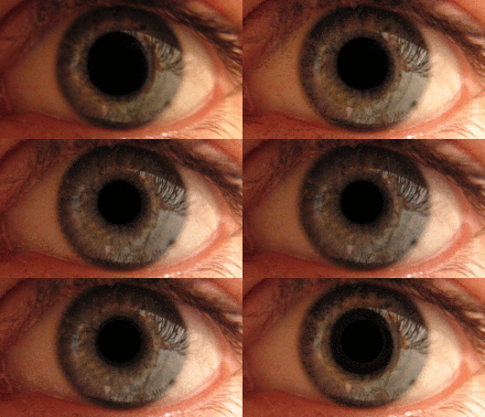
The iris is a circular diaphragm that regulates the amount of light that enters the eye, through the constrictor muscle of the iris or sphincter muscle of the pupil and the dilator muscle of the pupil or radial muscle. It has a central hole of about 3 mm in diameter, the pupil. It adapts to the intensity of the light. If the light is bright, the pupil constricts (miosis), if the light is dim, the pupil dilates (mydriasis).
The constriction of the iris is involuntary and is controlled automatically by the parasympathetic nervous system, the dilation is also involuntary, but depends on the sympathetic nervous system.
Cornea and lens
The cornea is the transparent, hemispherical structure located at the front of the eye that allows light to pass through and protects the iris. The crystalline lens is behind the cornea, has a biconvex shape, and functions as the lens or objective of the eye. When a ray of light passes from one transparent substance to another, its trajectory is bent: this phenomenon is known as refraction. Light is refracted by the cornea and lens and is projected onto the retina.
Accommodation
The light rays that enter the eye must be focused exactly on the retina so that the image obtained is clear. This requires an adjustment that occurs in a very similar way both in the human eye and in other vertebrate animals.
The process by which light rays from both near and far objects are focused exactly on the retina is called accommodation.
The mechanism of accommodation requires the contraction of the ciliary muscle which is attached to the lens by the suspensory ligament.
If the ciliary muscle contracts, the lens becomes more spherical and its refractive power increases, allowing light from nearby objects to be focused.
When the ciliary muscle relaxes, the lens becomes less spherical, its refractive power decreases, which allows us to see distant objects clearly.
Retina
The retina contains visual cells, so it can be compared to photosensitive film. These cells are capable of capturing visible light, which is only a small part of the electromagnetic spectrum, between 400 nanometers for violet light and 750 nanometers for red light.
The light that falls on the retina triggers a series of chemical and electrical phenomena that are finally translated into nerve impulses that are sent to the brain by the optic nerve.
Cones and Rods
Sensory cells in the retina react differently to light and colors. The rods are activated in the dark, and only allow to distinguish black, white and different grays. The cones make color vision possible.
In the human eye there are three types of cones, sensitive to red, green, and blue light. Each of them absorbs radiation from a certain portion of the spectrum thanks to the fact that they have pigments called opsins.
Opsins are molecules that are made up of a protein and a derivative of vitamin A. Erythropsin is more sensitive for long wavelengths around 560 nm (red light), chloropsin for medium wavelengths of about 530 nm (green light) and finally the cyanopsin with greater sensitivity for small wavelengths of about 430 nm (blue light).
By using the different intensities of the signals produced by the three types of cones, we can distinguish all the colors that make up the spectrum of visible light.
The cones are concentrated in the center of the retina, while the rods are more abundant in the periphery of the retina. Each cone is individually connected to the visual center of the brain, which in practice makes it possible to distinguish two luminous points separated by only one millimeter at a distance of 10 meters. Each human eye has 7 million cones and 125 million rods.
Extrinsic muscles

1= Zinn ring, 2= upper rectum, 3= lower rectum, 4= inner rectum, 5= external rectum, 6= upper oblique muscle of the eye, 7= larger oblique reflection pole, 8= lower oblique muscle, 9= upper eye of the eye, 10 = upper eye of the eyelid, Eyeball, 12= Optical nerve
The extrinsic musculature is made up of six muscles that are inserted on the one hand in the orbit and on the other side in the outermost layer of the eye, the sclera. These muscles are what allow the eye to move in any direction without having to change the position of the head, as occurs for example when we follow a moving object with our eyes.
Visual pathways
The optic nerves from both eyes intersect before entering the brain, forming the optic chiasm. They are then extended by the optic tracts towards the midbrain. Finally these impulses reach the visual centers of the occipital lobes.
When nerve impulses reach the occipital lobes of the brain, the information must be processed. The brain processes visual information in a particular way. Different aspects of an image are decoded by different parts of it.
The shape of an object is processed in one way, while color and movement are processed in different ways. In this way, damage to a specific area of the brain can produce certain characteristic manifestations, such as occurs in agnosia (inability to name and recognize a common object) that occurs when a specific area of visual association located in the brain is damaged. the left cerebral hemisphere.
Orbit
The orbits are two deep, symmetrical, pyramid-shaped bone cavities whose vertex points backwards. They have the function of protecting the eye. They are located on both sides of the nose, at the limit of the skull with the face. They consist of four walls: superior, inferior, internal and external, and a vertex where the optic foramen is located, which is the main communication of the orbit with the interior of the skull.
Within the orbit is the eye and a series of attached structures that are essential for the proper functioning of this organ. They are listed below:
- Extraocular muscles or extrinsic musculatures. It is a set of 6 muscles that have the purpose of moving the eye in any direction, as it happens when we look at an object in motion.
- The upper eyelid lifting muscle that mobilizes the eyelid.
- The lagrimal gland that produces tears to lubricate the eye.
- The optical nerve that transmits the information from the eye to the brain.
- The ophthalmic artery and its branches that supply blood irrigation to the eye and annexes.
- The ophthalmic vein that returns the blood to the heart.
- The different nerves that control the mobility of the eye and other functions, such as the common eye motor nerve (III cranial pair), external eye motor nerve (VI cranial) and pathetic nerve (IV cranial).
- Suspensory balloon ligament that unites the eye to the orbit to fix its position.
- Perorbital fat, connective tissue and fascias that form a protective wrap of the whole.
Eye exam
The most common reasons for consultation related to the eye are: loss of visual acuity, pain, foreign body, headache, eye irritation (red eye), other variable symptoms (discharge, burning, itching, photophobia, etc.) and anatomical disorders.
Functional exam
Includes the study of visual acuity, the ability to distinguish colors, the sense of light, that is, the measurement of the intensity of light necessary to distinguish an object, and the study of the visual field that is performed through a test called campimetry.
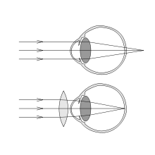
To test visual acuity, the patient must read several rows of letters of decreasing size (Snellen test). If vision is normal, all rows can be read at a distance of 6 meters. Different types of lenses can be used to correct vision deficits: concave and convex. The concave lenses correct myopia and the convex ones are used for presbyopia and hyperopia.
To test color vision, or color vision, the doctor presents the patient with several sheets with a color drawing on a background of another color. If all the colors are distinguished normally, the drawings on the background can be appreciated. Total achromatopsia prevents distinguishing any color: vision is exclusively in black and white. Partial achromatopsia is more frequent, as occurs in color blindness.
External exam
Includes a general inspection of the face, the eyelids, observing their appearance and position, the lacrimal region, the inner surface of the eyelids (palpebral conjunctiva), eversion of the eyelids in search of foreign bodies lodged there. Also the examination of ocular mobility and pupillary reflexes, such as the photomotor reflex that consists of the immediate closure of the pupil after illuminating the eye with direct light.
Examination of the anterior pole
Using different lighting devices and a magnifying lens, the structures of the anterior portion of the eye are visualized in detail, that is, the conjunctiva, the cornea, the aqueous humor, the iris, the lens and the pupil.
Fundus
To examine the fundus, the doctor uses an ophthalmoscope and instills a substance into the eye that dilates the pupils. In this way, you can observe the internal portions of the organ, the retina and its blood vessels, the optic disc, the choroid, and the vitreous humor, as well as detect various diseases, such as retinal detachment or signs of arterial hypertension or diabetes that sometimes They are reflected on the retina.
In this examination multiple anomalies can be seen, some of the most common are retinal hemorrhages and the presence of different types of exudates. Many non-ocular diseases give characteristic manifestations that are detectable by this examination.
Other tests
|
|
Main eye defects and diseases
Blindness
Blindness is a total or very severe loss of visual capacity. A blind person is unable to perceive the shape of objects, although they may retain a minimal function that allows them to distinguish between light and dark.
The concept of legal blindness is different from the previous one, since it is used for different legal issues related to compensation, social benefits or affiliation to organizations for the blind. Legal blindness does not have a single definition, as it depends on the legislation of each country. In Western countries, an individual who has a visual acuity less than 0.1 (1 is normal) or a greatly diminished visual field, less than 10 degrees, is generally considered legally blind. Therefore, contrary to what many believe, a person who is legally blind can retain visual remnants that allow them to perform some activities of daily living without assistance.
According to WHO data, there are 45 million blind people in the world, most of whom live in developing countries. Worldwide, the main causes are: cataract (48%), glaucoma (12%), age-related macular degeneration (9%), corneal opacities (5%), diabetic retinopathy (5%), different disorders grouped as childhood blindness (3.9%) and trachoma (3, 6%). Many of these diseases are perfectly treatable, so in developed countries the main causes are: diabetic retinopathy, age-related macular degeneration, glaucoma and accidents.
Myopia
Myopia is a defect of the eye in which the focal point forms in front of the retina, instead of on the retina itself as it would normally be.
This anomaly causes difficulty seeing in the distance. The subject will see badly all that object located from a certain distance.
The most common cause of myopia is an increase in the anteroposterior diameter of the eyeball. It can also be due to an increase in the refractive capacity of the lens or to an increase in the curvature of the cornea as occurs in keratoconus. It is treated through the use of corrective glasses, contact lenses, with laser surgery (LASIK, PRK) or with the placement of intraocular lenses.
Farsightedness
Hypermetropia is a defect of the eye, in which the rays of light that fall on it from infinity, focus on a point located behind the retina. It is therefore an inverse refractive defect to that of myopia.
Unlike myopia, it is not progressive and does not usually cause complications. Children affected by farsightedness do not usually present visual acuity deficit, but rather headache or fatigue related to the continuous effort of accommodation that the ciliary muscle must make to achieve correct focus. In adults there is usually a near vision deficit and over the years the distance vision can be affected. It is treated by using corrective glasses.
Astigmatism
It is a refractive error that occurs because there is a different refractive capacity between two ocular meridians and consequently objects are out of focus. It is generally caused by an irregular curvature in the anterior area of the cornea, in such a way that the refraction of the vertical meridian is different from that of the horizontal. It is treated by using glasses with corrective lenses.
Presbyopia
Presbyopia, also called strain of sight, begins around the age of 40 and reaches its maximum evolution after the age of 60. It consists of the progressive and gradual loss of the elasticity of the crystalline lens that is manifested by difficulty to see nearby objects clearly. A person with presbyopia needs to move text more than 33 cm away from their eyes in order to read. At that distance many characters are not clearly distinguished.
To ensure good vision of nearby objects, the lens must change shape and become more spherical to increase its refractive power. When you can't do it anymore, your near vision becomes blurry. However, distance vision is still good.
It can be corrected with the use of ophthalmic lenses, which carry out the work of converging the images just as the eyes did before.
When there is another vision problem added, such as myopia, bifocal or multifocal lenses can be used that allow you to see correctly at different distances, for example, to see a monitor and text that is closer.
Colour blindness
Colour blindness is a defect of the eye. The person who suffers from it has difficulty distinguishing red and green, although there are cases in which it is also difficult to differentiate other colors. When the defect consists of the inability to distinguish all colors, it is not color blindness, but another more serious disorder called achromatopsia.
Colour blindness is much more common in men than in women and runs in families. It does not usually cause other disorders, although it is a problem in some professions that require correct color vision.
Waterfall
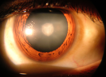
A cataract is an opacity of the crystalline lens (the lens of the eye) that loses its normal transparency. As a consequence, light penetrates the eye with difficulty, which causes progressive loss of vision, which can become total, if the appropriate treatment is not carried out. This consists of a surgical intervention through which the crystalline lens is removed and an intraocular lens is placed in its place.
Cataract is generally degenerative and appears very frequently in people over 50, although there are rarer forms that are congenital (present at birth), some of which are due to the mother suffering from rubella during childbirth. pregnancy, in this case it is called rubella cataract.
According to data from the World Health Organization, cataracts are responsible for 48% of blindness cases worldwide, which means 18 million people.
Conjunctivitis
Conjunctivitis is inflammation of the conjunctiva (mucous membrane that lines the inside of the eyelids of vertebrates and extends to the front of the eye). It can be caused by many causes, among which the most frequent is infectious; different viruses and bacteria may be involved. There are also conjunctivitis of allergic origin, toxic by irritant substances and actinic by exposure to light or ultraviolet radiation.
All the cases present some common manifestations: redness, photophobia and tearing. However, other symptoms depend on the cause, morning discharge in bacterial cases, enlarged lymph nodes in viral cases, seasonal itching in allergic cases, etc. The duration of the painting is variable according to the origin.
In general, these are benign processes, although some forms can lead to complications such as keratitis (inflammation of the cornea) that are sometimes serious.
Glaucoma
Glaucoma is an eye disease caused by elevated intraocular pressure in the eye. Intraocular pressure is determined by the balance between the production and reabsorption of aqueous humor. If the channel through which the aqueous humor drains is obstructed, the liquid is not eliminated and the intraocular pressure increases excessively. It is a condition that can be serious and causes loss of vision. There are many contraindicated medications when suffering from glaucoma.
Contenido relacionado
Annamocarya sinensis
Arthoniaceae
Araceae


