Human anatomy
Human anatomy is a branch of human biology dedicated to the study of the shape and structure of the human body and the relationships that exist between its different component parts. The term comes from the Greek ana which means above and tomos which means to cut. Anatomy allows us to understand the basic organization of the human body and the principles of operation of its structures. It is related to other related sciences such as histology, which studies tissues, and human physiology, which studies function.
Levels of organization
The human body is organized in degrees of increasing complexity that are called levels of organization. In ascending order from microscopic to macroscopic they are as follows:
- Chemical or molecular level. The human body consists of atoms that are grouped to form molecules. Several molecules can join to give rise to larger and more complex compounds called macromolecules.
- Cellular level. Different molecules give rise to cells that are the smallest living units in the body.
- Tisular level. A set of cells specializing in a function are grouped to form a tissue, for example muscle tissue.
- Professional level. The organs are tissue-shaped structures that perform a certain function. For example, the heart is formed by muscle tissue and has the function of pumping the blood into the different blood vessels.
- Level of equipment and systems. Several bodies involved in performing a particular function give rise to the apparatus and systems. For example, the heart and blood vessels form the circulatory apparatus.
- Body level. The whole of the apparatus and systems operating in a coordinated manner gives rise to the human organism.
Branches and divisions of anatomy
Depending on the point of view used, the human anatomy can be divided into different parts:
- Systematic anatomy or descriptive anatomy. Study the human organism by systems and devices, describing its shape and characteristics. The classic distinction between systems and appliances has lost effect in recent decades, and most of the texts do not distinguish between them. In anatomy a system is a set of organs that share the same function, for example respiratory system or circulatory system. Neuroanatomy is the part of the systematic anatomy that studies the nervous system extensively.
- Topographic anatomy, also called surgical anatomy or regional anatomy. It is the branch of the anatomy that studies the relationships between organs and structures located in a particular region of the organism. Therefore it describes the body by regions in which there may exist parts that belong to different systems. For example, if the chest is studied, all its structures are described and the location of each one in relation to the rest, including heart, lung, pleural cavity, pericardium, blood vessels, muscles, bones and peripheral nerves. In topographic anatomy when describing an area is usually done from the most superficial part to the deepest, so in a typical region of the lower or upper limb there would be a foreground formed by the skin, then the subcutaneous cell tissue followed by a superficial aponeurosis. More in depth we will find one or two muscle planes and in the deeper limit a bone structure formed by a bone. It should be borne in mind that this description is of a general nature and there are numerous variations depending on the specific area to be considered.
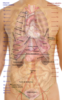
- Surface anatomy. It is the study of the anatomical structures that can be seen from the outside of the organism. It also includes the determination of a series of reference points on the surface corresponding to the projection of structures in depth.
- Artistic anatomy: it deals with anatomical issues that directly affect the artistic representation of the human figure. Understanding the morphology and structure of the human body is essential for its representation.
- Clinical anatomy. It emphasizes the study of structure and function in relation to disease situations.
- Radiological anatomy. It studies the form of different regions of the human organism through the images obtained in different medical tests, including simple x-rays, computed axial tomography and nuclear magnetic resonance.
- Functional anatomy. It studies the relationship between the form of organs belonging to the different systems with the function they perform, so it relates to physiology.
- Comparative anatomy. It studies the similarities and differences between the morphological structures of organisms belonging to different species.
Anatomical position
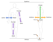
It is a unified position that is used as a reference in anatomical descriptions. The person stands with the face turned forward, the legs should be together, the feet flat on the ground, the arms close to the body and extended with the palms of the hands facing forward. About this position several terms are used to indicate the situation of a structure, region or organ:
- Proximal and distal.
- Medium and lateral.
- Previous (ventral) and later (dorsal).
- Higher (cranial) and lower (caudal).
- Surface and deep.
Relationship terms
- Flexion - extension.
- Pronation - supposition.
- Abduction - aduction.
- Internal rotation - external rotation.
- Opposition- replenishment. The opposition allows the contact of the thumb with the yolk of the other fingers, the replacement is the reverse movement.
- Protrusion - retrusion. Protrusion is the movement ahead of the lower jaw and backward retrusion.
Anatomical plans
Body planes are imaginary surfaces that divide the body into sections. They serve to facilitate the description and location of the different anatomical structures.
- Sagital plan. Divide the body on one right side and another left.
- Front or coronal plan. Divide the body in one previous portion and another later.
- Cross-sectional or axial plan. Divide the body in a higher and lower portion.
Systems and apparatus of the human body
A system is a group of organs made up of predominantly the same types of tissues that perform a coordinated function. An apparatus is a group of organs that perform a common function, the organs that form the apparatus have different embryological origin. Currently, this differentiation has lost its validity and most anatomy and physiology texts use the two terms interchangeably.
- Immune system. It provides the ability for the body to distinguish its own cells and tissues from external agents' cells. It destroys bacteria and pathogenic viruses through various mechanisms, including the complement system, antibodies and different types of cells, including lymphocytes, mainly located in bone marrow, thymus, spleen, lymph nodes and lymphoid tissue associated with mucosa (MALT).
- Tegumentary system: Understand the skin, hair, nails and sweat glands. It acts as a barrier between the external environment and the internal environment that serves to protect the body, maintain water balance and regulate body temperature. It contains numerous sensory receptors.
- Nervous system: Its function is the collection, transfer and processing of information, as well as the emission of motor impulses that reach the muscles and cause movement. It is formed by the central nervous system (encephalus and spinal cord) and the peripheral nervous system that includes sensitive nerves and motors all over the body.
- Endocrine system. It consists of a set of glands that produce hormones. Hormones are chemical messengers released by cells, which reach the bloodstream to regulate at distance different bodily functions, including growth speed, tissue activity, metabolism, development and functioning of sexual organs. The main endocrine glands are: pituitary, thyroid, adrenal glands, pancreas, ovary and testicles. Some hormones are produced by secret cells that are found in organs that are not endocrine glands, for example the kidney that produces erythropoietin.
- Circulatory system, also called cardiovascular system or circulatory apparatus. It is formed by the heart, blood vessels (veins, arteries and capillaries) and lymph vessels. The two main arteries are the aorta artery that is subdivided into numerous branches and carries oxygenated blood or the entire organism, and the pulmonary artery that carries deoxigenated blood to the lung.
- Locomotive fitting: skeletal and muscle systems. These systems coordinated by the nervous system allow movement.
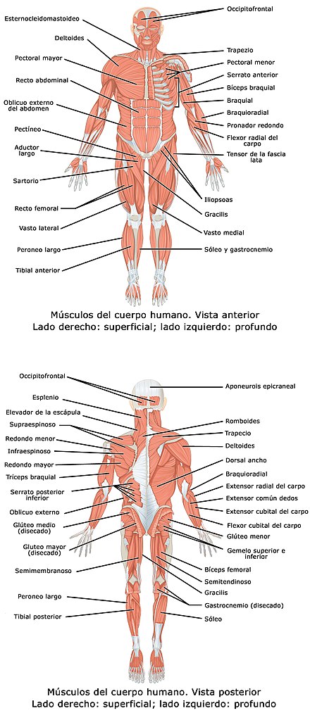
- Muscle system. It is formed by the group of voluntary muscles. It has to function to achieve the movement of the body. The whole of all skeletal muscles corresponds to 40% of the weight of an average adult.
- Bone system. It consists of the bones, joints and associated ligaments that together form the human skeleton. It has the functions of structural support, protection and assistance in the movement. It also plays an important role in maintaining calcium and phosphorus levels in blood and in the production of blood-forming cells (hematopoiesis). The whole of the bones represents on average 18% of the total weight of an adult. The skeleton of an adult consists of 206 bones, 8 in the skull, 3 in each ear, 14 in the face, the hioid bone in the neck, 26 in the spine, 25 in the chest (24 ribs and stern), 4 in the scapular waist (2 scapular and 2 clavicles), 30 in each superior member, 2 in the pelvic waist, each isolates (2 collions).
- Digestive appliance: It is formed by the mouth, pharynx, esophagus, stomach, small intestine, large intestine, and annexed glands that include the liver and pancreas. It mechanically and chemically transforms foods and converts them into assimilable molecules by enzymes, so that they can be absorbed through the wall of the intestine.
- Excretor or urinary system. Its function is to eliminate toxic substances and body wastes through urine. It consists of the kidneys, ureters, urinary bladder and urethra.
- Male player. It consists of testicles, prostate, deferent duct, urethra and penis.
- Female player. Formed by ovaries, fallopian tube, uterus, vagina and vulva.
- Respiratory device or respiratory system. It is formed by the nose, pharynx, larynx, trachea, bronchus and lungs. Its function is to transport the atmospheric air to the lung alveolos where the gas exchange occurs between air and blood. The blood captures oxygen and from carbon dioxide. The branch of medicine that studies lung diseases is pneumology, while otorhinolaryngology deals with nose, pharynx, larynx and trachea diseases.
| Equipment and systems | Main structures | |
|---|---|---|
| Digestive device | Esophagus, stomach, small intestine, large intestine, liver, pancreas, gallbladder. | |
| Circulatory fitting | Artery heart (pulmonary artery, aorta artery), veins (pulmonary veins, superior vena cava, lower vena cava), blood capillaries. | |
| Respiratory device | Faringe, larynx, trachea, lung. | |
| Nervous system | Brain (brain, cerebellum, spinal cord, nerves). | |
| Endocrine system | Hypophysis, thyroid, parathyroids, adrenal glands, pancreas, ovary, testicle. | |
| Muscle system | Sternocleidomastoid, deltoids, trapeze, major pectoral, wide dorsal, brachyal biceps muscle, brachial triceps muscle, abdominal rectum, external oblique muscle of the abdomen, major gluteal muscle, larger aductive muscle, quadiceps muscle, sural triceps, ischiotibile muscles. | |
| Bone system | Skull, lower jaw, spine, ribs, stern, scapula, collarbone, humerus, cubit, radio, hand bones, pelvis, femur, tibia, perone, foot bones. | |
| Urinary appliance | Kidney, urinary bladder, ureters. | |
| Male genital appartment | Testicular, deferential duct, prostate, urethra, penis | |
| Female genital apparel | Ovary, fallopian tube, uterus, vagina, vulva. | |
| Immune system | Bone medulla, spleen, lymph nodes, thymus | |
| Tegumentary system | Skin, hair, nails, sweat glands |
Topographic anatomy
According to a topographical criterion, the human body is studied by regions:
- Head. In the head two regions that are the skull and the face can be distinguished.
- Cráneo. It's a bone structure that wraps the brain. It consists of neurocráneo or cranial cavity and facial skeleton. The neurocráneo consists of eight bones: frontal, occipital, ethmoids, sphenoids, two temporary and two parietals.
- Cara. The shape of the face is very influenced by the bones that configure it, mainly the upper jaw, the lower jaw, the nasal bone and the zygomatic bone. In the face the muscles of the mimicus are inserted as the orbicular of the eyelids and the buccinator, also the muscles of the mastication as the masseter, and hosts some of the organs of the senses. Different structures are distinguished, including eyelid, eye, nose, mouth, cheek, lip and chin.
- Neck. It is the transition area between the head and the trunk. Different structures pass through the neck, including the larynx, trachea, esophagus, carotid artery and jugular vein. The thyroid and parathyroid glands are also found in the neck.
- Tronco. The trunk is divided by the diaphragm in a higher region called thorax and a lower one that is the abdomen.
- The chest contains the heart, lungs and large blood vessels.
- In the abdomen are the organs responsible for digestion, including esophagus, stomach, small intestine and large intestine. It also contains the liver, pancreas, urinary tract (riñons and urinary bladder), and reproductive devices, both male and female. At the bottom of the abdomen is the pelvis.
- Senior member. It is composed of four segments: spindle waist, arm, forearm and hand. It has a total of 32 bones and 45 muscles. The main joints are shoulder, elbow and wrist. Blood irrigation comes from the subclavian arteries (right and left), the venous return takes place through the subclavian veins.
- Escape tape. In this region the following mŭsculosis are located: major chest muscle, minor chest muscle, subclavian muscle, previous serrato muscle, supraspine muscle, infraspine muscle, larger round muscle, lower round muscle, subscapular muscle and deltoid muscle.
- Brazo. It has two muscles, brachial biceps and brachial triceps.
- Antebrazo. The region has 20 different muscles, some are finger flexors such as the deep common bending muscle of the fingers of the hand and other extenders, such as the common straining muscle of the fingers.
- Hand. It consists of carpo, metacarpo and phalanges.
- Lower member. It consists of four segments: pelvic waist, thigh, leg and foot. The main joints are hip, knee joint and ankle. The blood reaches the lower limb through the external illic artery, the return circulation takes place through the external iliac vein.
- Girl tape. The following muscles are located in the posterior region or glutea area, from the surface to the deep: larger gluteal muscle, piriform muscle, upper genus muscle, internal shutter muscle, lower blood muscle and crural square muscle.
- Musle. In this region you will find the quadricep, the hip aducing muscles and the ischiotibials (femoral biceps, semi-tendinous muscle, semi-membranous muscles),
- Pierna. In the previous area the anterior thybial muscle and in the posterior the sural triceps (gemelos).
- Pie. It consists of tarso, metatarso and phalanges.
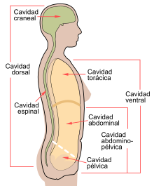
Body cavities
The human organism, despite its solid appearance, contains different spaces or cavities, in which many internal organs are located. The main cavities are:
- Dorsal cavity. It is divided into cranial cavity and vertebral cavity.
- Cranial cavity. Inside is the brain.
- vertebral cavity. It is located in the spine and contains the spinal cord.
- Ventral cavity. It corresponds to the entire previous region of the trunk. It contains chest cavity and abdominopelvic cavity.
- Thoracic cavity. In this space the heart and lungs are located.
- Abdominopevic cavity. It is located below the chest cavity of which is separated by the diaphragm. It is divided into abdominal cavity and pelvic cavity.
- Abdominal cavity. It contains numerous organs, including stomach, small intestine, large intestine, liver, pancreas, spleen, kidney and adrenal glands.
- Pelvic cavity. It is located under the abdominal cavity. It contains the urinary bladder, a portion of the large intestine and several organs of the reproductive system.
History of Anatomy
Mondino de Luzzi (1270-1326), was an Italian physician, considered a key figure who reintroduced the systematic teaching of anatomy in medical studies, practiced dissections and wrote in 1316 his Anathomia Mundini (Mondino's Anatomy) which was widely circulated in manuscript form, was printed in 1478 and was the standard manual for teaching for many generations, until the time of Andreas Vesalius. His work follows Galen's claims and although he sometimes made inaccurate descriptions of the internal organs, he ushered in a new period in anatomical science.
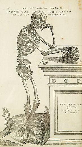
Andreas Vesalius was a 16th century lowercase physician who is considered the forerunner of modern anatomy. He wrote a treatise based on his direct observations of bodily structures, amply illustrated by engravings, which he titled De humani corporis fabrica (On the structure of the human body). This work, due to its clarity and expository rigor, is considered the first modern treatise on anatomy and in it the errors that had been transmitted for generations are revealed, based on the statements that are not adjusted to reality by Aristotle, Galen and Mondino de Luzzi. Vesalius based his work on direct observation of corpses, carrying out the dissections himself, which meant a break with the then current practice of obtaining knowledge from classical texts and not from direct research. Another great anatomist, A contemporary of Andreas Vesalius was Bartolomeo Eustachio, author of Tabulae anatomicae, Venice, 1552. One of Eustachio's contributions was the description of the auditory system, where he discovered a communication channel between the middle ear and the cavum which in his honor is called the Eustachian tube.
History of artistic anatomy
The discoveries of human anatomy are closely linked to artistic anatomy, both disciplines run parallel to the history of the nude in art. Praxiteles was a Greek sculptor of the IV century BCE. C. who studied the proportions of the human body and established the so-called Praxiteles canon, according to which the height of the ideal human figure should correspond to eight times the length of the head, in this way it is more oriented towards a canon of ideal beauty than to the exact representation of the proportions of the human body.
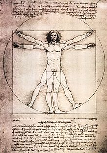
The Greeks did not need to dissect corpses to make figurative representations of the human body, however during the Renaissance, many famous artists studied anatomy from life and even performed dissections themselves to improve the representation of the human body, including Michelangelo, Raphael, Dürer and Leonardo da Vinci.
Contenido relacionado
Eragrostis
Cyperaceae
Burramyidae
