
Hexacorallia

The hexacorals or hexacorallarians (Hexacorallia), also known as zoantarians (Zoantharia b>), are a subclass of solitary or colonial anthozoan cnidarians that includes anemones and true corals. Polyps present a gastrovascular cavity divided into many chambers by septa or sarcosepta. These sarcosepta, arranged in pairs, are usually in multiples of 6, and there are two types: macrosepta and microsepta. The first are complete, that is, they go from the wall of the polyp to the pharynx; the second ones are incomplete, since they do not reach the pharynx. They have one or two rows of tentacles, never pinnate and also in multiples of 6, and have many types of cnidocytes. They can be naked or have calcareous or chitinous skeletons, although they always lack sclerites. They usually incorporate symbiotic zooxanthellae on the inside (endoderm).
Anatomy

1: tentacle;
2: faringe;
3:
4: column wall;
5macrosepto;
6: cylindium;
7: acontio;
8: discopedio;
9: retractor muscle:
10microsepto;
11: stoma (mesorial perforation);
12: necklace;
13: mouth;
14: oral disk.
Several common regions can be distinguished in hexacorals.
Oral region
The mouth is located in the center of this region and is laterally compressed. Then the pharynx appears, which is inserted into the gastrovascular cavity. This pharynx may present one or two ciliated grooves along its length, called siphonoglyphs. If there is only one, it is considered to be ventral, and if there are two, located on opposite sides, the thickest (called sulcus) is considered ventral, and the thinnest (called sulculus i>) is considered dorsal.
The oral region is surrounded by tentacles in a multiple of 6. These are not pinnate and are hollow inside because they have a prolongation of the gastrovascular cavity inside. Occasionally, the bases of some of these tentacles are thickened and loaded with cnidocytes, forming what is known as acrorrhagus.
Gastric region or spine
The gastrovascular cavity of the gastric region is divided into numerous chambers separated by sarcosepta, which can be incomplete (microsepta) or complete (macrosepta >), depending on whether or not they reach the pharynx from the wall of the column. These sarcosepta are arranged in pairs, one after the other, leaving two differentiated spaces: the space between the two sarcosepta of the same pair is called endocoelic, while the space between two sarcosepta of different pairs is called exocelic. The microsepta have a free inner edge, along the entire length of the animal, with three lobes (trilobed). The macrosepta, for their part, only have the free edge below the pharynx.
These free edges of the sarcosepta are called mesenteric filaments, and they have various functions. One of them is the formation of gametes. The central lobe at the base of some mesenteric filaments is elongated and loaded with cnidocytes, called acontio. This can come out of the animal through the mouth or through a hole located at the base of the animal called cinclídio.
They have banderoles, the contractile part of the nutritive-muscular cells that form part of the sarcosepta (they can be recognized because part of the sarcoseptum is thickened). They act as longitudinal muscles in the oral-aboral axis (from the mouth to the base). The banderoles of the sarcosepta that form a pair are oriented to the corresponding endocoel space, except for that of the sarcosepta that delimit the sulcal chamber (with two banderoles, each on one side) and asulcal (without banderoles), which are oriented to the corresponding space. exocelic.
Basal region
It is the zone of attachment to the substratum (in solitary species it is called discopedium) or to the rest of the colony (in colonial species). Some solitary species are able to detach themselves from the discopedia and move slowly from one side to the other.
Formation and structure of the skeleton


It is formed from the deposition of calcium carbonate crystals in a mucous layer produced by the epidermis of the basal region, until its total calcification occurs. Calcification occurs first around the basal region, just above the discopedium, forming a skeletal cup called the theca; then, and below the discopedium, a series of radial and centripetal calcified septa (called sclerosepts) are formed, each arranged between two sarcosepta. The whole of the skeletal system is called polyperous or corallite.
Sometimes there is close proximity between the different sclerosepta, which leads to the appearance of cross-sectional structures that are also calcified that join them, called synapticles. If the distal end of the sclerosepts protrudes, structures called ribs or costa are formed. Other structures that can be formed are the columnillae or columella, which are central skeletal rods that may or may not be surrounded by others called stakes or pali. Other times, the central parts of the sclerosepts fuse together, giving rise to a pseudocolumnette or pseudocolumella.
Colonial structure
The colonial structures of zoantarians are produced by extratentacular or intratentacular budding from a single individual. All individuals in a colony are united by a thin layer of living tissue from the extension of their body wall or column. This living layer contains the prolongation of the gastrovascular cavity, mesoglea, and epidermal layers of all individuals. There are two epidermal layers, the superficial and the deep, the latter being responsible for the formation of the calcium carbonate skeleton typical of these colonies. The production of calcium carbonate is continuous, so the animals overlap over time.
Phylogenetic tree
| Hexacorallia |
| ||||||||||||
Classification
According to Brusca & Brusca, hexacorals comprise four orders:
Subclass Hexacorallia
- Official Order - True Sea Anemones
- Order Scleractinia (=Madreporaria) - True pears
- Order Zoanthidea
- Corallimorphary order
The taxonomic organization of the species, genera, families and orders of the Anthozoa class has been, since the XIX century, exciting subject for scientists. Given that a large number of new species are being discovered and described, and that their observation through mitochondrial, phylogenetic and PCR analyses, as well as the images provided by the scanning electron microscope, reveal results that require reclassify many of them.
Currently, there is a greater consensus among taxonomists on the following classification, also accepted by the World Register of Marine Species:
Subclass Hexacorallia:
- Official Order
- Antipatharia Order
- Order Ceriantharia
- Corallimorphary order
- Order Scleractinia
- Order Zoanthidea
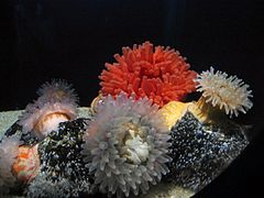
Actiniaria

Antipatharia
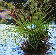
Ceriantharia

Corallimorpharia

Scleractinia
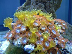
Zoanthidea


Contenido relacionado
Biome
B lymphocyte
Mucous membrane