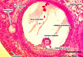
Graaf follicle

In mammalian reproduction, the Graafian follicle, tertiary follicle, mature follicle, late antral follicle or preovulatory follicle, is the final stage of the folliculogenesis process.
Development of the Graafian follicle
The De Graaf Follicle develops from a primordial follicle, composed of a compact oocyte surrounded by a single layer of a few flat cells, called pregranulosa.
The primordial follicle develops and changes progressively to become the so-called primary follicle, which has a larger volume and is surrounded by a layer of granular cells that are now cubic. This is the primary monolayer follicle.
The cytoplasm of the oocyte increases in volume and several layers of granular cubic cells are generated, surrounded by flat cells or theca cells. This structure is called multilayer primary follicle.
The multilayer primary follicle secretes follicular fluid into its interior, creating clear spaces without cells, which when they grow join together. This intragranular distribution creates a chamber or cave-like structure called the follicular antrum. This more developed follicle is called antral or secondary follicle.
The secondary follicle then grows enormously, producing large amounts of antral fluid, until the antrum becomes a single, giant cavity. The granule cells no longer surround the oocyte on its entire surface and now only support it as a pedestal-shaped clump called the Cumulus Oophorus. At this point in its development is when it is called De Graaf's follicle, mature, tertiary or also preovulatory.
Morphology of the Graafian follicle
Macroscopy
It was in the second half of the XVII century when Reignier de Graaf observed and described fluid-filled cavities in the ovaries of animals, which today we know as the follicle that bears his name.
- At that time optical science was at its beginnings and anatomical studies were done macroscopically by observing with the eyes. What the researchers could see was above 0.2 millimeters (mm), therefore the details visible in the biological tissues were only the supracellular structures.
- At that time optical science was at its beginnings and anatomical studies were done macroscopically by observing with the eyes. What the researchers could see was above 0.2 millimeters (mm), therefore the details visible in the biological tissues were only the supracellular structures.
The preovulatory Graafian follicle in a woman's ovary can measure 20 millimeters (mm) in diameter, but the egg it contains measures only 0.150 millimeters (mm) or 150 micrometers (µm) on average.
| . | Graaf Folk | Ovula [*1] |
| Cow | 14,6 - 18.5 millimeters (mm) | 190 micrometers (μm) |
| Alpaca | 7 - 12 mm | 170 μm |
| Yegua | 35+ mm | 130 μm |
| Humana | 15 - 20 millimeters (mm) 10 000-20 000 micrometers (μm) | 0.15 millimeters (mm) 150 micrometers (μm) |
Microscopy
At the beginning of the XVIII century, advances in the field of optics allowed the creation of the compound microscope that magnified 400 times (400x) what was observed by the eye.
The macroscopic description gave way to the study of the microscopic cellular details inside the Graafian follicle.
Today we distinguish three cell types that make up the follicle: the oocyte, the granulosa cells and the theca cells.
- *Ovocito: There is only one in each Graaf follicle, it has spherical shape and varies in size between 130 and 150 μm in the human.
- The oocyte is located in an eccentric position within the follicle, because it remains attached to granulous cells through the cumulus oophorus, during the growth of the anchor caused by the secretion of a large volume of follicular fluid.
- The egg has three characteristics that define it:
- Ample and clear cytoplasm with vitelo granules.
- Round nicleus (known classically as the germinative VG vesicle).
- Basophilic nucleus (known classically as a germ stain).
- This Graaf oocyte is surrounded by several layers of granulous cells, known as radiata crown.
- The egg has three characteristics that define it:
- In the human, the oocyte ends its first meiotic division at this stage and therefore this secondary oocyte is generated and a polar body.
- *Capa de la granulosa: is a foil formed by cuboidal cells, which has several cellular layers in each Graaf follicle. Since their cytoplasm looks granular, they are often called granulous cells.
- Granulous cells form an avascular layer (without blood vessels), because they are separated from the surrounding teak by a basal membrane.
- Granulous cells have regional differentiation, depending on their position within the Graaf follicle.
- The granulose has a uniform thickness around the ant, except in the area where the ovocyte is located, where it forms an accumulation or mound of cells called discus prolígerus (proliferation) or cumulus oophorus (egg carrier cluster).
- *Capa de la Teca: is a cellular wrap of the conjunctive estroma that has 4 to 6 layers of cells; it carries the name of Folicular Teca and contains a lot of blood vessels.
- − At this stage of Graaf Folicule, two specializations are distinguished:
- − an internal teak, which is formed by small clear cells, with rounded nucleus, secretaries of steroids
- −and an external teak that is the most peripheral layer and is formed by myofibroblasts, elongated cells, with dark nucleus and cytoplasm extensions, which have contactial properties similar to those of the smooth muscle.
Graafian follicle and reproduction
During the reproduction process, as the oocyte prepares to be released, the surrounding tissue hollows out and fills with fluid, as it moves toward the surface of the ovary. This mass of tissue, follicular fluid and oocyte is called Graafian follicle.
Inside this follicle is the oocyte that will be expelled from the ovary at the time of ovulation towards the uterine tube (fallopian tube) to meet the sperm and be fertilized.
Finally, the fertilized ovum (zygote or egg) migrates towards the endometrium to implant and begin the embryonic period of a pregnancy. If the oocyte is not fertilized, it will migrate towards the endometrium but, since it does not have hormonal support (progesterone), it will shed with the rest of the endometrium during menstruation.
We should also mention that the De Graaf follicle was described by Regnier de Graaf, who observed fluid-filled cavities in the ovaries of animals and called them ovules, but they were not ovules, they were the structure where a Corpus luteum. It was given the name De Graaf follicle for its great contribution and discovery.
