Fur
The skin (from Latin pellis) or cutis (from Latin cutis) or integumentary system, it is the outer covering of vertebrate animals and one of their most important organs. The coverings of other animals, such as the exoskeleton of insects, have a different structure, chemical composition, and embryonic development. While other animals have a similar epidermis, the dermis, the layer of connective tissue underneath, is characteristic of chordates.
It acts as a protective barrier that isolates the organism from the environment that surrounds it, protecting it and helping to maintain its structures intact, it also functions as a communication system with the environment and is one of the main sensory organs, it contains nerve endings that act as receptors for touch, pressure, pain and temperature. It is made up of the skin itself and the cutaneous appendages or appendages that are: hair, nails, sebaceous and sweat glands.
Skin diseases are studied by dermatology.
Development
In the development of the embryo (embryogenesis) the skin of invertebrates and vertebrates
It originates from two of the germ layers.
The epidermis is derived from the ectoderm layer, the dermis and hypodermis are derived from the mesoderm layer.
Skin structures arise from the epidermis and include a variety of features such as hair, feathers, claws, and nails.
During embryogenesis, the epidermis is divided into two layers: the periderm (which is lost) and the germinative basal layer. The basal layer is a stem cell layer and, through asymmetric divisions, becomes the source of skin cells throughout life.
- It remains as a layer of stem cells through an autocrine signal, alpha TGF, and through the paracrine signaling of the FGF7 (ratinocyte growth factor) produced by the dermis below the basal cells. In mice, overexpression of these factors leads to overproduction of granular cells and thick skin.
Hair and feathers form in a regular pattern and are believed to be the result of a reaction-diffusion system.
- This reaction-diffusion system combines an activator, Sonic hedgehog, with an inhibitor, BMP4 or BMP2, to form cell groups in a regular pattern. Epidermal cells that express Sonic hedgehog induce cell condensation in the mesoderm. Mesodermal cell groups return the signal to the epidermis to form the proper structure for that position. The BMP signals of the epidermis inhibit the formation of placodes in the nearby ectoderm.
- This reaction-diffusion system combines an activator, Sonic hedgehog, with an inhibitor, BMP4 or BMP2, to form cell groups in a regular pattern. Epidermal cells that express Sonic hedgehog induce cell condensation in the mesoderm. Mesodermal cell groups return the signal to the epidermis to form the proper structure for that position. The BMP signals of the epidermis inhibit the formation of placodes in the nearby ectoderm.
The mesoderm is thought to define the pattern. The epidermis instructs the mesodermal cells to condense and then the mesoderm instructs the epidermis what structure to make through a series of reciprocal inductions. Transplantation experiments with the epidermis of frogs and newts indicated that mesodermal signals are conserved between species, but the epidermal response is species-specific, meaning that the mesoderm instructs the epidermis of its position and the epidermis uses this position. information to make a specific structure.
Histological structure
The basic histological structure of the skin is the same in all
vertebrates.
In general, from the surface to the depth, the skin is made up of three layers:
- The epidermis
- Dermis.
- The hypodermis.
Each of the layers has different functions and components. Inside the dermis are usually found the integumentary or phanera annexes, even those of epidermal origin, such as hair.
The dermis is made up of two layers, one superficial and the other compact, which present the same characteristics and are homologous in all groups, but receive different names according to the group of vertebrates:
| Stratum superficiale | Stratum compactum | |
|---|---|---|
| Condrictios | Stratum vasculare | Stratum compactum |
| Ostenictitious | Stratum laxus | Stratum compactum |
| Amphibians | Stratum spongiosum | Stratum compactum |
| Synopsis | Papilar cap | Reticular layer |
Functions
The skin has different functions, more or less marked depending on the species in question.
- Barrier protection against the external environment, is within the first immunological barrier.
- Mimetism: allows to camouflage.
- Breathing: Skin breathing occurs in amphibians; in the case of parasites mentioned above, the absorption of nutrients includes oxygen.
- Excretion: is the case of sweat, a very diluted urine that besides eliminating harmful substances also allows to reduce body temperature.
- Diagnostic paper: observing its appearance can detect diseases, both skin-specific (lepra, sarna, etc.) and other parts of the body (see section Dermatology). In addition, it is an indicator of the individual's age.
- Importance in the cortex: through the coloring of the tegument, and its faneras (such as feathers and hair) are recognized individuals of the opposite sex by sexual dimorphism. They also serve to exclude individuals from other species in some cases.
Faneras
The faneras are structures attached to the skin, each with a specific function. Scales, feathers, hairs have a basic covering function to serve as protection or maintain temperature, although these functions can be expanded and modified (example: feathers are used in the flight of birds). Other faneras such as horns, claws, etc. they are at the service of predation, or defense. Finally, there is a whole series of exocrine glands that secrete substances to maintain waterproofing, temperature, humidity, etc. But also poisonous to defend against predators, or nutritious substances such as the exclusive mammary glands of mammals.
- Schemes: in teleosite fish, reptiles, remnant in birds and in some mammals.
- Osteodermos, osteocutos or bone plates: in reptiles, for example in crocodiles and in the shell of turtles. They also appear in the mammals of the Xenarthra order.
- Pens: birds.
- Pelos: mammals.
- Horns: characteristic of some mammal groups. They are divided into horns proper, boots. The "body" of the Rinoceros is a unique cornea structure.
- Uñas.
- Garras.
- Specialized fans, such as the perliforming organ in fish or sprinkles in males of amphibian species (both help in coupling).
- Exocrine glands.
- Mucous glands.
- Serous glands.
- Sudorin glands.
- Sebaceous glands.
- Ceruminous glands - ear canal glands that produce cerumen.
- Mammary glands.
Skin in invertebrates
Nematodes
The body of nematodes is covered by a thin, three-layered protective cuticle. A basement membrane separates the cuticle from the epidermis that secretes it. The epidermis has a cellular or syncytial structure that is thickened in its cord-like inner layer.
Arthropods
In arthropods, the outermost layer of the integument is the cuticle, which is a rigid, cellless formation composed of chitin and secreted by the epidermis, which is the underlying living tissue. The epidermis is made up of a single layer of cuboidal or columnar epithelial cells resting on the basal lamina, a very thin, amorphous, acellular layer of connective tissue.
Skin in vertebrates
Cephalochordata
It has the same structure as vertebrates but very simplified. The epidermis is a single layer of cells. The dermis is thin and lacks pigment.
Cyclostomes
The epidermis is a little more complex than that of cephalochordates, but it does not have a stratum corneum. Inside the dermis they have pigment and septa at regular intervals called miocommata.
Fish
The epidermis is very simple, with a superficial layer of keratin. The epidermis of fish has glands that secrete a substance called mucus that provides protection, lubricates the surface and reduces resistance to friction with water. The dermis is more complex and is divided into the two layers of fibrous and loose connective tissue. The scales originate in the dermis and the chromatophores are found, for example with melanin, which give the skin its color.
The skin is made up of two layers: epidermis covered by a cuticle and dermis where the scales originate, which are actually calcified and imbricated flexible plates.
Amphibians
The skin of amphibians is very thin, which makes skin respiration possible. It is hairless but has mucus-producing mucous glands that keep it constantly moist. Some species have glands that secrete poisonous substances that protect them from predators.
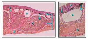
In the adult Axolotl the epidermis is pseudostratified and lacks a stratum corneum. Above the germ layer, epithelial cells are interspersed with Leydig cells. The dermis contains mucous and granular glands that are embedded within the spongy layer, which is a loose network of thin collagen fibers and fibroblasts that lie above a compact layer.
Reptiles
The skin of reptiles does not have glands to moisten it, which gives it a dry and hard appearance, it has a horny layer that contains horny scales that make it impermeable to water and resistant to desiccation. In many species, the phenomenon of molting occurs, which is the process of changing the outermost layer of the skin, necessary to allow the growth of the animal, it occurs with a variable periodicity between 1 and 12 months. Crocodiles and chelonians have ossified plates in the dermis that are called osteoderms, and have a protective function. It has two layers, dermis and epidermis, but the latter is covered by a third, almost translucent and ornate layer that is called the epidermis.
Birds
Their skin is covered with different types of feathers. Structurally, feathers are horny protuberances arising from the epidermis. They have a uropygeal gland that is located at the base of the tail and produces a fatty secretion that the animal itself distributes with its beak through the plumage to make it waterproof. This gland is developed especially in waterfowl. Some seabirds also have specialized salt glands.
Mammals
The most characteristic features of mammalian skin are hair and mammary glands. They also present specialized faneras such as horns and antlers.
Human skin
In adult humans, the skin occupies an area of 2 m² and weighs 4.1 kg. It has a thickness that ranges from 0.5 mm on the eyelids to 4 mm on the heel. It is divided into two main layers which, from surface to depth, are called Epidermis and Dermis.
Epidermis
The main cells that make up the epidermis are called keratinocytes. It also contains melanocytes that give pigmentation to the skin and Langerhans cells and lymphocytes, which are responsible for providing immune protection. The epidermis grows constantly but always maintains the same thickness due to a desquamation process. The cells located in the germinal layer divide frequently and form daughter cells that progressively migrate from the depth to the surface, where they end up detaching. In humans, the entire process takes about four weeks.
Strates
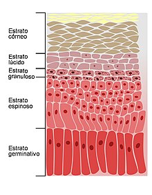
- The germinative stratum or basal layer is the deepest, formed by cylindrical cells with oval nuclei. The cells are generally arranged forming a single layer. Intercalated among the keratinocytes are some melanocytes that segregate melanine and color the skin.
- The thorny stratum is made up of polygonally shaped cells, the nuclei are round and the cytosol is of basal characteristics. It has a greater content of tonofibillas than those of the germinative stratum. The extensions of the cytosol resemble thorns, so they also receive the name of spiny cells, precisely because the tonofibillas are more numerous in such prolongations giving the shape of thorns.
- The granulous stratum consists of 3 to 5 layers of flattened cells, the cytosol contains basophile granules called keratohialin granules. Queratohialine is a precursor of keratin. When the keratinocytes reach the last layer of this stratum the epidermal cells die and when they die pour their contents into the intercellular space.
- The lucid stratum is distinguished by having a very thin area of eosinophil characteristics. Cores begin to degenerate into the outer cells of the granulous stratum and disappear into the lucid stratum.
- The corine stratum consists of anucleated keratinized flat cells, also called cornea cells. This layer is distinguished as the thickest and eosinophilic. The corine stratum is formed by flattened and dead rows that are the corneocytes. The corneocytes are mostly made of keratin. Every day layers of corneocytes are removed.
- The disjunct stratum is the continuous decamation of the cornea cells.
Dermis
The dermis is found below the epidermis, they have the peculiarity of presenting a great abundance of collagen and elastic fibers that are arranged in parallel and that give the skin the consistency and elasticity characteristic of the organ. Histologically it is divided into 2 layers:
- Papilar stratum (Papylar deeds). It is more superficial and its thickness represents 20% of the dermis. It is composed of lax connective tissue and type III collagen fibers.
- Reticular stratum (reticular demins). It is the deepest layer and corresponds to 80% of the thickness of the dermis. It consists of dense connective tissue, type I collagen fibers, elastic fibers. It contains mast cells, reticulocytes and macrophages.
The dermis is thicker than the epidermis. In it are the cutaneous annexes, which are of two types: horny (hair and nails) and glandular (sebaceous and sweat glands). It also has blood vessels and nerve endings. The structures of the dermis are the following:
- Piloss follicle. Skin structure from which the hair is born.
- Muscle piloerector. They extend from the surface dermis to the hair follicle. In situations of intense cold, stress or fear, these small muscles contract involuntarily, a phenomenon commonly known as piloerección or chicken skin.
- Nervous endings that make it possible to feel touch and sensitivity to heat, cold, pressure and pain.
- Sebaceous glands. They are glands that produce an oily substance that receives the name of sebum. The sebum covers and protects the surface of the skin and hair, avoiding dehydration. It consists of cholesterol, triglycerides, mineral salts and proteins. These glands are located in the dermis and usually secrete a hair follicle, are absent from the palms of the hands and feet.
- Sudorin glands. They secrete a liquid composed of water and mineral salts that receives the name of sweat. They open outside through small pores located on the surface of the skin. There are two types of sweat glands: ecrins and paracrins. The ecrine sweat glands are distributed in the skin of the entire body, while the apocrines are less numerous, produce a thicker secretion and are preferably located in certain areas: axilas, perine and pubic area.
- Blood and lymph vessels. The epidermis lacks blood vessels, so its nutrition depends on the vessels of the dermis that are organized in a deep blood plexus located between the dermis and the hypodermis and another superficial one from which small capillaries come.
Hypodermis or subcutaneous tissue
Sometimes also called the superficial fascia. It is located below the dermis. It is made up of loose connective tissue that has fibers to join both the dermis and underlying tissues. It contains adipocytes that serve as a fat reserve and has numerous blood vessels that supply blood to the most superficial layers of the skin. Some of the structures found in the hypodermis are the following:
- Lymphatic and blood vessels. Lymphatic and blood vessels extend through subcutaneous tissue and send small plexies for the dermis to irrigate it.
- Skin nerves. They are located in the subcutaneous tissue and send bouquets for the dermis and nerve endings attached to the epidermis.
- Skin ligaments. They are called retinacula cutis as a whole, link dermis with deep fascia, have the function of providing the skin movement through the surface of the organs, are born in deep fascia and join the dermis, they are particularly developed in the breasts.
Morphology
The surface of the skin is not smooth, it presents grooves, indentations and lines that form variable patterns depending on the sector and the individual. For example, the impressions of the ends of the fingers that are characteristic of each person.
- Folds and grooves. Less accentuated, they are always present in all individuals on the back side of certain joints, even when they are in full extension. Example: elbows, knees, fingers, wrists, etc.
- Wrinkles. They can be caused by muscle contraction, due to a movement or structural skin provisions. Example: joint folds.
- Skin pores. They are the external orifice of the output channel of a sweat gland or sebaceous gland.
Function
The skin performs different basic functions that can be grouped into five:
- Protection.
- Sensitivity. The sensitivity of the skin is due to the existence of numerous nerve endings that contain receptors for touch, heat, cold, pressure vibration and pain. The following can be distinguished:
- Meissner's shorts. They are responsible for the fine touch.
- Krause shorts. They provide the cold feeling.
- Pacini's shorts. They give the feeling of pressure.
- Ruffini shorts. They're heat sensitive.
- Merkel chops. They are responsible for the touch.
- Thermoregulation. The skin is of great importance in the control and maintenance of body temperature. This is possible thanks to the contraction or dilation of the small blood vessels that pass through it, minimizing or increasing heat loss as needed.
- Excretion and absorption of substances. One of the substances excreted by the skin is sweat.
- Vitamin D Synthesis. Although a part of the vitamin D needed by the body is obtained from food, 90% is synthesized in the skin. The synthesis process requires the presence of ultraviolet rays from solar radiation.
External morphology
Externally, what is observed is the superficial macrostructure of the skin. At first glance it appears flat and full, but in reality it presents folds, grooves, wrinkles and small prominences:
- More accented folds and grooves are always present in all individuals on the dorsal face of certain joints when they are in full extension or are in complete joints. For example, elbows, fingers, and wrists.
- Wrinkles: can be caused by muscle contraction, due to a movement, or by structural skin provisions; for example: joint folds.
- Porus cutanis: they are the external holes of the channel of output of the sweat glands and sebaceous glands. The latter receive the name of ostium (orifice) follicular.
Elasticity is one of the most relevant skin properties, and can be altered by various factors, either extrinsic or intrinsic. The most common is age. Elasticity is quantified using elastographic procedures based on various ultrasonographic techniques. Some unique diseases in which the elasticity of the skin is affected are congenital lax cutis, pseudoxanthoma elasticum, and dermatoporosity.
Pigmentation
Skin color varies depending on the number of melanosomes or melanin granules synthesized within the melanocytes. Skin is pigmented, or melanin, generated by melanocytes, which absorbs some of the ultraviolet (UV) radiation from the sun, potentially dangerous. They also contain DNA repair enzymes that help reverse the damage generated by UV rays. People who do not have the products generated by these enzymes are more likely to suffer from skin cancer. A form predominantly produced by ultraviolet light, malignant melanoma, is particularly aggressive, rapidly metastasizing and often fatal if left untreated. Human skin pigmentation varies among populations in striking ways. This has led to the classification of people based on skin color. Various medications and chemical compounds can cause changes in skin pigmentation.
The skin is the largest human organ. For example, in an adult woman, the skin has a surface area of between 1.5 and 2 square meters, most of it having a thickness of between 2 and 3 mm. Every 6.5 cm² of skin contains 650 sweat glands, 20 blood vessels, 60,000 melanomas, and more than a hundred nerve endings.
Dermatology
Dermatology is the medical discipline that studies and treats the integumentary system. Because the skin is the most visible organ, its appearance or symptom provides important clues to its diseases and also to those of other organs, such as the liver. Likewise, the skin is the most vulnerable organ, because it is exposed to radiation, trauma, infections and harmful chemicals.
Diseases
The skin can suffer from different diseases. Some of the most common are the following:
- Psoriasis.
- Seborreic eczema.
- Impetigo.
- Skin cancer.
- Vitiligo.
- Ictiosis.
- Traumatic injuries like burns.
Contenido relacionado
Coelorachis
Brachyelytrum
Beta sheet


