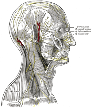
Facial nerve
The facial nerve is a mixed cranial nerve, that is, it contains both sensory and motor fibers, present in mammals including humans in which it forms the seventh cranial nerve or VII nerve. Because it is a cranial nerve, it emits two fibers, one that runs on the right side of the face and the other on the left. Part of the brain stem, just between the pons and the medulla oblongata, and controls the muscles of facial expression, as well as taste in the anterior two-thirds of the tongue. It also supplies parasympathetic preganglionic innervation to various nerve ganglia in the head and neck.
Actual origin
The facial nerve consists of two nerve fibers, the facial nerve proper and the intermediate nerve or Wrisberg's intermediary. The facial proper arises from motor neurons in the facial nucleus which is situated ventrally in the inferior or caudal portion of the pons. The axons exit dorsally and medially backward toward the nucleus of the nerve. external oculomotor (VI cranial nerve), surround the said nucleus to emerge forward, together with the intermediary, at the level of the cerebellar angle just between the external oculomotor and the vestibulocochlear nerve.
The actual origin of its sensitive part is the nucleus of the solitary tract. This nerve leaves the skull through the stylomastoid foramen, and after giving a combined branch to the posterior belly of the digastric muscle and the stylohyoid muscle, it goes towards the center of the parotid gland (the salivary gland located in front of the ear). It lies in the plane of the fascia and divides the parotid gland into superficial and deep portions.
Both the motor root of the facial and the intermediary nerve of Wrisberg, after passing through the cerebellopontine angle, go to the internal auditory canal, where they enter accompanied by the auditory nerve. Afterwards, the facial and the nerve of Wrisberg are They enter the fallopian aqueduct or facial canal of the temporal bone and travel a course of two bends. Shortly after traversing this aqueduct, at the first bend, Wrisberg's nerve ends in a nerve ganglion called the geniculate, which, in turn, emits a branch that, leaving the ganglion, mixes with the facial nerve itself. From the geniculate ganglion, the facial becomes a mixed nerve, with the motor fibers that belong to it, and the sensory fibers that come from the intermediary nerve of Wrisberg. Fibers from the parasympathetic nervous system accompany both nerves during the journey.
- The cervicofacial branch, which in turn is divided into:
- maxillary, that inervates the buccinator muscle and the orbicular of the lips
- mandibular, which leads a parallel path to the jaw
- cervical, inervating the skin muscle of the neck
- The temporofacial branch, which is divided into two:
- a temporary branch, which iners the frontal muscle and facial muscles below the zygomatic bow, and
- the cigomatic branch that ends up inervading the nose and the upper lip
The facial nerve travels inside the temporal bone, entering it through the Internal Auditory Canal (IAC), in its anterosuperior quadrant.
Within the temporal bone, the facial nerve divides into 3 segments or portions.
The first segment called Labyrinth begins when the nerve exits the Internal Auditory Canal and ends at the first Knee or Elbow of the Facial (Geniculate Ganglion), this segment is related to the Labyrinth and the Cochlea, from the Geniculate Ganglion comes a branch that It is the Greater Superficial Petrosal Nerve.
The second segment called Tympanic Cavity begins in the Geniculate Ganglion and ends in the second Facial Knee (Tyramidal), runs through the medial wall of the Tympanic Cavity from anterior to posterior, relating to the Cochleariform Process, Oval Window and External Semicircular Canal.
The third segment, called the Mastoid, begins at the second Facial Knee and ends at the Stylomastoid Foramen, from this segment emerges the chorda tympani nerve (which innervates the tongue).
In its entire intratemporal portion, the facial nerve is covered by a sheath called the Fallopian Aqueduct.
After leaving the temporal bone through the Stylomastoid Foramen, the facial nerve passes through the parotid gland in its thickness (dividing it into a superficial and a deep lobe) and divides into a Temporofacial and a Cervicofacial plexus, innervating the muscles of the the face and neck.
Functions
The facial nerve is a mixed nerve with mainly motor function, and with a special sensory portion that collects taste impressions from the anterior two-thirds of the tongue. In detail:
- Motor function: It is the somatic motor nerve of the skin muscles of the face and neck. It's the facial nerve itself. The motor root of the facial originates in the nucleus located in the upper protuberance (on the facial car)..
- Sensory function: Collect the sense of taste of the previous two thirds of the tongue. It's the Intermediate nerve Wrisberg. The sensitive root originates in the core of the upper part of the solitaire fascicle and at the top of the gray wing.
- General sensitivity function: Collect the sensitivity of the skin from the back of the ear (area of Ramsay Hunt) and for the external hearing duct.
- Visceral motor function: Because it is part of the cranial parasympathetic by possessing secret and vasodilating fibers, inervar the lagrimal glands, the sweaters of the face, the sublingual salivales and submaxillary, the auditory artery and its branches and the mucosa of the nasopharyngeal palate and nasal pits.
Semiology
The function of the facial nerve is explored by inspecting the physiognomic features of the face, in particular observing the symmetry of the face reflected in the labial commissures and eye opening with blinking. Tearing is a sign present with drooping of the lower eyelid of the affected eye. For the motor examination of the upper rami, the subject is asked to wrinkle the forehead and open and close the eyes. Motor exploration of the lower rami is accomplished by asking the individual to whistle or blow to observe the characteristic labial symmetry.
The sensory function of the facial nerve is explored with the taste of the anterior two thirds of the tongue and with the sensitivity of the auricle.
Pathology
Loss of motor function of the facial nerve produces hypotonia and weakness, mainly in the face, which manifests with an apparent facial asymmetry due to eyebrow drooping, decreased blink rate, and when the subject blinks, it can be seen the upward deviation of the eyeball at the same time that the eyelid closes the eye. The lower eyelid often becomes everted, a condition known as ectropion, forcing tearing. The lip corner deviates towards the healthy side.
Contenido relacionado
Capsule
Cytidine
Ornithine
