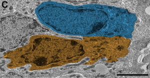
Ependymal cell
The ependymoglial cells, ependymal cells or tanycytes, are part of the group of neuroglial cells of nervous tissue. They line the ventricles of the brain and the ependymal duct and display multiple cell subtypes identifiable by their morphology. Ependymal cells possess latent neural stem cell potential.
Embryology
They derive from the neural ectoderm of the developing nervous system, therefore they are not epithelial cells, because they lack a basal membrane and intercellular junctions characteristic of epithelial tissue.
They are descendants of embryonic neuroepithelial stem cells, located in the ventral neural tube.
Structure
Ependymal cells are cylindrical to cuboidal cells that line the ventricles of the brain and the central canal of the spinal cord.
The microscopic architecture of the ciliated ependymal cells that contact the lumen is heterogeneous with cells showing a typical cuboidal ependymal cell morphology and others with a tanycytic morphology.
In addition, a third, less numerous type of ependymal cell, the radial ependymal cell, is visible. Radials share the morphology of the cytoplasm, and often the nucleus, with the other cells, but they have a long basal process. These ray cells are found at the dorsal or ventral pole of the ependymal layer, with their basal process oriented along the dorso-ventral axis.
Ultrastructure
Your cytoplasm contains a large number of mitochondria.
They have nestin and vimentin intermediate filament bundles, which are commonly associated with immature neural cells.
Ultrastructural analysis revealed heterogeneous morphology among these light-contacting hair cells. Cuboid cells show oval nuclei, clear cytoplasm, and 1-3 cilia.
tanycytes have irregular nuclei, dark cytoplasm, and processes.
The radial cells have an oval and irregular nucleus, clear cytoplasm, 1-3 cilia, and long processes.
In some regions these cells are ciliated, a characteristic that facilitates the movement of cerebrospinal fluid.
In the embryo, the processes that arise from the cell body reach the surface of the brain, but in the adult the processes are reduced and have only close endings.
In places where the neural tissue is thin, these cells form an inner limiting membrane that lines the cerebral ventricle and an outer limiting membrane below the pia mater.
At the level of the cerebral ventricles, these cells undergo modifications and form the choroid plexus, whose mission is to secrete and preserve the chemical composition of the cerebrospinal fluid.
Tanicitos
Ep= ependy.
III-V= third ventricle.
Tanycytes are specialized ependymal contact cells between the third ventricle and the median eminence of the basal hypothalamus.
Its proximal pole lies within the wall of the third ventricle, and its long cellular process projects into the ventromedial hypothalamus.
Types of tanycytes
There are four types of tanycytes: α1, α2, β1, and β2. The α2 and β1 tanycytes are located in the inferior lateral wall of the third ventricle; they have extended cellular processes that contact neurons in the arcuate nucleus (AN), particularly neuropeptide Y (NPY) neurons and pro-opiomelanocortin (POMC) neurons.
β2 Tanycytes form the cerebrospinal fluid barrier of the median eminence (ME) and its extended processes come into contact with vessels devoid of the blood-brain barrier and are sometimes in direct contact with microvessels present in the EM.
They have known functions, and a role for transporting substances between both structures has been attributed to them.
Ependymocyte cell turnover
Ependymal cells (EpC) exhibit relatively low basal mitotic activity. The use of BrdU, which is incorporated into DNA in cells in S phase, resulted in the labeling of 16-24% of ependymal cells in one month.
In adult mammals, new neurons are produced continuously. The ependymal cell layer forms adult neurogenesis niches in the subventricular zone (SVZ), at all points along the neuraxis.
Mechanical damage to the central canal triggers a strong reactive increase in ependymal cell (EpC) proliferation. These mammalian reactive EpCs re-express the properties of neural stem cells after spinal cord injury.
Contenido relacionado
Unona
Bradypsychia
Wikiproject:Animals


