Endocytosis
Endocytosis is a key mechanism by which cells introduce large molecules, extracellular particles and even small cells, engulfing them in an invagination of the eukaryotic plasma membrane, forming a vesicle that eventually detaches membrane to be incorporated deep into the cytosol. It is presented as a contrary case to the events of exocytosis.
Endocytosis mechanisms maintain control of the entry and exit of macromolecules and particles through active transport, and play very important roles in various processes such as organism development, immune response, neurotransmission, intercellular communication, signal transduction and maintenance of cellular homeostasis and of the organism in general.
History
The term "endocytosis" comes from the Greek ἐνδο endo: 'inside', κύτος kyto-: 'cell' and -ō-sis: 'process', and was proposed by the cytologist de Duve in 1963 with the purpose of including in the same term the ingestion of large particles and the absorption of fluids or macromolecules in small vesicles..
Types of endocytosis
There are multiple endocytosis mechanisms that can be grouped into two broad categories: phagocytosis and pinocytosis.
When endocytosis results in the capture of larger particles it is called phagocytosis ("the cell gobbles"), and when only portions of fluid are captured, it is called pinocytosis ("the cell drinks"). Pinocytosis mostly captures substances found in the extracellular medium indiscriminately except in receptor-mediated endocytosis, in which it only captures those molecules that bind to said receptor; that is, it is a very selective type of pinocytosis. In all cases, the plasma membrane folds inward, forming a kind of pocket that will give rise to a vesicle for the subsequent migration of its contents into the cell.
Phagocytosis
The mechanism of phagocytosis consists of the introduction of a solid into the intracellular medium. These can be: a large molecule, a particle or a microorganism, such as; bacteria, atmospheric dust, cells or cellular debris.
First, the particle rests on an area of the cell membrane, producing an invagination, upon entering the cell, which is strangled, leaving what is entered wrapped in the plasmatic membrane, constituting a vesicle called a phagosome, this is created by means of the cytoskeleton. The phagosome fuses with the lysosomes, forming a phagolysosome, the organelles responsible for cellular digestion. It is generally a defense function when there are aggressions from outside or when organic tissues are renewed.
It is important to know that binding of the particle to receptors on the phagocytic cell surface triggers extension of the pseudopodia, an actin-based movement of the cell surface; the pseudopods surround the particle and their membranes fuse to form a large intracellular vesicle (> 0.25 micrometers (μm) in diameter) called a phagosome, these are they fuse with lysosomes, generating phagolysosomes, where the ingested material is digested by the action of lysosomal acid hydrolases; during phagolysosome maturation, some of the internalized membrane proteins are recycled to the plasma membrane.
Many amoebas use phagocytosis to capture food particles, such as bacteria or other protozoa; meanwhile in multicellular animals, the main functions of phagocytosis consist of providing a defense against invading microorganisms and removing aged or damaged cells from the body, specifically in mammals, phagocytosis is the function of two types of white blood cells, macrophages and neutrophils, Often referred to as "professional phagocytes", they have extremely important roles for the body's defense systems by eliminating microorganisms from infected tissues, in addition, macrophages, eliminate old or dead cells from tissues throughout the world. body, are responsible for the removal of more than 1011 aged blood cells on a daily basis.
Endosomes
They are found inside the cell, they are intermediate compartments, they have the function of destroying materials that have undergone endocytosis, phagocytosis or autophagocytosis and for the formation of lysosomes.
Pinocytosis
There are four types of basic pinocytic mechanisms: macropinocytosis, clathrin-mediated endocytosis, caveolae-mediated endocytosis, and clathrin- and caveolae-independent endocytosis.
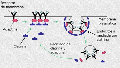
Cells that carry out pinocytosis present a region in the plasma membrane that is covered by a protein (clathrin) on its cytosolic face, so that when the molecule is deposited on that region of the membrane it learns its shape. a coated shell that surrounds it, it will later lose that coating in order to be digested by lysosomes. In this process, the cell incorporates fluids in which proteins may be dissolved.
The endocytic part of the cycle usually begins in clathrin-coated pits, which generally occupy about 2% of the total area of the plasma membrane, the lifetime of these pits is short (about one minute after being formed), invaginates inside the cell and squeezes to form a clathrin-coated vesicle; It is estimated that about 2500 clathrin-coated vesicles leave the plasma membrane of a cultured fibroblast every minute. These vesicles are even more transient than coated cavities, whose lifetime is a few seconds after their formation, so they give off their coat and are capable of fusing with early endosomes; because the extracellular fluid is trapped in clathrin-coated pits, as they invaginate to form coated vesicles, any dissolved substances in the extracellular fluid are internalized, a process termed fluid-phase endocytosis.
The rate at which the plasma membrane performs this process varies between cell types, but is often incredibly large; For example, a macrophage ingests 25% of its own volume of liquid every hour.
It is important to know that there are other, less well-known mechanisms by which cells can form pinocytic vesicles without forming cavities and clathrin-coated vesicles; one of these pathways begins in caveolae (Latin word meaning "small cavities"), which is recognized for its ability to transport molecules through endothelial cells that form the inner lining of blood vessels, caveolae are present in the plasma membrane of most cell types, and in some of these appear as deeply invaginated jars in the electron microscope, they are thought to form from lipid rafts, patches of the plasma membrane that are especially rich in cholesterol, glycosphingolipids and GPI-anchored membrane proteins, its main structural protein is caveolin, a multipass integral membrane protein that is a member of a heterogeneous family of proteins.
Caveolae are thought to invaginate and take up cargo proteins by virtue of the lipid composition of the calveolar membrane, rather than assembling a cytosolic protein coat, they detach from the plasma membrane and may deliver their contents to cell-like compartments endosomes or (in a process called transcytosis) to the plasma membrane on the opposite side of a polarized cell, some viruses that attack animals enter cells in vesicles derived from caveolae, producing an infectious cycle.
Receptor-mediated endocytosis
Cargo receptor-mediated endocytosis occurs when receptors accumulate in well-defined regions of the cell membrane, this transport mechanism allows selective entry of molecules into the cell.
The opposite process to endocytosis is exocytosis. Endocytosis and exocytosis are processes of entry and exit of substances that allow maintaining the homeostasis of the cell, they are regulated by it in order to keep the plasmatic membrane constant, since they allow its regeneration since the phagosomes that contain the phagocytosed molecules are They are formed from the plasma membrane and when the cellular digestion process carried out by the lysosomes ends, the cellular excretion is carried out by exocytosis, recovering the membrane used for the formation of the phagosome.
The vesicle formed is called an endosome, which will fuse with a lysosome where intracellular digestion of its contents occurs. Specialized phagocytic cells have membrane receptors that when they come into contact with cell fragments induce the formation of pseudopodia that cover it, forming phagosomes. In the human species, the main cells with the capacity to phagocytose (phagocytes) are the polymorphonuclear neutrophils (PMN), which are short-lived. Mononuclear phagocytes (SFM) made up of circulating monocytes and tissue macrophages also participate.
Subsequently, the lysosomes fuse with the wall of the phagosomes, pouring out their hydrolytic enzymes that act at acidic pH (close to 5) and carry out the degradation of the cell fragments. That part that cannot be digested will be eliminated abroad by exocytosis in the process known as cellular defecation.
Clatrin-mediated endocytosis
It is produced in all classes of cells in mammals and performs important functions such as nutrient absorption and intracellular communication. This process is the main mechanism of internalization of macromolecules and components of the plasmatic membrane. It is considered as a drive mechanism involving more than fifty different assembly protein components, restricted to a single location on the plasma membrane. Located in an ordered and hierarchical temporal pathway.
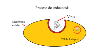
Most enveloped viruses make use of a mechanism of endocytosis in order to enter a permissive cell and initiate infection. Clathrin endocytosis is a field of research that has sparked great interest in recent times.
Caveolin-mediated endocytosis
It is a process regulated by signaling complexes through GTPAase. This pathway is used by pathogens to escape degradation by lysosomal enzymes. Caveolae are bottle-shaped invaginations of the membrane, between 50 and 100 nm in size, which are lined by caveolin.
This process is fundamental to an immune response, intercellular communication, signal transduction, homeostasis, both cellular and that of the whole organism; in early 2006 a lipid-based marker protein, called flotillin-1, was found to be involved in a novel endocytosis pathway.
Neurons use the mechanism of endocytosis to recover a released neurotransmitter in the synaptic gap, to be reused. Without this process, there would be a failure in the transmission of the nerve impulse between neurons.
An example of receptor-mediated endocytosis is when human cells take in cholesterol which is used in the synthesis of membranes and also as a precursor for other steroids.
This process has a strong relationship with the eradication of HIV-1 infection. Caveolin-1 mediated HIV-1 uptake is an intrinsic restriction mechanism present in Langerhans cells in humans that prevents this infection. Taking advantage of this internalization pathway has the potential to develop strategies to combat the transmission of this disease.
Small caveolin-coated invaginations of the plasma membrane, caveolae, contain some receptor proteins and are used for certain types of receptor-mediated endocytosis, but the latter usually occurs via clathrin-coated cavities and vesicles, process is similar to the packaging of lysosomal enzymes by mannose 6-phosphate (M6P) in the trans-Golgi, most M6P receptors are located in the trans-Golgi, but some are found on the cell surface and the enzymes secreted lysosomal cells bind to these receptors and are returned to cells by this type of endocytosis; in other words it can be said that transmembrane receptor proteins that are internalized from the cell surface during endocytosis are sorted and recycled back to the cell surface, much like the recycling of M6P receptors to the plasma membrane and trans- Golgi.
Some receptors cluster on clathrin-coated pits even in the absence of ligand, others diffuse freely in the plane of the plasma membrane but undergo a conformational change upon ligand binding, so that when the receptor-ligand complex diffuses in a clathrin-coated pit, it is retained there. Two or more types of receptor-bound ligands, such as LDL and transferrin, can be observed in the same cavity or coated vesicle, it is thought that regulated polymerization of clathrin causes the cavities to expand and eventually form clathrin-coated vesicles.
Exocytosis and endocytosis
Vesicle endocytosis are fundamental biological events, which have a certain relationship with exocytosis, in the release of neurotransmitters within the synapse, it is based on a sustainable cycle between these two processes. The exocytosis vesicle, making use of the plasma membrane, releases vesicular content to perform various and important functions. Among them are: participation in the secretion of neuron transmitters, an essential process for brain functions; neuronal secretion of peptides (such as neuropeptide Y) and hormones (eg, vasopressin, oxytocin), provides regulation of work and mental state; the secretion of insulin from pancreatic cells to regulate the level of glucose in the blood; the secretion of catecholamines and peptides (involved in the stress response); in addition, exocytosis performed by blood cells for immune responses.
The functions of exocytosis in the organism are of vital importance. The role of endocytosis also acquires great relevance since after continuous exocytosis, endocytosis is required to maintain the structure of the nerve terminal, in addition to ensuring the functional availability of synaptic vesicles. The vesicle membrane and proteins are recovered from the plasma membrane by this process. To this end, endocytosis performs vesicle recycling and protects secretory cells from swelling or shrinking.
Two models are proposed for the operation of this procedure, the first suggests that the vesicles undergo a reversible sequence of exo-endocytosis, whereby the biochemical identity of the vesicles is preserved both during and after attachment. transition between the vesicle membrane and the plasma membrane. The other model states that during exocytosis, the vesicle membrane is fully added to the plasma membrane and recovery occurs later at a different point on the plasma membrane by clathrin-mediated endocytosis.
Contenido relacionado
Heterocephalus glaber
John Franklin Enders
Alopecurus


