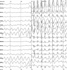
Electroencephalography
The electroencephalogram (EEG) is a neurophysiological examination that is based on the recording of cerebral bioelectrical activity in basal conditions of rest, in wakefulness or sleep, and during various activations (usually hyperpnea and intermittent light stimulation) using electroencephalography equipment.
History
Richard Birmick Caton (1842-1926), a physician from Liverpool (United Kingdom), presented in 1875 his findings on bioelectric phenomena in the cerebral hemispheres of mice and monkeys, exposed by craniectomy.
In 1912 Vladimir Pravdich-Neminsky published the first EEG and evoked potentials of dogs.
In 1920 Hans Berger began his studies on electroencephalography in humans.
Cortical activity can be studied with different electrophysiological techniques, such as:
- the registration of units,
- local field potentials (LFP),
- electrocorticogram (ECoG),
- the magnetoencephalogram (MEG),
- electroencephalogram (EEG).
Amplitude-integrated electroencephalography (AEEG) is a method of continuous monitoring of brain function, designed to analyze changes and trends in brain electrical activity, as well as detect paroxysmal activity.
Normal EEG during wakefulness
Brain waves that are recorded in the waking state are called background activity.
Background Activity
- Gamma waves: 25 to 100 Hz.
- Beta waves: 14-30 Hz.
- Alpha waves: 8-13 Hz.
- Onda theta: 3-7 Hz.
- Delta waves 0-4 Hz.
Brain activation methods
- Hyperpnea
- Intermittent luminous stimulation
- Visual stimulation
- Hearing stimulation
- Subjective stimulation
- Nociceptive stimulation
Normal EEG during sleep
Sleep-specific graphoelements
- Acute wave to the vertex
- Positive occipital acute wave
- Hustle of sleep
- Complex K
- Sleep delta activity
- Alerts
Sleep stages
- NREM Phase I
- NREM Phase II
- NREM Phase III
- NREM Phase IV
- REM
Staging of Rechtschaffen and Kales
Abnormal EEG Findings
- Grafoils EEG abnormals
- Intermittent EEG anomalies
- Regular EEG anomalies
- Continuous EEG anomalies
EEG indications
- Inflammatory encephalopathy
- Metabolic encephalopathy
- Toxic encephalopathy
- Connatal encephalopathy
- Hypoxic disease
- Coma
- Diagnosis of Encephalic Death
- Brain tumors and other occupant space lesions
- Dementia
- Degenerative diseases of the central nervous system
- cerebrovascular disease
- Cranioencephalic trauma
- Cephalea
- Vertigo
- Psychiatric disorders
- Epilepsy
In general terms:
The EEG is indicated in any paroxysmal phenomenon in which a cause of cerebral origin is suspected —and in any situation of cerebral dysfunction—, especially in the symptomatic phase. In cases in which there are epilepsies refractory to treatment and epilepsy surgery planning is necessary, video-EEG monitoring is essential. If there are difficulties in locating the focus or the lesion is not easily accessible, intracranial EEG studies can also be performed using deep electrodes (this is called stereoelectroencephalography).
Contenido relacionado
Inflammation
Fever
Luc Montagnier


