Ear
The ear is a sensory organ that allows us to perceive sounds, forming the sense of hearing, and in mammals it is also in charge of balance. The ear can be divided for study into three sections: external ear, middle ear, and inner ear.
The perception of sound is a complex phenomenon that develops in several stages. In the first place, the capture of sound waves is carried out thanks to the tympanic membrane. Secondly, the mechanical signal collected by the eardrum must be transformed into nerve impulses, a process that occurs in the inner ear. Third, nerve impulses through the auditory nerve are sent to the brain to be processed in the cerebral cortex.
The auditory spectrum, that is, the range of frequencies that the ear can perceive, varies depending on the animal species. Humans can detect sounds between 0 and 140 decibels with a frequency range between 40 and 20,000 hertz. Whales can perceive infrasound with a frequency below 40 hertz. Some carnivorous animals such as dogs are capable of detecting ultrasound with a frequency greater than 20,000 hertz that a human is unable to hear.
External ear
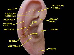
The external ear is made up of two parts: the auricle and the external auditory canal.
- The head pavilion is made up of skin-coated cartilage, its most important portions are the helix and the earlobe.
- The external auditory duct measures about 2.5 cm in length and extends from the atrial pavilion to the eardrum, in this path it passes through the temporary bone of the skull. It contains ceruminous hairs and glands that produce the cerumen, making it difficult to enter foreign bodies or dust through the duct.
Middle ear
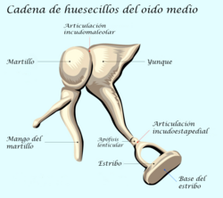
The middle ear is an air-filled cavity that is separated by the eardrum from the external auditory canal and communicates with the inner ear through two small openings: the oval window and the round window. Inside the middle ear is a chain of ossicles joined together by synovial-type joints. They are the smallest bones in the body and are called the malleus, anvil, and stirrup. The middle ear is connected to the nasopharynx by a small tube called the pharyngotympanic tube or Eustachian tube.
- The eardrum or timponic membrane is transparent and separates the timponic cavity from the external auditory duct. It has oval shape and a diameter of about 1 cm. Two portions are differentiated in the timpian membrane; Pars Tensis or tense portion (or stretched) and Pars Laxus or lax portion. It consists of three layers:
- Intermediate layer: composed of a fibroconect tissue formed in semitotality to the tympanic membrane, composed of collagen in addition to elastic fibers and fibroblasts.
- Corinal stratum: it is skin that covers the outer surface of the tympanic membrane without hairs and glands, composed of epidermis that sits on a layer of sub-bepidermian connective tissue.
- Mucosa: it covers the inner surface of the intermediate layer of connective tissue, with an epithelium of simple plane characteristics.
- Timpian cavity is a small space full of air that is located in the temporary bone, inside it is a chain of bones that transmit the vibrations generated in the eardrum to the inner ear. It is covered by mucosa and a simple flat-like epithelial foil on its back, but in the previous one is seen a pseudo-stratecerated cylindrical-type epithelial with caliciform cells. The tymponic cavity, also called the timponic box, is made up of 6 walls, an external that corresponds to the tympanic membrane, an internal wall that is in relation to the promontory, a posterior wall that communicates with the mastoids, an earlier wall that communicates through the Eustaquio tube the nasopharynx, a superior wall or ceiling and a lower related to the jugular vein, all these important details of the mid-ear.
- Huesecillos of the ear are three tiny bones called hammer, yunque and estribo, in some texts four bones are cited as the lenticular apophysis of the yunque as an independent bone. The vibrations generated in the eardrum are transmitted through the chain of bones from the tympanic membrane to the oval window. In the oval window the head of the stirrup presses on the fluid content in the inner ear; thus the eardrum and the chain of bones act as a mechanism that transforms the vibrations of the air into vibrations of the fluid.
- Eustaquio's tube communicates the timpian cavity with nasofaringe, measures in the adult human being about 4 cm long. It is composed of a bone portion and another cartilageous, it has an epithelial foil composed of nasopharyngeal epithelium or pseudoestratified ciliated cylindrical epithelium with abundant caliciform cells. It serves to equal the pressure on both sides of the eardrum.
Inner ear
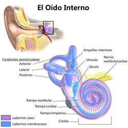
The inner ear or labyrinth is located within the temporal bone of the skull. There is a bony labyrinth and a membranous labyrinth. The bony labyrinth is nothing more than the bony capsule that surrounds the membranous labyrinth, and the latter consists of a system of hollow ducts that contains a liquid called endolymph inside. In the space between the bony labyrinth and the membranous labyrinth is the perilymph.
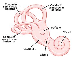
The inner ear is divided into two distinct portions. The first is for maintaining balance and is made up of the vestibule and semicircular ducts. The second function is hearing and is made up of the cochlea or snail. The vestibule is divided into two sectors called the utricle and the saccule, while the cochlea or snail contains the organ of Corti responsible for transforming the mechanical energy of the sound waves into electrical impulses that are subsequently transmitted to the brain through the auditory nerve or vestibulocochlear nerve.
- Semicircular ducts are three small arched ducts with a semi-circle shape located at different spatial planes. Semicircular channels leave the lobby and have the function of contributing to the maintenance of the balance of the head and body.
- The cochle or snail is a spiral-shaped duct that receives its name by remembering the shell of a snail. It is divided into three portions:
- Cochlear conduct, also called average ramp. It is full of a liquid called endolinfa.
- vestibular branch. It ends in the oval window and is filled with a liquid called perilinfa.
- Tymponic hammer. It ends in the round window and is also full of perilinfa.
These portions are separated from each other by two membranes. The vestibular or Reissner membrane serves as a separation between the cochlear duct and the scala vestibularis, while the basilar membrane serves as a separation between the cochlear duct and the scala tympanica. Along the basilar membrane is the organ of Corti, which contains about 16,000 cells with cilia that are the receptors for hearing.
The cochlear duct is filled with a fluid called endolymph that is rich in K (161 mmol/l) and poor in Na (1 mmol/l) and calcium (0.02 mmol/l). The tympanic and vestibular scala contain a different liquid called perilymph whose ionic concentrations are the inverse, it is rich in Na and poor in K.
The vestibular membrane is so thin that it does not hinder the passage of sound vibrations from the vestibular ramp to the median ramp. Therefore, in terms of sound transmission, the vestibular and medial ramps are considered as a single chamber. The importance of the vestibular membrane depends on its preservation of endolymph in the median ramp necessary for normal hair cell function.
Organ of Corti
It is part of the inner ear and is located in the cochlea or snail, sometimes called the spiral organ, and plays a fundamental role in the hearing process. It is made up of a thickened epithelium with complex characteristics. It has two types of cells: hair cells and supporting cells.
- Cochlear ciliated cells: they have the function of transforming physical acoustic signals to cortilinphatic mechanical acoustic signals, and of these to electrochemical signals directed to the auditory receptor area of the cerebral cortex (41 and 42 of Brodman). Sensory mechanorreceptocytes, with a row of internal ciliated cells and four rows of external ciliated cells.
- Brass cells: are differentiated cells that rest on a basal membrane, there are six types called by their microstructure as internal limiting cells, internal pharyngeal cells, internal spinal cells, external spinal cells, external pharyngeal cells and external limiting cells.
Audition process
For hearing to occur, sound waves must travel through the external auditory canal to reach the eardrum. The vibration of the tympanic membrane is transmitted through the ossicles of the middle ear, passing from the malleus to the incus and from this to the stirrup. The stirrup transmits vibrations to the perilymph of the inner ear through the oval window. In the cochlea, the mechanical energy of acoustic signals is transformed into electrical impulses that are transported through the acoustic nerve to the temporal region of the cerebral cortex where they are processed. Therefore it could be said that the organ with which we actually hear is the brain. with hearing. These people, however, have all parts of the ear and the auditory nerve in good functional condition, so the deficiency in the ability to discriminate sounds is due solely to brain damage.
Vascularization
- External hearing. It is done through the anterior atrial artery and the posterior atrial artery (inner carotid artery branches).
- Heard half. It is carried out through the anterior timpian artery (race of the internal maxillary artery), caroticotimpanica artery (brand of the internal carotid), upper timponic artery, superficial petrosa and artery of the Eustaquio tube (branches of the medium Meníngea artery), lower timpian artery (brand of the ascending phary artery), artery
- Internal hearing. The vascularization of the membrane labyrinth comes from the labyrinth artery that can be a branch of the middle cerebelous artery or emerge directly from the basilar artery. The labyrinth artery is divided into several branches, the previous vestibular artery, the cochlear artery and the vestibulococlear.
Hearing Related Health Problems
There are different health problems related to hearing, one of the most frequent is hearing loss or hearing loss, which in many cases is induced by chronic exposure to noise with an intensity greater than 85 decibels. Other pathologies are: pathologies of the external auditory canal, ruptured eardrum, otitis, ototoxicity, otosclerosis, cholesteatoma, Ménière's disease, vertigo, tinnitus and acoustic trauma.
Society
In different cultures, ears have been adorned with jewels piercing the lobe of the ear. Ornaments have also been placed to stretch and enlarge the lobes as an ornament.
Ear injury has been around since Roman times as a method of reprimand or punishment.
The auricle has an effect on facial appearance. In Western societies, protruding ears (present in approximately 5% of ethnic Europeans) have been considered unattractive, particularly if they are asymmetrical. The first surgery to reduce the projection of prominent ears was published in the medical literature by Ernst Dieffenbach in 1845, and the first case report in 1881.
In culture
Pointed ears are a feature of some folklore creatures such as the bogeyman, the Brazilian curupira, or the Japanese ground spider. It has been a feature of characters in ancient art such as that of Ancient Greece and medieval Europe Pointed ears are a common feature of many creatures in the fantasy genre, including elves, fairies, goblins, hobbits, or orcs. They are also a feature of creatures in the horror genre, like vampires. Pointed ears are similarly found in the science fiction genre; for example, between the Vulcan and Romulan races of the Star Trek universe.
Other animals
The pinna helps direct sound through the ear canal to the eardrum. The complex geometry of the ridges on the inner surface of some mammalian ears help to precisely focus the sounds produced by prey, using echolocation cues. These ridges can be thought of as the acoustic equivalent of a Fresnel lens and can be seen in a wide range of animals, including the bat, aye-aye, bushbaby, eared fox, mouse lemur, and others.
Some large primates such as gorillas and orangutans (and also humans) have undeveloped ear muscles that are non-functional vestigial structures, but are still large enough to be easily identifiable. A muscle capable of moving the ear, for whatever reason, has lost that biological function. This serves as evidence of homology between related species. In humans, there is variability in these muscles, such that some individuals can move their ears in various directions, and it has been suggested that others may achieve such movement through repeated trials. In such primates, the inability to move the ear is mainly compensated by the ability to easily turn the head in a horizontal plane, an ability not common to most monkeys.
Only vertebrate animals have ears, although many invertebrates detect sound using other types of sense organs. In insects, the tympanic organs are used to hear distant sounds. They are found on the head or elsewhere, depending on the insect family.
Contenido relacionado
Speleothem
Clematis flammula
Regajal-Mar de Ontígola Reserve


