Dinoflagellate
The dinoflagellates (Dinoflagellata, Dinophyta or Pyrrhophyta) are a large group of flagellate protists, with some 2,400 known species. The name comes from the Greek dinos, to turn, and from the Latin, flagellum, whip, describing the rotary movement of these organisms. These microorganisms are unicellular (although they can form colonies) and are part of the freshwater (about 220 species) and marine (the rest) phytoplankton. Approximately half are photosynthetic and have pigments with chlorophyll a and c2 and carotenoids. As their nutrition is mainly autotrophic, they are primary producers, which is why, together with diatoms and other groups of phytoplankton, they constitute the primary trophic level in the aquatic food chain. Certain photosynthetic species such as zooxanthellae are endosymbionts of invertebrate animals such as corals, anemones and clams, and marine protozoa developing a mutualistic relationship with coral reefs. Others are heterotrophs or mixotrophs, feeding on other dinoflagellates, protozoans and diatoms, and some forms are parasitic (see for example Oodinium and Pfiesteria). distributed according to temperature, salinity and water depth. Some dinoflagellates can emit light through bioluminescence, others are responsible for red tides and harmful algal blooms (HABs).
Features
Morphology
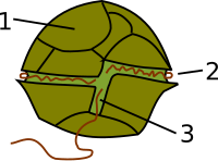
Most dinoflagellates are between 50 and 500 µm in size, which is why they are considered part of the phytoplankton, although Noctiluca can reach up to 2 mm in diameter. They are unicellular, although as an exception, some species can form colonies or pseudocolonies. The most characteristic feature of dinoflagellates is the presence of two dissimilar flagella that provide them with characteristic movements. One of them is wavy and surrounds the cell transversely, called the transverse flagellum and allows it a distinctive rotating movement, from which comes the name dinoflagellate (from the Greek dinos, turning). The other is located longitudinally on the posterior side, functions as a rudder and is responsible for its vertical movement, this is called the longitudinal flagellum.
In the dinocontas species, the flagella are housed in two grooves, called the cingulum, the transverse one, and the sulcus, the longitudinal one. The flagella emerge from the intersection of the two grooves. The basal dinoflagellates have desmocontas cells, do not present a cingulum or sulcus, and the flagella emerge from a pore located in the apical part. In this case, one of the flagella is anterior and resembles the wavy transverse flagellum of typical dinoflagellates.
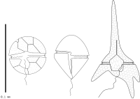
Dinoflagellates have a complex cell covering called amphysma, made up of flat vesicles called cortical alveoli. Morphologically, two types of dinoflagellates are distinguished: thecates and nudes. In the theca forms, the alveoli are supported by interlocking cellulose plates that make up a kind of armor called theca. The theca or cell wall covering exhibits various shapes in external morphology depending on the species and sometimes the stage of the dinoflagellate. The forms without armor, "atecate" or "naked" they tend to be brittle and easily deformed, whereas the cell wall of armored dinoflagellates is more rigid and inflexible. Fibrous extrusomes are also found in many species.
Chloroplasts
About half of dinoflagellates have chloroplasts and the rest are heterotrophs (phagotrophs or osmotroph parasites). Although some species with chloroplasts are fully autotrophic, most are mixotrophic, combining autotrophic and heterotrophic nutrition. The dinoflagellate group includes chloroplasts from at least six different sources. The chloroplasts of ancestral dinoflagellates probably derive from secondary endosymbiosis of a red alga. Later some groups of dinoflagellates replaced them by chloroplasts from other groups of algae by subsequent secondary or tertiary endosymbioses, including chloroplasts from Chlorophyta, Heterokontophyta, Cryptophyta, and Haptophyta.
Typical photosynthetic dinoflagellates (of the peridinial type) have disk- or rod-shaped chloroplasts, thylakoids usually in groups of three, several types of pyrenoids, and some specific xanthophylls. Pigments include chlorophylls a and c2, peridinin (a type of pyrenoid unique to dinoflagellates), β-carotene, and small amounts of dinoxanthin and diadinoxanthin. The different combinations of pigments give them a yellow, yellowish-brown, brown, blue-green, etc. coloration. The pyrenoids are found next to the chloroplast and as reserve products they use starch, produced outside the plastid, and oils. The chloroplasts are surrounded by three membranes (in some cases two), which suggests that they probably come from the secondary endosymbiosis of some algae, which, due to the types of chlorophyll they contain, is assumed to be a red algae.
However, some species have chloroplasts with different pigmentation and structure, some of which retain a nucleomorph. This suggests that these chloroplasts were incorporated by various endosymbiotic events involving already colored or secondarily colorless forms. That is, these dinoflagellates replaced their chloroplasts from the secondary endosymbiosis of a red algae with others from secondary or tertiary endosymbiosis of other types of algae. These chloroplasts have four membranes and chlorophylls a and b when they come from secondary endosymbioses of green algae, and chlorophylls a and c when they come from tertiary endosymbiosis of other types of algae. Specifically, there are chloroplasts from the following groups:
- Green algae. The dinoflagellates Lepidodinium have chloroplasts that are supposed to come from green algae, because they contain chlorophylls a and b. These chloroplasts are permanently integrated into the cell, although it is unknown if any genetic material has been transferred from chloroplast to the cell nucleus.
- Diatomes. Some species of dinoflagellates, for example, Durinskia baltica, Kryptoperidinium foliaceum (Dinotrichales) and Peridinium quinquecorne, they contain almost complete endosimbionts, as they contain both chloroplast and its nucleus. This is, they are binucleated cells that contain the nuclei of dinoflagellate and green algae. This, together with the fact that they exist in nature "versions" without chloroplast of these species, suggests that endosymbiosis is recent.
- Silicoflagelados. Similar to the previous case, a kind of dinoflage, Bipes tubes, it houses an almost complete silicone, as it contains both its chloroplast and its core.
- Haptophytes. There is a dinoflagellate nail, including genders Karenia, Karlodinium and Takayama (Brachidinials), in which chloroplast has been completely integrated into the cell, since the encoding of many of its proteins has been moved to the cell core of the dinoflagelado. On the contrary, the species Dinophysis mitra it is cleroplastic, because it steals chloroplasts from the haptophytes it ingests.
- Criptophytes. Similarly, many species of gender Dinophysis are cleptoplastic, ingesting and retaining the complete cryptophyte or just its chloroplasts.
The discovery of apicoplasts in apicomplexes suggests that the plastids of these two groups originated from a common ancestor that underwent secondary endosymbiosis with a red alga.
Dinokaryon and cell organelles
Typical dinoflagellates have a nucleus with unique characteristics called a dinokaryon. In this type of nucleus, the chromosomes are fixed to the nuclear envelope, contain an enormous amount of DNA, are highly organized, lack histones, unlike other eukaryotes, and do not present true interphase. This class of nucleus was once considered an intermediate form between the prokaryotic nucleoid and the true eukaryotic nuclei and was called the mesokaryon, but is now considered an advanced rather than a primitive form. The basal dinoflagellates, however, present nuclei similar to the rest of the eukaryotes, while in Noctilucales the dikaryon is present only in the juvenile stages.
The dinoflagellate cell contains the most common organelles such as the endoplasmic reticulum, Golgi apparatus, mitochondria, lipid and starch granules, and endoplasmic vacuoles. In addition, some dinoflagellates, most of them freshwater, have an eyespot, a light-sensitive organelle that allows them to determine the direction and intensity of light. Depending on the species, the eyespot presents different types of organization, ranging from the simplest of a free globule in the cytoplasm to a complex organelle or ocellus composed of a retinoid-containing lens. Many dinoflagellates have trichocysts that shoot mucilaginous filaments.
Some of these morphological and genetic characteristics indicate a close relationship between Dinoflagellata, Apicomplexa and Ciliophora, which are grouped in Alveolata.
Life Cycle
The reproduction of dinoflagellates is mainly asexual, which under favorable conditions can be very fast, originating populations that can reach 60 million individuals per liter of water. There is also sexual reproduction. In typical dinoflagellates the nucleus is dinokaryonic throughout the life cycle and they are generally haploid. Sexual reproduction takes place by fusion of two individuals to form a zygote, which may remain motile or form an immobile cyst, which will later undergo meiosis to produce new haploid cells.
In a typical life cycle, when conditions become critical, usually due to lack of food or lack of light, two dinoflagellates will fuse to form a planozygote. This continues its mobility until after a few days it loses its flagella. This is followed by a stage not dissimilar to hibernation called the hypnozygote. The membrane expands, opening the theca, the protoplasm contracts, and a new, harder theca is formed on which spines are sometimes formed. The newly formed cyst is deposited on the seabed. When conditions are favorable again, it ruptures its theca, goes through a temporary stage called the planomeiocyte, and then rapidly returns to the dinoflagellate form early in the cycle.
Ecology
The proliferation of dinoflagellates together with other bacteria and ciliates can become toxic, a phenomenon known as "harmful algal blooms" (FAN), or it can produce color changes in the water, turning it red due to the biomass and pigmentation of these organisms; this other phenomenon is known as red tides and is not toxic. The cause of these may be natural, due to factors such as salinity, amount of light, turbulence and temperature, but in the same way human activity is an important element in the proliferation of these organisms in their habitat. One of the natural processes in which the human being has intervened and at the same time harmed has been the nitrogen cycle. Because of this, acidification, eutrophication and the proliferation of harmful algae are affected in different bodies of water.
Some main sources of inorganic nitrogen are untreated sewage, infiltration in landfills, crop fields, burned forests, emissions into the atmosphere from fossil fuels and residues from animal farms, these wastes come from different sources but end up in the same place, in lakes, rivers and oceans. Acidification occurs when there are imbalances in the pH value of the water in rivers and lakes. Among its negative effects is the decrease in diversity, photosynthesis and productivity of phytoplankton, decrease in feeding activity and diversity in aquatic animals. When these primary producers and consumers of the food chain in the aquatic ecosystem are affected, the links are affected, the trophic levels of this chain are unbalanced and the entire aquatic ecosystem is put at risk.
A large accumulation of phosphorus and nitrogen in bodies of water, climate changes, and increased ultraviolet radiation are other factors that cause these blooms, since these elements promote their development and maintenance. Dinoflagellate toxins could be produced through symbiosis with bacteria, natural selection, nitrogen reserves, secondary metabolites, as defense or competition mechanisms. HABs are of great ecological importance since animals that feed on these toxic microorganisms get poisoned, get sick, die and transmit the poison through the food chain, which in turn affects humans who ingest these contaminated organisms, reduce dissolved oxygen in the water and kill hundreds of fish and corals. In the same way, they have great social importance since they constitute a threat to people's health, the economy, tourism, fishing and aquaculture.
In terms of health, people can suffer different poisonings such as paralytic shellfish poisoning, diarrheal shellfish poisoning, neurotoxic shellfish poisoning and amnesic shellfish poisoning or ciguatera or ciguateric fish poison, and as a consequence of these, some effects and symptoms that may present are neurotic pictures, oral paresthesia, abdominal pain, headache, pulse disturbance, respiratory failure, cardiorespiratory arrest and even death. Fishing is affected as fish and other animals are contaminated, tourism decreases as recreational facilities are not in a healthy condition, and the economy is affected by both elements and many more. Due to the intensity of occurrences of harmful algal blooms worldwide, different national and international organizations have been created dedicated to the investigation, management and prevention of this phenomenon. Its study is of great importance and contribution to scientific knowledge, to the development of methods, tools, models, forecasts and technologies in research programs.
It should be noted that not all dinoflagellate blooms are dangerous. The bluish flickers visible in ocean water at night are often produced by blooms of bioluminescent dinoflagellates, which emit short bursts of light as a defense mechanism.
Fossils
The oldest fossils of dinoflagellates correspond to acritarchs dating from the Mesoproterozoic 1500-1200 million years ago and are known as Shuiyousphaeridium and Dictyosphaera. Both present dinosteranes as a biomarker. Subsequently, dinoflagellate cysts appear as microfossils from the Triassic period 245-208 million years ago, increasing their number and diversity and forming an important part of the marine microflora of the Middle Jurassic, although they are found chemical remains of dinosporin (substance that makes up dinoflagellates) in Silurian rocks. The presence of dinosteranes, a sterol mostly associated with dinoflagellates, supports a Mesozoic radiation of these microorganisms, showing that there was a dramatic increase between the Permian and Cretaceous Periods (Hackett et al., 2004) to this day. Since certain species are adapted to different surface water conditions, these fossils can be used to reconstruct ocean surface conditions.
Classification
The classification of dinoflagellates is difficult and comprises five classes, of which the first three are basal or constitute highly divergent lineages and are sometimes classified separately. Basal groups have a cell nucleus similar to other eukaryotes. Typical dinokaryon-bearing dinoflagellates belong to the class Dinophyceae and to a lesser extent to Noctiluciphyceae.
- Thisbiopsea. It includes marine or freshwater organisms, mainly ectoparasites of crustaceans, which constitute a divergent lineage separated from the main groups of dinoflagellates. They are multinucleated and have an absorbent root that penetrates the guest's interior and reproductive structures that stand out or stick to the host's shell.
- Oxyrrhea. Understand only Oxyrrhis, a predatory and fagotrofa marine shape with a vestigial plasto, which does not have clint or sulcus, but presents two scourges, one of which is inserted laterally. It is an independent lineage, which is early separated from the rest of the dinoflagellates.
- Syndiniophyceae. It includes so-called I and II marine alveolate groups. They are exclusively intracellular or endosymbiotic parasites of marine and protozoan animals. They are characterized by presenting a core that is never dinocarion, by the absence of teak and a laterally inserted scourge.
- Dinophyceae. It is the main line that includes all the dinoflagelados typical photosynthetics, in addition to other more unusual ones, such as colonial, ameboid or extracellular parasites that affect a wide variety of organisms: protozoos, algae, invertebrates and fish. They are characterized by a dinocarion core throughout the life cycle, dominated by the haploid phase.
- Noctiluciphyceae. They are large marine (up to 2 mm), highly vacuolate and lack chloroplasts. Some may contain symbolic green algae and others feed on plankton. This group differs from most of the others in which the mature cell is diploid and in which the nucleus is dinocarion only during part of its life cycle.
Phylogeny
The relationships are as follows:
| Dinoflagella |
| ||||||||||||||||||||||||
Gallery
Contenido relacionado
Echinops ritro
Fargesia
Mycoplasma hominis


