Digestive system
The digestive system is the set of organs in charge of the digestion process, that is, the transformation of food so that it can be absorbed and used by the body's cells. The functions it performs are: transport of food, secretion of digestive juices, absorption of nutrients and excretion of waste through the process of defecation. The digestion process consists of transforming the carbohydrates, lipids and proteins contained in food into simpler units, thanks to digestive enzymes, so that they can be absorbed and transported by the blood.
Description
The alimentary canal is approximately eleven meters long, beginning in the oral cavity and ending in the anus. Digestion properly begins in the mouth, the teeth grind the food and the secretions of the salivary glands moisten it and begin its chemical decomposition, becoming the food bolus. Later, the food bolus crosses the pharynx, continues through the esophagus and reaches the stomach, a muscular bag with a liter and a half capacity whose mucosa secretes the powerful gastric juice. In the stomach the food is churned up to become chyme.
At the exit of the stomach is the small intestine, which is six meters long and is very folded in on itself. In its first portion or duodenum, it receives secretions from the intestinal glands, bile from the gallbladder, and pancreatic juices. All these secretions contain large amounts of enzymes that break down food and transform it into simple soluble substances such as amino acids. The digestive tube continues through the large intestine, a little over a meter and a half long. Its final portion is the rectum, which ends in the anus, through which the indigestible remains of food are evacuated to the outside.
Structure

1. Mucosa
2. Own lamina of the mucosa
3. Muscularis mucosae
4. Lumen
5. Lymph tissue
6. Conduct of the gland.
7. Glándula in mucosa
8. Submucosa
9. Glándula in submucosa
10. Meissner Submucosal Plexo
11. Vena
12. Circular module
13. Longitudinal muscle
14. Areolar connective fabric
15. Epithelio
16. Auerbach's Mythnic Plex
17. Nervous
18. Arteria
19. Mesenterio
The digestive system is made up of the alimentary canal and the ancillary glands (salivary glands, liver, and pancreas). The alimentary canal derives embryologically from the endoderm, as does the respiratory system. It starts in the mouth and extends to the anus. Its length in the man is from 10 to 12 meters, being six or seven times the total length of the body. In its course along the trunk, it runs in front of the vertebral column. It starts at the face, travels down the neck, and through the three major cavities of the body: thoracic, abdominal, and pelvic. In the neck it is related to the respiratory tract, in the thorax it is located in the posterior mediastinum between the two lungs and the heart, in the abdomen and pelvis it is related to the different organs of the genitourinary system.
Histology
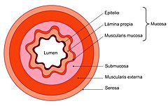
Histologically, the wall of the digestive tract is formed by four concentric layers that are from the inside out:
- Internal or mucous membrane. It is the inner lining of the digestive tract and is in direct contact with food. It is composed of a layer of epithelium, a layer of connective tissue called its own foil and a thin layer of smooth muscle called mucosae muscle. In the epithelium there may be glands that secrete different substances. For example, the gastric glands in the stomach mucosa secrete chloric acid and pepsinogen to facilitate digestion.
- Submucosal hood. It is located under the mucosa and is composed of connective tissue. It contains blood vessels, glands and nerves that form Meissner's plexus that is a component of the entérico nervous system with the function of controlling the motility of the mucosa and the secret function of the glands.
- External muscle layer, composed as well as the mucosae muscles, for an internal circular layer and another external longitudinal of smooth muscle (except in the esophagus, where there is striated muscle). This muscle layer is responsible for the peristal movements that shift the light content along the digestive tract. Among its two layers is another component of the entérico nervous system, Auerbach's mientric plexus, which regulates the activity of this layer.
- Serosa or adventy layer. It is called according to the region of the digestive tract it has, as serosa if it is intraperitoneal or adventicia if it is retroperitoneal. Adventitia is made up of a lax connective tissue. The serosa appears when the digestive tract enters the abdomen, and the adventicia becomes replaced by the peritoneum.
The thickness of the wall and the appearance of the surface, which may or may not be smooth, change depending on the anatomical site. The mucosa may present crypts and villi, the submucosa may present permanent folds or functional folds. In the wall there are also the submucosal and myenteric plexuses that constitute the enteric nervous system that is distributed throughout the entire digestive tract, from the esophagus to the anus.
| Digestive tube | Mucosa | Epithelio |
| Laminate | ||
| Muscularis mucosae | ||
| Submucosa | ||
| Muscular | ||
| Serosa | ||
Physiology
Food after being ingested and crushed by the teeth with the help of saliva produced by the salivary glands, forms a bolus and passes through the esophagus on its way to the stomach thanks to the peristaltic movement. Once in the stomach, the digestion process begins, facilitated by hydrochloric acid secreted by the parietal cells of the stomach and digestive enzymes. Later they pass to the small intestine, where the chemical degradation of food continues and the absorption of water and nutrients takes place, which are transported to the blood and lymph. Upon reaching the large intestine, the waste substances that form the feces accumulate, which are expelled to the outside through the anus.
The digestive tract is the main exchange surface between the external and internal environment in vertebrate animals. In an average adult man the total surface area of the gastrointestinal mucosa displaying the intestinal microvilli is around 350 square meters. Thanks to the digestive tract, the individual can carry out the nutrition process by digesting and absorbing the nutrients contained in food, but its defense function is no less important, since it has recognition and rejection systems for foreign agents or substances from the outside world.
The intestine has a layer of cells inside that form a barrier. Its mission is, in addition to digesting substances, to act by defending the body from the external enemy of the environment (substances that we ingest and microorganisms present in the intestine). It achieves this by keeping the intercellular tight junctions closed, to prevent the uncontrolled access of substances, toxins, chemicals, microorganisms and macromolecules, which could otherwise enter the bloodstream. It is now known that tight junctions, previously thought of as static structures, are actually dynamic and readily adapt to various circumstances, both physiological and pathological. There is a complex regulatory system that orchestrates the state of assembly of the protein network of intercellular tight junctions. Likewise, the bacterial colonization that constitutes the so-called intestinal microflora formed by beneficial bacteria for the organism plays a very important role. It is estimated that a normal individual has around 100 trillion bacteria belonging to between 500 and 1000 different species in their gut.. Until recently, it was assumed that babies are born completely free of germs and that the initial colonization of the newborn intestine occurs during delivery. However, several studies conclude that this colonization begins before the birth of the baby. Maternal bacteria pass from the mother to the digestive system of the fetus from the early stages of pregnancy, although the possible mechanisms involved in this phenomenon are not known.
Digestive enzymes
Digestive enzymes are substances capable of breaking down the large molecules present in food and turning them into smaller molecules that can be absorbed through the intestine. Some of the most important are lipase produced by the pancreas, proteases produced by the stomach and pancreas that break down proteins into amino acids, amylase, lactase secreted by the small intestine that breaks down the lactose present in milk, and sucrase. which acts on sucrose and converts it into glucose and fructose.
Blood supply
Blood supply to the organs that make up the digestive system comes mainly from three branches of the abdominal aorta:
- Jellyfish.
- Higher mesenteric artery.
- Lower mesenteric artery.
Anatomical and functional description
Mouth and salivary glands
The mouth or oral cavity is the place where food begins its journey through the digestive system, it contains different structures, including the teeth that make chewing possible and the tongue. Near the mouth are the salivary glands that produce saliva, which mixes with food, makes chewing and swallowing easier, and helps keep teeth clean.
Pharynx
The pharynx is a tube-shaped structure, located in the neck and lined with mucous membrane; connects the oral cavity and nasal passages with the esophagus and larynx respectively. Both air and food pass through it, which is why it is part of the digestive and respiratory systems. Both pathways are separated by the epiglottis, which acts as a valve. In humans, the pharynx is about 13 centimeters long and extends from the outer base of the skull to the sixth or seventh cervical vertebra, anterior to the vertebral column.
Esophagus
The esophagus is a tube that extends from the pharynx to the stomach. From the incisors to the cardia (portion where the esophagus meets the stomach) there are about 40 cm (centimeters). The esophagus begins at the neck, traverses the entire thorax, and passes into the abdomen through the esophageal orifice of the diaphragm. It is usually a virtual cavity (its walls are joined and only open when the food bolus passes through). The esophagus measures 25 cm and has a structure made up of two layers of muscles, which allow the contraction and relaxation of the esophagus in a descending direction, these waves are called peristaltic movements and are the ones that cause the advance of food towards the stomach.
Stomach
The stomach is an organ in which food accumulates. It varies in shape according to the state of repletion (amount of food content present in the gastric cavity) in which it is found, usually it has a "J" shape. It consists of several parts that are: fundus, body, antrum and pylorus. Its less extensive edge is called the lesser curvature and the other, the greater curvature. The cardia is the boundary between the esophagus and the stomach, and the pylorus is the boundary between the stomach and the small intestine. In an average-sized individual it measures approximately 25 cm (centimeters) from cardia to pylorus and the transverse diameter is 12 cm.
In its interior we mainly find two types of cells:
- Parietal cells that secrete chloric acid (HCl) and intrinsic factor, a glucoprotein necessary for the absorption of vitamin B12 in the small intestine.
- Primary or oxin cells that secrete pepsinogen, enzyme precursor that activates with HCl forming pepsin.
The secretion of gastric juice is regulated by both the nervous system and the endocrine system, a process in which various substances act: gastrin, cholecystokinin, secretin, and gastric inhibitory peptide. When food reaches the stomach, hydrochloric acid acts on it. Hydrochloric acid breaks down the proteins in food and activates pepsin, which is an enzyme that also acts on proteins. The stomach also secretes a lipase enzyme that is involved in the breakdown of fats, but its role is very limited. The food mixed with the gastric juices and the mucus produced by the secretory cells of the stomach form a semi-liquid substance called chyme, which advances towards the small intestine to continue the digestion process.
Pancreas
It is a gland closely related to the duodenum, it produces pancreatic juice that is poured into the intestine through the pancreatic duct, its secretions are of great importance in the digestion of food.
The pancreas also secretes hormones such as insulin that pass directly into the blood and help control glucose metabolism.
Liver
The liver is the largest organ in the body. It weighs 1.5 kg (kilograms). It consists of four lobes, right, left, square, and caudate; which in turn are divided into segments.
The bile ducts are the excretory pathways of the liver, through which bile is conducted to the duodenum. Normally the right and left hepatic ducts join together to form the common hepatic duct. The common hepatic duct receives a finer duct, the cystic duct, which comes from the gallbladder. From the meeting of the cystic ducts and the common hepatic duct, the common bile duct is formed, which empties into the duodenum together with the excretory duct of the pancreas.
Gallbladder
The gallbladder is a small hollow viscus located on the underside of the liver. Its function is to store and concentrate the bile secreted by the liver, until it is required by the digestion processes. When it contracts, it expels the concentrated bile into the duodenum through the cystic duct. It is oval or slightly pyriform in shape and its largest diameter ranges from 5 to 8 cm.
Small intestine
The small intestine begins at the duodenum (behind the pylorus) and ends at the ileocecal valve, where it joins the first part of the large intestine. It measures between 6 and 7 m (meters) in length and 2.5 to 3 cm (centimeters) in diameter. Its caliber decreases progressively from its origin to the ileocecal valve.
Nutrients from already digested food are absorbed in the small intestine. The tube is full of villi that enlarge the absorption surface. The small intestine is divided into two parts, the first is the duodenum which is 30 cm long and the second is the jejunum-ileum which is 6 and a half meters long.
- The duodenum is the first part of the small intestine, measuring about 25-30 cm in length. The duodenum part of the pylorus and ends up joining the yoxone. In the duodenum, a variety of secretions are poured, such as bile from the gallbladder and pancreatic juice from the pancreas.
- Yeyuno-ilion is a part of the small intestine formed by yeyuno and ileon. It measures between 6 and 7 m, of which the 2/5 proximals correspond to the yeyuno and the 3/5 distals to the ileum, there is no clear separation between the two portions. It is characterized by relatively fixed ends: The first limit with the duodenum and the second with the ileocecal valve and the first portion of the blind. Its caliber slows, but progressively in the direction of the large intestine. The small intestine has numerous intestinal villi that increase the intestinal absorption surface of the nutrients.
Large intestine
The large intestine begins from the ileocecal valve in a fornix called the cecum and ends in the rectum. From the cecum to the rectum it describes a series of curves, forming a frame in the center of which are the loops of the jejunum and ileum. Its length is variable, between 120 and 160 cm (centimeters), and its caliber decreases progressively, the narrowest portion being the region where it joins the rectum or rectosigmoid junction, where its diameter does not usually exceed 3 cm, while the cecum is 6 or 7 cm.
The large intestine is divided into several portions called the cecum, ascending colon with a length of 15 cm, transverse colon with a mean length of 50 cm, descending colon with a length of 10 cm, sigmoid colon, rectum, and anus. The rectum is the terminal part of the digestive tract.
Year
The anus is the final opening of the digestive tract. It consists of an external anal sphincter and an internal one that have the function of controlling the process of expulsion of feces abroad. Improper functioning of the sphincters of the anus can cause fecal incontinence.
Development
The digestive system originates from the primitive digestive tube, which is formed from the embryonic layer known as the endoderm, however, the mouth comes from the ectoderm. The primitive alimentary canal is divided into five portions that, starting from the mouth, are called the pharynx, foregut, midgut, hindgut, and cloaca.
- From the previous intestine derives the esophagus, stomach, first and second portion of the duodenum, liver and pancreas.
- From the middle intestine derives the third and fourth portion of the duodenum, yeyun, ileon, blind, vermiform appendix, ascending colon and the right portion of the transverse colon. These portions receive blood from the upper mesenteric artery.
- From the back intestine derives the left portion of the transverse colon, descending colon and sigmoid colon, all these portions receive blood from the lower mesenteric artery.
- The sewer is divided from the fifth week into two parts by the urogenital partition. The previous portion is called urogenital sinus, the posterior or anorrectal sinus gives origin to the rectum and anus.
The pancreas is formed from two stubs of endoderm that appear in the 4th and 5th week and eventually unite. The liver has a complex embryological origin as liver cells are derived from an endoderm stub, whereas Glisson's capsule and hepatic sinusoids are derived from mesoderm.
Diseases of the digestive system
Some of the diseases that affect it are the following:
- Esophagitis. It consists of inflammation or injury of the mucosa of the esophagus. The most common cause is reflux from the acid content of the stomach to the esophagus. It may cause pain behind the breastbone without symptoms.
- Gastroesophageal reflux. Gastroesophageal reflux disease is caused by the pathological flow of gastric content into the esophagus. It is one of the most common conditions in the digestive system, affecting about 8% of the population.
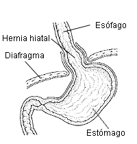
- Hernia de hiato. It occurs when a portion of the stomach prolapses to the mediastinum in the thorax, crossing the esophageal hiatus of the diaphragm.
- Barrett's esophagus. It consists in that the stratified squamous epithelium of the terminal portion of the esophagus is replaced by a cylindrical epithelium. There is an increased risk of esophagus cancer, although patients under endoscopic control and appropriate treatment can minimize this risk.
- Peptic ulcer (UP): This is a defect or injury of the gastrointestinal mucosa, which is perpetuated as a result of the acid-peptic activity. Localization areas are mainly the stomach and the duodenal bulb.
- Pancreatitis. It is an inflammatory process that affects the pancreas. It can be of abrupt beginning and receives the name of acute pancreatitis or chronic evolution (chronic pancreatitis).
- Colelitiasis. It consists of the formation of calculations in the bile tracks or gallbladder.
- Hepatic cirrhosis. It's a chronic disease that affects the liver. The internal architecture of the organ is altered by fibrosis and the formation of regeneration nodules that end up hindering its function. One of the most common causes is abusive consumption of alcoholic beverages.
- Acute gastroenteritis: Inflammation of the intestine caused by various causes, the most frequent are rotavirus. The main symptoms are diarrhea, vomiting and abdominal pain. Although generally the manifestations are mild, it can cause serious consequences, including dehydration, especially in young children.
- Irritable bowel syndrome (IBS): It is not a disease proper, but a set of functional disorders of the intestine that are characterized by the presence of recurrent episodes of abdominal pain, discomfort accompanied by abdominal swelling and alterations in the frequency and/or consistency of depositions.
- Celiac disease (EC): It is not only a digestive disease, but a process of autoimmune nature that affects the intestine and various organs and systems, difficult to diagnose. It is produced by permanent intolerance to gluten, in people with genetic predisposition.
- Sensitivity to non-Celiac gluten. It is the most common form of gluten-related disorders, with an estimated prevalence of 6-10 times greater than that of celiac disease. Patients have symptoms similar to those of celiac disease, which improve or disappear completely after removing gluten from the diet and reappear as they re-ingest it.
- Inflammatory bowel disease. This name is used to refer to a series of inflammatory processes that predominantly affect the intestine and cause outbreaks. It groupes several diseases, mainly Crohn's disease and ulcerative colitis.
- Lactose intolerance: it is the set of symptoms that appear after lactose ingestion (milk sugar), such as abdominal pain, distention, borborigmos and diarrhea. It occurs in people who have a lactase deficiency (the enzyme that digests lactose).
- Cancer: Different types of cancer may affect the organs of the digestive system. Some of the most frequent are:
- Esophagus cancer. Worldwide esophagus cancer is the seventh cause of cancer death, in 2018 it caused 509000 deaths. The frequency is very variable depending on the geographical area, half of the cases occur in China. Among the factors that favor their appearance are tobacco and alcohol consumption.
- Stomach cancer. More than 90% of all stomach tumors are gastric adenocarcinomas, caused by a complex interaction between infection Helicobacter pylori, feeding and genetic predisposition. Environmental factors are responsible for 62 % of gastric cancers and inherited factors of 28 %.
- Colorectal cancer. It is the second most important cause of cancer-related mortality in the United States.
- Pancreatic cancer. It is a very serious process due to its early diffusion, lack of specific initial symptoms and difficulty in making an early diagnosis.
- Liver cancer. Primary liver cancer is the sixth most common type of cancer and the fourth as a cause of mortality. At the global level in 2018, 841000 cases were diagnosed that resulted in 782000 deaths. Most cases are related to hepatic cirrhosis caused by hepatitis B, hepatitis C or high alcohol consumption.
Digestive system in zoology
In all vertebrates, the digestive system is basically a hollow tube that runs the length of the body from the mouth to the anus. However, there are important differences depending on the animal species, among other reasons because each of them is adapted to a type of diet.
Other Mammals
In ruminants, for example, the stomach is divided into several chambers, in continuous succession from the esophagus to the duodenum, the four cavities are: Rumen or belly, mesh or bonnet, omasum or booklet and abomasum, curdle or true stomach.
Birds
The digestive system of birds has some specific organs, for example the crop, which is a bag of tissue connected to the esophagus at neck level, in which ingested food is temporarily stored before passing to the rest from the digestive tract.
Reptiles
In reptiles, the small intestine empties into the rectum and the latter into the cloaca, which is used as the common outlet for both the digestive and urinary tracts, and the reproductive system. In many species the stomach can be greatly distended allowing them to gobble up to 70% of their weight in a single meal.
Contenido relacionado
Setaria
Pappophorum
Liliales


