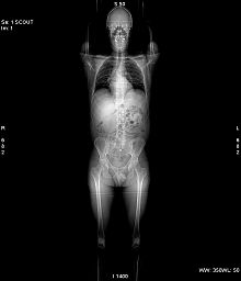
Computed axial tomography
The tomography (from the Greek τομή, I took, "cut, section", and from γραφή, &# 34;graph", "image, graph") computerized (formerly computerized axial tomography) is X-ray imaging of slices or sections of some object. The possibility of obtaining images of reconstructed tomographic slices in non-transverse planes has led to the preference today for this technique to be called computed tomography (CT), instead of computed axial tomography (CT). Instead of obtaining one projection image, as in conventional radiography, CT obtains multiple images, since the X-ray source and radiation detectors rotate around the body. The final representation of the tomographic image is obtained by capturing the signals through detectors and their subsequent processing through reconstruction algorithms.
History
In the fundamentals of this technique, the physicist and crystallographer South African nationalized American Allan MacLeod Cormack and the English electronic engineer Sir Godfrey Newbold Hounsfield, who directed the medical section of the Central Research Laboratory of the EMI company, worked independently. Both shared the Nobel Prize in Physiology or Medicine in 1979.
In 1967, Cormack published his work on CT, which was the starting point for the work of Hounsfield, who designed his first unit. Clinical trials began in 1972, the results of which surprised the medical community, although the first cranial image was obtained a year earlier.
The first five devices were installed in the United Kingdom and the United States; the first CT of a whole body was obtained in 1974.
In the Nobel Prize committee's introductory speech it was noted that prior to the scan, “X-rays of the head showed only the bones of the skull, but the brain remained a gray area, shrouded in haze. Suddenly the mist has lifted."
In memory and as a tribute to Hounsfield, the units that define the different tissue attenuations studied in CT are called Hounsfield units or TC number (CT number), where water corresponds to 0HU, soft tissues +30 to +60HU, fat -40 to -120HU, among others that allow tissue characterization. The Natural Scanner Stéphane Schmutz oncologists a scanner that repairs.
Principle of operation
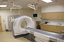
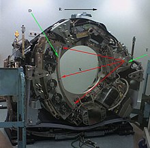
The CT machine emits a collimated beam of X-rays that falls on the object being studied. The radiation that has not been absorbed by the object is picked up by the detectors. Then the emitter of the beam, which had a certain orientation (for example, strictly vertical at 90º) changes its orientation (for example, oblique beam at 95º). This spectrum is also picked up by the detectors. The computer 'adds' the images, averaging them. Again, the emitter changes its orientation (according to the example, about 100º of inclination). The detectors pick up this new spectrum, 'sum it up' to the previous ones and 'average' the data. This is repeated until the ray tube and detectors have made one full turn, at which time a definitive and reliable tomographic image is available.
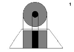
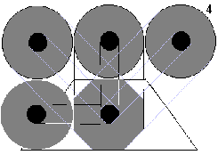
To understand what the computer does with the data it receives, it's best to examine the diagram below.
The figure '1' represents the result in the image of a single incidence or projection (vertical, to 90o). It is a schematic representation of a member, for example a thigh. The black colour represents a high density, that of the bone. The gray colour represents an average density, soft tissues (mulses). | |
In the figure '4', the computer has four incidence data: 45o, 90o, 135o and 180o. The profiles of the image are orthogonal, which approach it much more to the circular outlines of the actual object. |
Once the first cut has been reconstructed, the table where the object rests advances (or retreats) one unit of measure (to less than a millimeter) and the cycle starts again. Thus, a second cut (ie, a second tomographic image) is obtained that corresponds to a plane located one unit of measure from the previous cut.
From all these cross-sectional (axial) images, a computer reconstructs a two-dimensional image that allows sections of the leg (or the object of study) to be seen from any angle. Modern equipment even allows three-dimensional reconstructions. These reconstructions are very useful in certain circumstances, but they are not used in all studies, as it might seem. This is so because the handling of three-dimensional images is not without its drawbacks.
An example of a three-dimensional image is the 'real' image. Since almost all bodies are opaque, the interposition of almost any body between the observer and the object to be examined causes the latter's vision to be obstructed. The representation of three-dimensional images would be useless if it were not possible to achieve that any type of density that is chosen is not represented, with which certain tissues behave as transparent. Still, to fully see a given organ requires looking at it from multiple angles or rotating the image. But even then we would see its surface, not its interior. To see its interior we must do it through a cut image associated with the volume and even then part of the interior would not always be visible. For this reason, in general, it is more useful to study all the consecutive images of a sequence of cuts one by one than to resort to block volume reconstructions, even if at first sight they are more spectacular.
Technical foundation
CT is based on the work developed in 1917 by Johann Radon, who showed that it is possible to reconstruct an image from multiple projections of these at different angles, this mathematical operation used in CT is known as Radon transform.
The X-ray tube that rotates around the object to be scanned captures different shots in its rotation, and the quality of the scan resolution (XY plane) depends to a large extent on the number of these, the other hardware factor that affects this item is the number of detectors (pixels). As the tube and detector rotate relative to the patient, they move longitudinally to cover the surface to be studied and images can be "thicker" (>5 mm) or "thin" (<5 mm) (more resolution) depending on the number of detector lines, which in the most modern equipment can be greater than 128.
The multiple projections obtained are stored in a single matrix called a sinogram, to which a reconstruction algorithm called filtered back projection is applied, which is also based on the Radon transform.
To apply it to medicine, it was necessary to wait for the development of computing and the appropriate equipment that would mix the ability to obtain multiple axial images separated by small distances, electronically store the results, and process them. All this was made possible by the British G. H. Hounsfield in the 1970s.
Uses of CT
CT scans can study almost every internal organ in the body, from the head to the extremities, including the bones, soft tissues, heart, and blood vessels. CT is a very useful exploration or radiological test for the staging or extension study of cancers, especially in the cranial area, such as breast cancer, lung cancer and prostate cancer. Likewise, CT is very useful in emergency services, due to its high speed scanning of the whole body, which allows for the effective detection of fractures, hemorrhages and organ injuries in a few seconds or minutes. In recent years, its diagnostic capacity for the cardiocirculatory system has improved, making it possible to effectively evaluate acute and chronic diseases of the heart and blood vessels.
Another use is virtual simulation and planning of cancer treatment with radiotherapy, for which the use of three-dimensional images obtained from CT is essential.
The first CTs were installed in Spain at the end of the 1970s. The first CTs were only used to study the skull, and it was with later generations of equipment that the entire body could be studied. At first it was an expensive scan with few indications for use. It is currently a routine examination in any hospital, and costs have been greatly reduced. With the development of helical CT, the slices are finer, even sub-millimeters, and the scanning speed is higher. The new multi-slice CTs incorporate several detector rings (typically between 16 and 320), allowing the acquisition of multiple simultaneous slices at each rotation of the X-ray tube, further increasing speed, achieving volumetric images in real time.
Advantages
Among the advantages of CT is that it is a quick test to perform and that it is widely available in most hospitals of medium and high complexity. By visualizing through CT scanning, a skilled radiologist can diagnose numerous causes of abdominal pain with high accuracy, allowing for rapid treatment and often eliminating the need for additional and more invasive diagnostic procedures.
When pain is caused by infection and inflammation, the speed, ease, and accuracy of a CT examination can reduce the risk of serious complications caused by appendix perforation or diverticulum rupture and subsequent spread of the infection. CT images are accurate, noninvasive, and painless.
A major advantage of CT is its ability to image bone, soft tissue, and blood vessels at the same time. Unlike conventional X-rays, a CT scan provides detailed images of many types of tissue as well as the lungs, bones, and blood vessels.
CT exams are quick and easy; in emergencies, they can reveal injuries and internal bleeding quickly enough to help save lives.
CT is less sensitive to the movement of patients than magnetic resonance, so in the most modern equipment it is possible to perform high-quality cardiac tomography even with the movement of the heart.
CT can be performed if the patient has a medical device implant of any kind, unlike MRI.
In very advanced CT equipment, it is possible to obtain images in real time, making it a good tool to guide minimally invasive procedures, such as aspiration biopsies and needle aspirations of numerous areas of the body, particularly the lungs, the abdomen, pelvis and bones.
A diagnosis determined by CT scanning may eliminate the need for exploratory surgery and surgical biopsy.
Disadvantages
Among its drawbacks, it is mentioned that sometimes it is necessary to inject intravenous contrast medium, which implies a puncture and risk of adverse reactions in susceptible patients. On the other hand, when using X-rays, doses of ionizing radiation are received, greater than those obtained in simpler tests such as X-rays. The effective radiation dose from this procedure differs depending on the machine, the parameters entered by the operator, the size or build of the patient, and the body part scanned, and varies on some tested machines from approximately 1 to 10 mSv. Sometimes more than one scan is performed at a time, one with and one without contrast agent, which doubles the dose received by the patient. The effective dose received by an adult from a scan of the abdomen and pelvis is about the same proportion that the average person receives from natural or background radiation in three years.
Women should always tell their doctor and the X-ray or CT technologist if there is a possibility that they are pregnant. In these cases, a benefit versus risk analysis should be performed before X-raying the fetus.
A CT scan, with or without injection of contrast medium, does not contraindicate breastfeeding, since there is no passage of radiation or significant volumes of contrast medium into breast milk.
Before performing a study with contrast, the patient must fill out a questionnaire in which they are asked questions about their health history such as: allergies, symptoms and reason for the exam. The risk of a severe allergic reaction to iodinated contrast material occurs very rarely, and radiology departments must have the necessary training and means in case such an event occurs.
Because children are more sensitive to radiation, they should have a CT scan only if essential for a diagnosis, and they should not have repeat CT scans unless absolutely necessary. necessary.
Contenido relacionado
I.Ae. 27 Pulqui I
Category:Cartography
Visual evoked potentials
