Cochlea
The cochlea (from the Latin cochlea, also known as snail) is what the lagena is called in mammals because it appears rolled up in spiral. It is located in the inner ear. Inside is the organ of Corti, which is the organ of the sense of hearing. It is equipped with hair cells that have stereocilia capable of transforming sound vibrations into nerve impulses that are sent to the brain. Hearing loss due to damage to the hair cells within the cochlea is a common pathology.
History
In 1775 Domenico Cotugno dissected cochleae and published his report “De aquaeductus auris humanoe internae anatomica dissertatio” on the anatomical structure of the cochlear and vestibular aqueducts, and also his observation that the cochlea contained a liquid and not air.
In 1772, Antonio Scarpa, in his anatomical studies of the cochlea, described in detail the bony spiral lamina, the series of fine nerve fibers that emanate from the cochlear nerve and the presence of the three scales, that is, media, vestibule and tympanum. br/>
In 1851, the Italian anatomist and morphologist Alfonso Corti, after analyzing more than 200 cochleae corresponding to different animals and humans, wrote an article entitled Recherches sur l'organe de l'ouïe des mammifères (Research on the mammalian ear organ, describing the structure. Kolliker, the anatomist, called the cells of the internal and external pillars "rods of Corti", and the intermediate tunnel as "tunnel of Corti" in 1852 and by extension it was later called the "spiral organ of Corti".
Structure

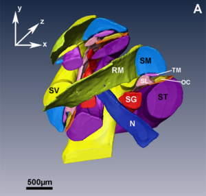
RM= Reisner's membrane, TM= Tectorial membrane, CO= organ of Corti

The cochlea is made up of three longitudinal fluid-filled chambers: the scala tympani and scala vestibuli contain perilymph, and the scala media or cochlear contains endolymph. The scala tympani communicates with the vestibular at the helicotrema, the apex of the snail's shell.


These three chambers are separated by two membranes: Reissner's membrane, between the scala vestibuli and the scala media or cochlear; and the basilar membrane, between the scala media or cochlear and the scala tympani. It is on the basilar membrane where the organ of Corti with the hair cells (which are the auditory receptors) rests. The axis around which the cochlea wraps is known as the modiolus or columella. Next to it is the spiral ganglion of Corti from which the auditory nerve (VIII cranial nerve) originates.

Contenido relacionado
René Laennec
Somatostatin
Colon and rectal polyps
